Fundamentals of Biochemistry - A. A. Anisimov 1986
Carbohydrates
Carbohydrate transformations associated with respiration and fermentation
6.9.1. Glycolysis. Stages and Reactions of Glycolysis. Glycolysis is an anaerobic process resulting in The breakdown of one glucose molecule into two molecules of lactic acid. This process releases energy, which the Organism stores in the form of ATP. The reactions of glycolysis take place in the Cytosol without the consumption of oxygen. Under anaerobic conditions, glycolysis serves as the sole energy-supplying pathway in animals, plants, and many Bacteria.
During the aerobic degradation of glucose, one of the End products of glycolysis—pyruvic acid—is oxidized to CO2 and H2O in The Tricarboxylic Acid Cycle (see Section 6.9.5). The reactions of this cycle occur in the Cell/35.html">Mitochondria (or corresponding Membrane structures in bacteria) with the participation of oxygen.
The complete chain of glycolytic reactions was elucidated through the work of L. A. Ivanov, S. P. Kostychev, A. N. Lebedev, G. Embden, J. O. Parnas, and O. Meyerhof by the mid-1930s. These reactions are catalyzed by a group of eleven well-characterized Enzymes that are easily extracted from Cells and catalyze reactions in vitro. Glycolysis proceeds in two stages.
The First stage is the preparatory, or capture, phase. At this stage, various hexoses are funneled into glycolysis. Although glucose is primarily oxidized during glycolysis, other hexoses such as fructose and mannose can also enter the pathway. In the process, the inert hexose molecules are activated and phosphorylated at the expense of ATP, converting them into glucose-6-phosphate. This phase culminates in the Formation of glyceraldehyde-3-phosphate. The crucial role of phosphoric acid in the anaerobic breakdown of glucose was first established in the studies of Russian biochemists L. A. Ivanov (1901, 1906) and A. N. Lebedev (1905).
The Second Stage is the oxidative phase. Glyceraldehyde-3-phosphate is oxidized to pyruvic acid (Pyruvate) or lactic acid (lactate). The energy of oxidation is captured in ATP, and reducing equivalents of NADH are generated.
The diagram (Fig. 6.12) illustrates the complete pathway of glycolytic reactions. It begins with the phosphorylation of D-glucose using ATP. This is the initial triggering reaction of glycolysis and one of its key regulatory steps. The neutral D-glucose molecule is converted into negatively charged glucose-6-phosphate:
Class="center">
This reaction is catalyzed by Two Types of enzymes that differ in their sugar Specificity: hexokinase and glucokinase. Hexokinase plays the primary role, functioning in the vast majority of cells. It is capable of catalyzing the phosphorylation not only of D-glucose but also of many other hexoses, such as D-fructose, D-mannose, and D-glucosamine.
Glucokinase phosphorylates exclusively D-glucose and possesses a lower affinity for it than hexokinase. Glucokinase is found in The Liver and becomes active only when Blood glucose concentrations are very high. Both Kinases require divalent cations (Mg2+ or Mn2+) that bind to ATP to form the true substrates, MgATP2- or MnATP2-. The hexokinase reaction is accompanied by a significant decrease in Free energy, which ensures the irreversibility of this reaction within The Cell. The reaction rate is inhibited by its product, glucose-6-phosphate.
The second reaction is the isomerization of glucose-6-phosphate into fructose-6-phosphate:

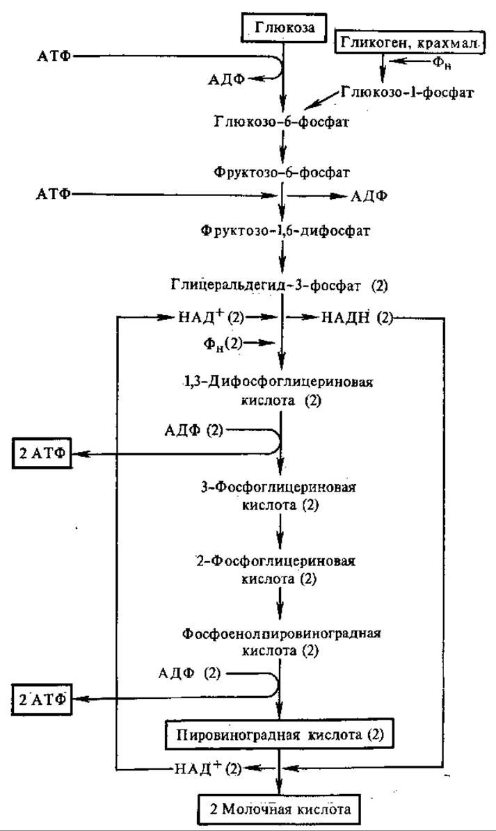
Fig. 6.12. Reactions of glycolysis.
The initial substrates and end products of glycolysis are enclosed in boxes; numbers in parentheses indicate the number of molecules
The third reaction is the phosphorylation of fructose-6-phosphate to yield fructose-1,6-diphosphate. This is the second triggering reaction of glycolysis, which consumes a second molecule of ATP:

The reaction is irreversible due to a substantial decrease in free energy. It is mediated by the enzyme Phosphofructokinase in the presence of Mg2+ ions, which are apparently required to form the true substrate, i.e., MgATP2-. This is the slowest step in glycolysis, determining the overall rate of the process, and serves as the primary site of Metabolic Regulation. Phosphofructokinase is an allosteric, or regulatory, enzyme. It is inhibited by ATP and citric acid, and activated by ADP and AMP. During cellular rest, when the ATP/ADP ratio is high, The activity of phosphofructokinase drops, slowing down glycolysis. During functional cellular activity, The amount of ATP decreases, and glycolysis is accelerated.
The fourth reaction—the Cleavage of fructose-1,6-diphosphate—represents a reversed aldol Condensation (see Section 6.5.1):

The fifth reaction is the isomerization of triose phosphates. Of the resulting triose phosphates, only one—glyceraldehyde-3-phosphate—feeds directly into the subsequent steps of glycolysis. Dihydroxyacetone phosphate is converted into glyceraldehyde-3-phosphate in a reversible reaction:
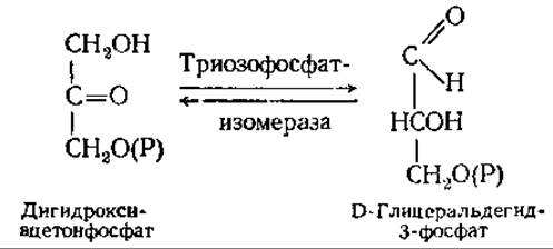
The formation of phosphotrioses marks the completion of the first stage of glycolysis. Through two phosphorylation steps and a cleavage, a single glucose molecule has been transformed into two molecules of phosphotriose.
The second stage of glycolysis comprises redox and phosphorylation reactions that generate ATP. At this stage, both molecules
of phosphotriose—representing the two halves of the original glucose molecule—are oxidized. Therefore, in all subsequent reactions, a coefficient of 2 should precede the substrate formula.
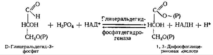
The sixth reaction, which is the central event of glycolysis, is a redox process. The aldehyde group of glyceraldehyde-3-phosphate is oxidized to the COOH group of 1,3-diphosphoglyceric acid, while NAD+ is reduced to NADH. This releases energy, which drives the Formation of the high-energy bond in 1,3-diphosphoglyceric acid. Thus, this reaction achieves substrate-level phosphorylation. The overall reaction equation is as follows:
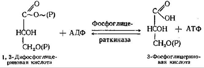
The seventh reaction: the energy-rich phosphoryl group of 1,3-bisphosphoglycerate (at the C-1 position) is transferred to ADP, yielding ATP;
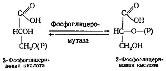
The eighth reaction: the phosphate group of phosphoglycerate is shifted from position 3 to position 2:
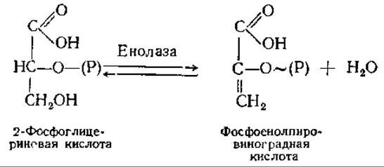
The ninth reaction involves an intramolecular redox process: the oxidation state of C-2 in 2-phosphoglycerate increases, while that of C-1 decreases. This results in the formation of phosphoenolpyruvate, a high-energy compound. Thus, substrate-level phosphorylation also occurs here. Formally, the reaction represents the elimination of a Water molecule:
The tenth reaction: The transfer of the phosphoryl group, along with its high-energy bond, from phosphoenolpyruvate to ADP:
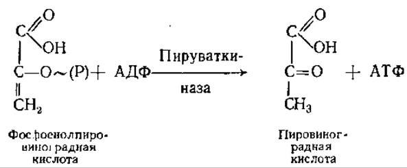
This reaction is strongly exergonic, making it virtually irreversible within the cell.
In the eleventh reaction, pyruvate (PA) is reduced to lactate in the presence of NADH:
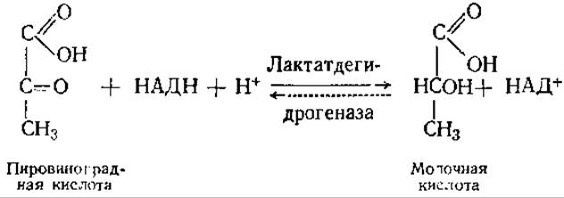
The equilibrium strongly favors the formation of lactic acid. The coenzyme NAD Functions as an electron carrier. In glycolytic reactions, its electron donor is glyceraldehyde 3-phosphate (reaction 6). If the NADH produced during The oxidation of glyceraldehyde 3-phosphate were not oxidized, glycolysis would grind to a halt because all available NAD+ would be reduced to NADH. This is prevented by the action of Lactate dehydrogenase.
The eleventh reaction brings The Glycolytic Pathway to a close. At this stage, glycolysis is regulated by lactate dehydrogenase Isoenzymes (see Section 3.8). Lactic acid represents a metabolic dead end of sorts. Under aerobic conditions, it can only be converted back into pyruvate by reversing the lactate dehydrogenase reaction. Under anaerobic conditions, lactic acid serves as the final product of glycolysis, such as in Muscles during intense physical exertion. In this scenario, large amounts of lactic acid are released from the muscles into the bloodstream. In the liver, and partially within the muscles themselves, roughly one-fifth of this lactic acid is oxidized to CO2 and H2O during the aerobic phase of Respiration. The remaining four-fifths are resynthesized into Glycogen.
The NADH produced in the Cytoplasm during glycolysis cannot cross the mitochondrial membrane. Under aerobic conditions, it transfers its electrons and protons to the mitochondrial Respiratory Chain (see Section 7.2) via the glycerophosphate shuttle. In the cytoplasm, NADH reacts with dihydroxyacetone phosphate to form glycerol 3-phosphate, which easily crosses the mitochondrial membrane. This reaction is catalyzed by cytoplasmic glycerol 3-phosphate dehydrogenase (NAD+-dependent):
Dihydroxyacetone phosphate + NADH + H+ ⇄ Glycerol 3-phosphate + NAD+
Inside the mitochondrion, glycerol 3-phosphate is oxidized back to dihydroxyacetone phosphate, this time mediated by a flavin enzyme—mitochondrial glycerol 3-phosphate dehydrogenase:
Glycerol 3-phosphate + FAD Dihydroxyacetone phosphate + FADH2
The coenzyme FADH2 feeds electrons and protons into the respiratory chain. Here, a pair of H atoms yields only two ATP molecules, rather than three, as is typical when a pair of electrons enters the respiratory chain via NADH. The glycerophosphate shuttle provides a unidirectional electron transport mechanism exclusively into the mitochondria. It is particularly prominent in liver cells and insect flight muscles. In those same liver cells, as well as in other Tissues, the malate-aspartate shuttle operates reversibly, transporting hydrogen both into and out of mitochondria. In this mechanism, cytoplasmic NADH reduces oxaloacetate to malate, which readily crosses into the mitochondrion, where it is oxidized back to oxaloacetate, transferring hydrogen to the respiratory chain.
Energy balance of glycolysis. During glycolysis, each 6-carbon D-glucose molecule is split into two 3-carbon molecules of lactic acid. In the initial phase of glycolysis, two ATP molecules are consumed to activate the substrate (via the hexokinase and phosphofructokinase reactions). In the second phase, ATP is generated in two Reactions Catalyzed by phosphoglycerate kinase and pyruvate kinase. Since each glucose molecule yields two molecules of glyceraldehyde 3-phosphate, a total of four ATP molecules are produced. However, because two are consumed in The first phase, the net yield of glycolysis is only two ATP molecules per glucose molecule. The efficiency of this process is easy to calculate. The energy released is:
Glucose → 2 Lactic acid + 2H+; ∆G0' (pH 7) =
= − 196 kJ ∙ mol-1
This energy is captured in two ATP molecules:
ADP + H3PO4 ATP + H2O; ∆G0' (pH 7) =
= + 34.5 kJ ∙ mol-1; 34.5 kJ∙mol-1 × 2 = 69 kJ ∙ mol-1
Thus, the energy efficiency of glycolysis, or its yield, is approximately 35%. Consequently, only one-third of the energy released during the oxidative breakdown of glucose to lactic acid is stored by the organism in the high-energy bonds of ATP.
The primary role of glycolysis as the anaerobic phase of respiration lies in extracting free energy from respiratory substrates (CARBOHYDRATES) and accumulating it in an easily usable form—ATP molecules. Additionally, glycolysis reactions yield numerous highly reactive compounds that are utilized in Various metabolic pathways. The Significance of glycolysis is especially profound in tissues and Organs where oxygen availability is limited or where sudden and sharp surges in ATP consumption may occur. For instance, in The cardiac Muscle, The rate of ATP consumption can increase 10-fold, and in active Skeletal Muscle, more than 100-fold. In such cases, the demand for ATP is partially met by the aerobic phase of respiration, but this is constrained by oxygen supply. Under these circumstances, skeletal muscles (and to a lesser extent, cardiac muscles) acquire supplementary amounts of ATP through glycolysis.
In plants, glycolysis occurs in certain tissues even under sufficient oxygen supply. Such single-phase "Anaerobic respiration" (or "aerobic Fermentation") is particularly characteristic of the embryonic tissue of seeds and the inner tissues of plant fruits due to oxygen deficiency within them. Glycolysis serves as the main catabolic pathway of glucose in homoermentative lactic acid bacteria and Yeasts.
The glucose residues of glycogen can also undergo anaerobic breakdown, a process known as Glycogenolysis. The incorporation of glycogen into the anaerobic degradation pathway is carried out by three enzymes. Glycogen phosphorylase and amylo-1,6-glucosidase (isoamylase) break down glycogen into glucose-1-phosphate. Subsequently, phosphoglucomutase converts the latter into glucose-6-phosphate, which enters the standard glycolytic pathway. Because glycogenolysis does not consume ATP to form glucose-6-phosphate, three molecules of ATP are accumulated per glucose molecule rather than two. However, this does not imply that glycogenolysis is energetically more advantageous than glycolysis, since ATP is consumed during the synthesis of glycogen from glucose.
6.9.2. Fermentations. Many microorganisms (yeasts, Molds, bacteria) derive energy through fermentation. This is the most primitive mechanism of energy generation. Various Types of fermentation share many stages with glycolysis, a fact established by Academician S. P. Kostychev (1907–1912). One of the end products of glycolysis—pyruvic acid (PA)—is the starting compound for both the aerobic breakdown of glucose and various fermentation processes (Fig. 6.13). The subsequent conversion reactions of pyruvic acid differ across various microorganisms, leading to the formation of characteristic fermentation products such as alcohols and acids.
The fundamental difference between fermentations and the anaerobic breakdown of glucose versus aerobic respiration is that in fermentation, The Role of the final oxidant—the electron and hydrogen acceptor—is played not by oxygen, but by various Organic compounds. Fermentation and the anaerobic degradation of carbohydrates are internal oxidation-reduction processes that result in energy storage and the regeneration of oxidized NAD+. This regeneration is absolutely essential for the continuation of glycolysis and fermentation, as the intracellular pool of NAD+ is limited.

Fig. 6.13. Possible pathways of pyruvic acid conversion:
TCA — tricarboxylic acid cycle; products of potential pyruvate conversions are enclosed in frames
Yeasts, particularly strains of Saccharomyces cerevisiae, are characterized by Alcoholic Fermentation, during which sugars are broken down with a high yield of ethanol and carbon(IV) oxide: С6Н12О6 = 2СО2 + 2С2Н5ОН. The conversion of pyruvic acid into the end products of alcoholic fermentation involves two reactions:

A characteristic feature of this reaction is its complete irreversibility. Pyruvate decarboxylase requires Mg2+ and the coenzyme Thiamine diphosphate (TDP).

Another common type of fermentation is Lactic acid fermentation. Homofermentative lactic acid fermentation produces lactic acid from glucose with a nearly 100% yield. The reactions of this process are identical to those of glycolysis. In heterofermentative (mixed) lactic acid fermentation, besides lactic acid, significant quantities of other products are formed, including acetic acid, ethanol, and carbon(IV) oxide. The chemistry of this process is entirely different, being based on the oxidation of glucose via the Pentose Phosphate Pathway (see Section 6.9.3).
Lactic acid fermentation takes place during the ensiling of forage for livestock and the pickling of cabbage. The resulting lactic acid prevents the growth of putrefactive bacteria and molds, thereby acting as a preservative.
Propionic acid is a product of carbohydrate fermentation by propionic acid bacteria. Alongside it, acetic acid and carbon(IV) oxide are formed. For every molecule of pyruvic acid oxidized to acetic acid and CO2, There are two molecules of pyruvic acid converted into propionic acid.
Acetic acid bacteria (genus Acetobacter) oxidize ethanol to acetic acid. These bacteria are also capable of oxidizing other alcohols, including saturated polyhydric alcohols. This phenomenon is termed incomplete oxidation.
6.9.3. The Pentose Pathway, or Pentose Phosphate Cycle of Carbohydrate Oxidation. In the 1930s, the research of V. A. Engelhardt, O. Warburg, F. Lipmann, and D. Dickens proved that, In addition to glycolysis, cells possess another pathway for carbohydrate breakdown: the stepwise oxidative degradation of hexoses into pentoses and other shorter-chain sugars. Pentose phosphates play a pivotal role in the reactions of this cycle, which is why it is called the pentose pathway or pentose phosphate cycle. The first reaction of this pathway is the oxidation of glucose-6-phosphate to 6-phosphogluconic acid (6-phosphogluconate), hence The pentose phosphate pathway is also referred to as the phosphogluconate pathway. Because the pathway's reactions oxidize glucose monophosphate, it is frequently called the Hexose monophosphate pathway (HMP pathway).
The pentose pathway consists of two phases, with all reactions taking place in the cytoplasm, nuclei, and mitochondria. The first phase is oxidative: glucose-6-phosphate is oxidized to pentose phosphates. The second phase is non-oxidative, representing the interconversion of three-, four-, five-, six-, seven-, and eight-carbon sugar phosphates, which ultimately regenerates glucose-6-phosphate.
The first reaction of the oxidative phase is the dehydrogenation of glucose-6-phosphate, catalyzed by glucose-6-phosphate dehydrogenase, which specifically utilizes NADP+ as an electron acceptor:

The reaction equilibrium is strongly shifted to the right; the lactone spontaneously hydrolyzes to the free acid. However, cells contain a specific enzyme, gluconolactonase, that catalyzes this Hydrolysis:
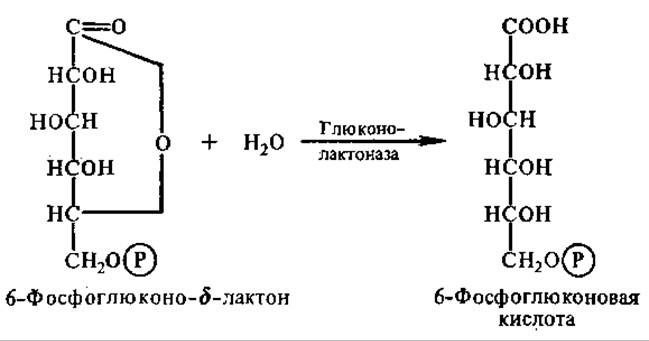
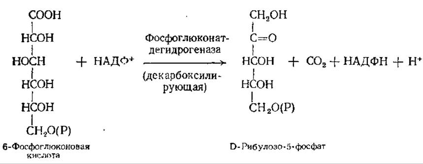
In the subsequent oxidative reaction, 6-phosphogluconic acid is both dehydrogenated and decarboxylated. This reaction is accompanied by the formation of another NADPH molecule:
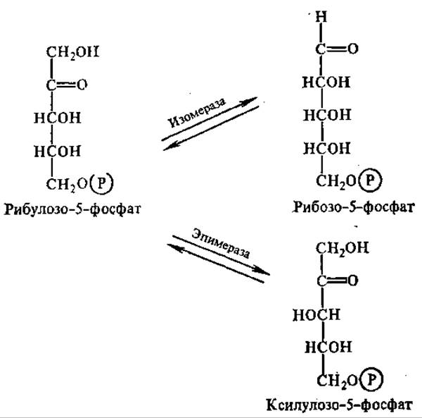
Catalyzed by the enzyme ribulose-phosphate 3-epimerase, ribulose-5-phosphate can be reversibly converted into its 3-epimer, xylulose-5-phosphate. Assisted by a specific ribose-phosphate isomerase, ribulose-5-phosphate can also undergo reversible conversion into its aldoisomer, ribose-5-phosphate:
Under certain conditions, the phosphogluconate pathway terminates at this point, and the overall net result is described by the equation
Glucose-6-phosphate + 2NADP+ → Pentose-5-phosphate + CO2 +
+ 2NADPH + 2H+
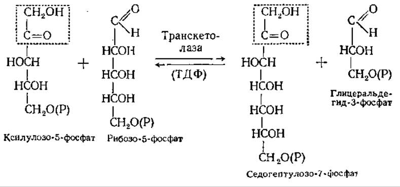
Under other conditions, the non-oxidative phase of the pentose phosphate pathway takes place. This phase proceeds under anaerobic conditions. The key reactions of this stage are carried out with the participation of specific enzymes: transketolase and transaldolase. Transketolase catalyzes the transfer of a glycoaldehyde group (CH2OH—CO—) from xylulose-5-phosphate to ribose-5-phosphate, with thiamine diphosphate (TDP) acting as the intermediate carrier of the glycoaldehyde group:
Transaldolase acts on the products of the transketolase reaction. It catalyzes the transfer of a dihydroxyacetone group from sedoheptulose-7-phosphate to glyceraldehyde-3-phosphate, resulting in the Formation of fructose-6-phosphate:

The second product of the reaction is a phosphoric ester of a four-carbon sugar, erythrose-4-phosphate. It is utilized in the second transketolase reaction:

Through the pentose phosphate cycle, Complete oxidation of glucose-6-phosphate to CO2 can occur. In this process, 6 molecules of glucose-6-phosphate yield 5 molecules of glucose-6-phosphate and 6 molecules of CO2 (Fig. 6.14). The overall reaction equation is as follows:
6 Glucose-6-phosphate + 7H2O + 12NADP+ → 5 Glucose-6-phosphate +
+ 6CO2 + 12NADPH + 12H+ + Pi

Fig. 6.14. The pentose phosphate cycle of glucose oxidation: the numbers in squares indicate the number of molecules
After canceling out common terms, we obtain:
Glucose-6-phosphate + 7H2O + 12NADP+ → 6CO2 + 12NADPH +
+ 12H+ + H3PO4
Such complete oxidation of glucose-6-phosphate does not occur in all cells. More commonly, the pentose pathway transitions into the glycolytic pathway at one of its stages. There are exactly three points of intersection between the pentose pathway and glycolysis: at the level of glucose-6-phosphate, fructose-6-phosphate, and glyceraldehyde-3-phosphate. These metabolic intersections enable the generation of a wide array of diverse metabolites.
Work by D. F. Williams, M. G. Clark, and S. E. Severin has demonstrated that pentose phosphate conversions in the non-oxidative phase can be even more complex. Following the first transketolase reaction, the sequence continues as follows:

The final transketolase reaction serves as further Evidence of the close interrelation between the glycolytic and pentose phosphate carbohydrate pathways. It demonstrates that erythrose-4-phosphate can be formed via a transketolase reaction from the initial products of glycolysis—glucose-6-phosphate and fructose-6-phosphate. Consequently, as many researchers believe, glycolysis and the pentose pathway are capable of switching from one to the other. Erythrose-4-phosphate acts as the regulator of this process. When pentose phosphates are abundant, erythrose-4-phosphate participates in the transketolase reaction, leading to the formation of glucose-6-phosphate and fructose-6-phosphate. Conversely, when hexose phosphates are abundant, erythrose-4-phosphate enters the aldolase reaction, replenishing the sedoheptulose-1,7-bisphosphate pool.
The primary Significance of the pentose pathway lies in its generation of cytoplasmic reducing equivalents in the form of NADPH, which is required in large amounts for The Biosynthesis of numerous substances, particularly Fatty acids and Steroids. Therefore, the activity of the pentose cycle is high in the liver, Mammary Glands, adipose tissue, and adrenal cortex. Another function of this pathway is to supply pentose phosphates for the synthesis of Nucleic Acids and NUCLEOTIDES. In addition, in A number of microorganisms, this pathway allows pentoses to be oxidized as well as converted into hexoses.
The pentose pathway can also serve as a source of C1 and C7 sugars, which are essential for the cell to synthesize vital metabolites. The generated NADPH typically does not participate in Oxidative Phosphorylation occurring in mitochondria; therefore, in terms of energy production, the pentose phosphate pathway does not play a significant role in most organisms. According to the prevailing view, the pentose phosphate pathway originally evolved presumably to supply prokaryotic organisms with pentoses. The evolutionary emergence of just three new enzymes (glucose-6-phosphate dehydrogenase, gluconolactonase, and phosphogluconate dehydrogenase) solved Structure/149.html">The problem of pentose synthesis. Having arisen to fulfill a narrow specific task, the pentose phosphate pathway in certain microorganisms acquired an additional function: energy supply under anaerobic conditions. For instance, in some obligate Heterofermentative lactic acid bacteria (Leuconostoc mesenteroides), it serves as the sole pathway for carbohydrate fermentation, as these bacteria lack key glycolytic enzymes. The capacity to harness energy emerged with the evolutionary development of two new enzymes: phosphopentose epimerase and transketolase. This led to the formation of a novel biochemical mechanism—the pentose phosphate cycle—in which respiratory substrates are completely degraded to CO2.
6.9.4. The Entner–Doudoroff Pathway. A small group of microorganisms, primarily bacteria of the genus Pseudomonas, generate energy via a specific metabolic pathway known as the Entner–Doudoroff pathway. It links to the pentose phosphate pathway via 6-phosphogluconic acid and to glycolysis via glyceraldehyde 3-phosphate. The first two reactions of this pathway are identical to the initial steps of the pentose phosphate pathway. What makes the Entner–Doudoroff pathway unique are the next two reactions: the formation of ketodeoxyphosphogluconic acid and its subsequent aldolytic cleavage into two trioses (Fig. 6.15). If glyceraldehyde 3-phosphate is further oxidized through glycolytic reactions to pyruvate, the cleavage of a single glucose molecule via the Entner–Doudoroff pathway yields one molecule of ATP, one molecule of NADH, and one molecule of NADPH.
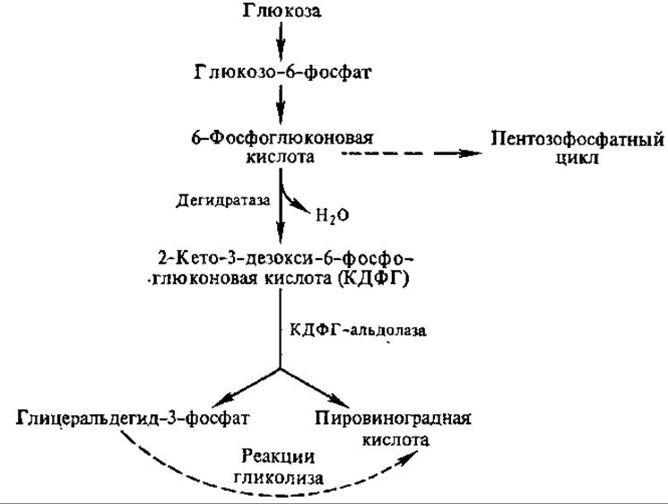
Fig. 6.15. Reactions of the Entner–Doudoroff pathway
The Entner–Doudoroff pathway likely evolved as an offshoot of the pentose pathway, given that their first two reactions are identical. It has been hypothesized that this pathway emerged due to the high demand of prokaryotes for pyruvate, serving as the shortest route for its formation (requiring only four reactions, whereas glycolysis produces pyruvate after nine).
6.9.5. The Tricarboxylic Acid Cycle (Krebs Cycle). Oxidation of Pyruvate to acetyl-coenzyme A. The tricarboxylic acid (TCA) cycle incorporates acetyl groups derived from carbohydrates, fats, and Amino Acids. It is within the TCA cycle that pyruvate, produced during glycolysis, is completely degraded into the end products: CO2 and water. This cycle was discovered and investigated by H. Krebs in 1937, which is why it is also commonly referred to as the Krebs cycle. All reactions of the cycle take place within the mitochondrial matrix. The direct reactions of the cycle are preceded by a preparatory phase—the oxidation of pyruvate to acetyl-coenzyme A (acetyl-CoA). This is a complex process whose overall equation can be represented as follows:
ПВК + НАД+ + КоА Ацетил-КоА + НАДН + Н+ + СО2
This process is accompanied by a significant decrease in Free Energy and is practically irreversible.
The oxidative decarboxylation of pyruvate is catalyzed by the pyruvate dehydrogenase system1. It consists of at least three enzymes that utilize five Coenzymes: thiamine diphosphate (TDP), lipoamide 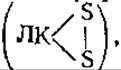 coenzyme A (CoA-SH), FAD, and NAD+.
First, a reaction occurs between pyruvate and the TDP bound to pyruvate dehydrogenase, catalyzed by the enzyme pyruvate dehydrogenase. Decarboxylation takes place, yielding α-hydroxyethyl-TDP and CO2. The hydroxyethyl group remains attached to the enzyme's surface while reacting with the disulfide form of Lipoic Acid (LA), which is bound to the second enzymatic protein of the system—dihydrolipoyl transacetylase. A redox reaction occurs wherein the Disulfide Bonds of lipoic acid are reduced to two SH-groups; one of these forms an ester bond with the acetyl group generated from the oxidation of the hydroxyethyl group. The acetyl group is then transferred to the SH-group of CoA to form acetyl-CoA.
Acetyl-CoA detaches from the enzyme surface, leaving behind reduced LA(SH)2. The oxidation of the reduced lipoamide is mediated by the third enzyme of the system, dihydrolipoyl dehydrogenase, which contains a redox-active FAD coenzyme. FADH2 is then oxidized by NAD+ present in the medium. This completes the entire process, returning all coenzymes to their initial states, ready for the next cycle.
In animal cells, the pyruvate dehydrogenase system has a molecular mass of approximately 9×10⁶. It comprises 30 molecules of pyruvate dehydrogenase, 60 molecules of dihydrolipoyl transacetylase, and 10 molecules of dihydrolipoyl dehydrogenase.
Reactions and Enzymes of the tricarboxylic acid cycle. The acetyl-CoA produced via the Oxidative Decarboxylation of pyruvate enters the tricarboxylic acid cycle. It condenses with a molecule of oxaloacetic acid (OAA, or oxaloacetate) to yield citric acid. This reaction is catalyzed by the enzyme citrate synthase, which is found in animals, microorganisms, and various plants.
Citric acid is converted into cis-aconitic acid and subsequently into isocitric acid. These reactions are catalyzed by a single enzyme, aconitate hydratase (Fig. 6.16). Next, isocitric acid is dehydrogenated and decarboxylated by isocitrate dehydrogenase to yield CO2 and α-ketoglutaric acid. NAD+ serves as the coenzyme for mitochondrial isocitrate dehydrogenase, and the enzyme requires Mg2+ or Mn2+ ions while being allosterically activated by ADP. Enzyme activity is strongly inhibited by ATP and NADH, the latter acting through competition with NAD+. Consequently, under any metabolic conditions that increase the cellular concentration of ADP, the rate of isocitric acid oxidation automatically rises. This in turn accelerates the entire TCA cycle, as this specific reaction typically acts as the rate-limiting step of the overall process. Conversely, an elevated ATP concentration and the accumulation of NADH lead to the downregulation of isocitrate dehydrogenase and a slowdown of the cycle's reactions.
1 The term "complex," previously used in this context, is no longer recommended by the new Enzyme Nomenclature rules (1979).
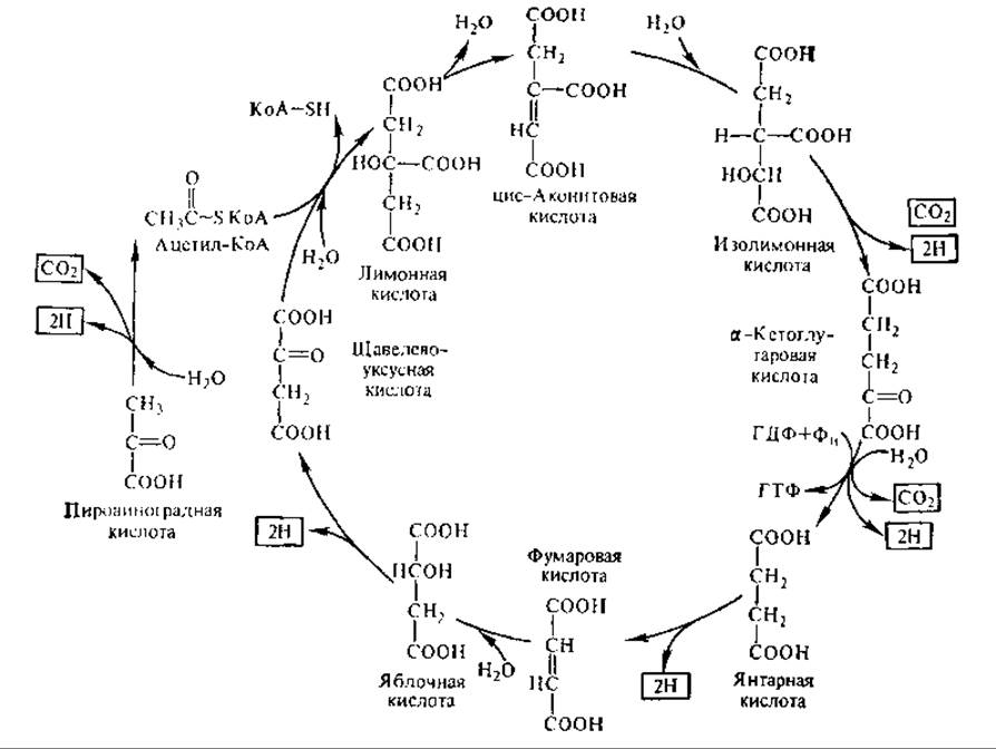
Fig. 6.16. The tricarboxylic acid cycle (end products are boxed)
The next reaction is the oxidative decarboxylation of α-ketoglutaric acid to succinic acid. The overall equation for this reaction is:
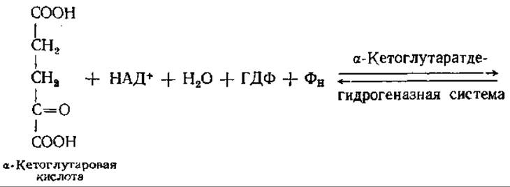
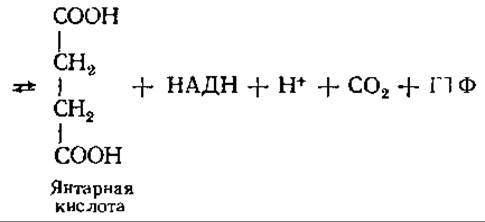
This is a complex, multi-step reaction that shares similarities with the pyruvate oxidation pathway. The mechanisms of both reactions are virtually identical, with both utilizing thiamine diphosphate, LA, CoA, NAD+, and FAD as Cofactors.
The α-ketoglutarate dehydrogenase system resembles the pyruvate dehydrogenase system in both Structure and properties. The initial stage of α-ketoglutaric acid oxidative decarboxylation involves oxidation and decarboxylation:

The final product of this reaction, succinyl-CoA, is a high-energy thioester in which one of the COOH groups of succinic acid (succinate) participates in forming the ester bond. In the subsequent step, succinyl-CoA loses its CoA moiety, and the released energy is conserved within the phosphate bond of GTP:
Сукпинил ~ КоА + ФH + ГДФ Янтарная кислота + ГТФ + KoA-SH
This reaction is catalyzed by the enzyme succinyl-CoA synthetase and represents a substrate-level phosphorylation: the oxidation energy stored in succinyl-CoA is transferred to GTP. GTP can then transfer its terminal phosphoryl group to ADP, yielding ATP according to the reaction: GTP + ADP⇄GDP + ATP. This reaction is catalyzed by nucleoside diphosphate kinase.
Next, succinic acid is oxidized to fumaric acid. This reaction is catalyzed by succinate dehydrogenase, a flavoprotein whose protein molecule is covalently bound to FAD, which acts as a hydrogen acceptor in this process. Unlike other TCA cycle enzymes located in the mitochondrial matrix, succinate dehydrogenase is an integral protein of The inner mitochondrial membrane. Through succinate dehydrogenase, electrons and protons from FADH2 directly enter the respiratory chain.
In the subsequent reaction, which takes place in the matrix, fumaric acid undergoes reversible Hydration catalyzed by fumarase (fumarate hydratase) to form L-malic acid (L-malate). In the final reaction of the TCA cycle, NAD-dependent malate dehydrogenase catalyzes the oxidation of L-malic acid to oxaloacetate (OAA), which then enters a new cycle of reactions.
Energy balance of the tricarboxylic acid cycle. During one turn of the TCA cycle, a pyruvic acid molecule is completely oxidized to CO2 and H2O. A total of 3 molecules of CO2 are produced in the following reactions: during the oxidative decarboxylation of pyruvate, during the decarboxylation of isocitric acid, and during the oxidative decarboxylation of α-ketoglutaric acid. Three water molecules enter the cycle: in the oxidative decarboxylation reactions of pyruvate and α-ketoglutaric acid, and in the fumarase reaction. Overall, five pairs of hydrogen atoms are removed during pyruvate oxidation: one pair from succinic acid is transferred to FAD, and four pairs are transferred to NAD+ to form NADH (during the oxidative decarboxylation of pyruvate, α-ketoglutaric, and isocitric acids, as well as during the oxidation of malic acid). It is easy to see that molecular oxygen is not directly involved in the TCA cycle; oxidation is accomplished through The addition of water and dehydrogenation (see Section 6.16).
The overall balance equation for the oxidation of pyruvate—the end product of the anaerobic phase of respiration—in the TCA cycle is as follows:
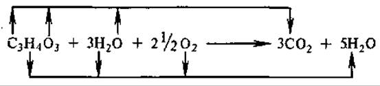
As shown in the diagram, all of the substrate's carbon is oxidized to carbon dioxide (IV), which incorporates oxygen from both the substrate and water. Atmospheric oxygen is consumed after the direct reactions of the Krebs cycle, specifically for the oxidation of hydrogen derived from pyruvate and water that added to the substrate at specific Stages of the cycle.
The reduced coenzyme NADH enters the mitochondrial matrix and diffuses to the inner mitochondrial membrane. Here, it binds to NADH dehydrogenase and is oxidized by it, releasing electrons and protons into The electron transport (respiratory) chain. The energy of oxidation is stored as the transmembrane electrochemical proton gradient ΔμH+, which drives the synthesis of ATP from ADP and H3PO4 via oxidative phosphorylation (see Section 7.3).
One can calculate the energy yield of glucose oxidation via pyruvate during respiration. When pyruvate is oxidized, four pairs of hydrogen atoms are transferred to NAD+. The oxidation of one molecule of NADH yields three molecules of ATP. One pair of hydrogen atoms is oxidized via FAD, yielding two molecules of ATP. One molecule of ATP is produced via substrate-level phosphorylation. In total, 3 × 4 + 2 + 1 = 15 molecules of ATP. Consequently, the oxidation of 1 mole of pyruvate yields 15 moles of ATP. It should be noted that each glucose molecule produces two pyruvate molecules, so a coefficient of 2 must be applied: 15 ATP molecules × 2 = 30 ATP molecules.
In addition, the anaerobic phase (glycolysis) produces another two molecules of ATP and two molecules of cytoplasmic NADH. Depending on the mechanism by which reducing equivalents are transported into the mitochondrial matrix and subsequently into the respiratory chain (see Section 6.9.1), an additional 4 or 6 molecules of ATP are generated. Thus, during the aerobic oxidation of 1 mole of glucose, the cell obtains up to 38 moles of ATP—significantly more than during glycolysis. In this process, 34.5 × 38 = 1311 kJ of free Energy is stored in the high-energy bonds of ATP. Overall, the complete oxidation of 1 mole of glucose to CO2 and H2O releases approximately 2872 kJ of energy. Therefore, during the aerobic breakdown of glucose, more than 45% of the released free energy is conserved in high-energy bonds.
The tricarboxylic acid cycle has been found in animals, plants, and microorganisms. It can operate not only under aerobic conditions but also under anaerobic conditions—for instance, in photosynthetic bacteria, provided that a suitable hydrogen acceptor is present. Anaerobic Variants of the TCA cycle are also known in animal cells. Under oxygen deprivation in mammalian tissues, NADH can be consumed to reduce fumaric acid to succinic acid. Phylogenetically ancient blue-green Algae lack a complete cycle.
Carbohydrates are not the only compounds processed in the Krebs cycle. Acetyl-CoA derived from the Oxidation of Fatty acids can condense with oxaloacetate. Many amino acids, through Transamination reactions, can be converted into cycle metabolites and thereby enter the pathway. Conversely, intermediate products of the cycle can be removed to serve as precursors for Amino Acids and other biologically important substances. Thus, the tricarboxylic acid cycle is an amphibolic metabolic pathway (see Section 1.2). Its functions are linked not only to catabolic processes but also to anabolic processes, for which it supplies precursor molecules. The tricarboxylic acid cycle is not merely the aerobic breakdown of glucose, but the focal point where many metabolic pathways intersect.
6.9.6. Glyoxylate cycle. There are special enzymatic mechanisms that replenish the pool of TCA cycle intermediates. The glyoxylate cycle performs this specific function. It has been found in the cells of higher plants, molds, and certain bacteria (Pseudomonas, E. coli). Key Enzymes of this cycle have not yet been detected in animal cells, leading to the consensus that it does not operate in them. The cycle was discovered by H. Krebs in 1957.
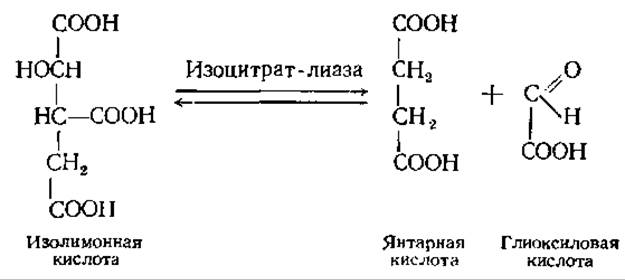
The glyoxylate cycle is a modified tricarboxylic acid cycle. In this pathway, the enzymes isocitrate dehydrogenase and α-ketoglutarate dehydrogenase are replaced by isocitrate lyase, which cleaves isocitric acid into succinic and glyoxylic acids:
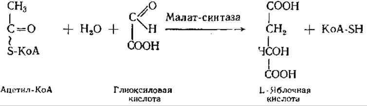
Another key enzyme of the cycle is malate synthase. It catalyzes the condensation of acetyl-CoA with glyoxylic acid:
Aside from these two enzymes specific to the glyoxylate cycle, a number of standard TCA cycle enzymes are also involved.
The initiating reaction of the cycle is driven by the Krebs cycle, which produces oxaloacetate. Catalyzed by citrate synthase, acetyl-CoA condenses with oxaloacetate to form citric acid. Through the action of aconitase (aconitate hydratase), citrate is converted first into cis-aconitic acid and then into isocitric acid. The latter is cleaved by isocitrate lyase into succinic and glyoxylic acids. With the participation of the second cycle-specific enzyme, malate synthase, glyoxylic acid condenses with a second molecule of acetyl-CoA to yield malic acid, which is then oxidized by malate dehydrogenase to regenerate oxaloacetate (Fig. 6.17).
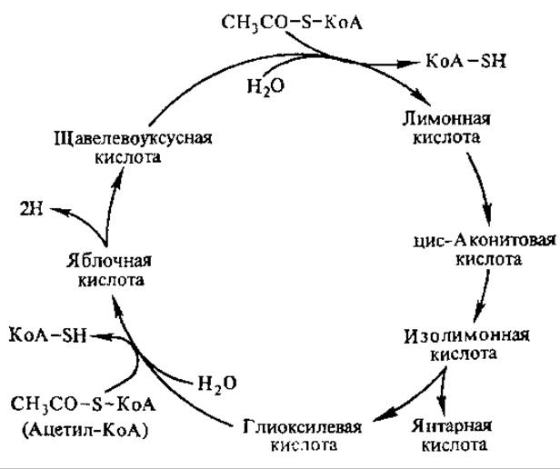
Fig. 6.17. Reactions of the glyoxylate cycle
Thus, with each turn of the cycle, two molecules of acetyl-CoA are incorporated, and one molecule of succinic acid is produced, which can enter Gluconeogenesis, other biosynthetic pathways, or the TCA cycle. Glyoxylate cycle metabolites such as malic and isocitric acids can also feed into the latter. As a result of glyoxylate cycle reactions, two hydrogen atoms can enter the respiratory chain, generating three molecules of ATP. This is particularly important for organisms and physiological conditions where acetyl groups and other two-carbon compounds serve as the sole energy source.
The glyoxylate cycle is characteristic, for example, of microorganisms that utilize two-carbon compounds as energy and carbon sources. Acetic acid and ethyl alcohol are directly incorporated into the TCA cycle as acetyl-CoA, whereas more reduced compounds like Glycine and glycolic acid cannot be utilized directly in it; instead, they are oxidized to glyoxylic acid via the glyoxylate cycle.
In plant tissues, the glyoxylate cycle facilitates the conversion of fats into carbohydrates and other cellular components. This process is especially active in germinating oilseeds. Reserve triacylglycerols are broken down, and the oxidation of their constituent fatty acids yields acetyl-CoA. This enters the glyoxylate cycle reactions to produce succinic acid, which is subsequently converted into carbohydrates via TCA cycle reactions and gluconeogenesis. In plants, the enzymes of the glyoxylate cycle are localized in Microbodies called glyoxysomes.
Last update: 06/08/2026
Editorial and Educational Adaptation: This material has been compiled based on the primary/original source text. The project team performed an editorial review, corrected technical inaccuracies, structured sections, and adapted the content for an educational format.
What was processed:
- elimination of formatting defects (OCR errors, structural breaks, corrupted characters);
- editorial organization of content;
- standardization of terminology in accordance with academic sources;
- verification of factual statements against the original source text.
All mentions of the author, publication year, and origin of the primary text have been preserved in accordance with the source.