Fundamentals of Biochemistry - A. A. Anisimov 1986
Carbohydrates
Regulation of Blood Glucose Homeostasis
Monosaccharides absorbed from the intestine (primarily glucose) are transported via the portal vein primarily to the Liver, where a portion of glucose is used to build up Glycogen reserves. The glycogen content in the liver of humans and animals depends on dietary habits; the higher the carbohydrate intake, the more glycogen accumulates in the liver. The Synthesis and Breakdown of glycogen are catalyzed by different Enzymes and controlled independently. A fraction of glucose is utilized by the liver itself to generate energy required for its numerous metabolic reactions. In addition, a certain amount of glucose in the liver is converted into fats. For instance, under a normal mixed diet, only 3–5% of glucose is converted into glycogen (its average content in the liver ranges from 5 to 7%), 30% of glucose is channeled into fat synthesis, and 60–70% is oxidized to CO2 and H2O. Under a high-carbohydrate diet, 10% of glucose is stored in the liver as glycogen, 40% is converted into fats, and 50% is oxidized.
Blood, Lymph, and CEREBROSPINAL FLUID contain sugar in the form of glucose, the level of which is regulated with high precision. The glucose concentration in arterial and capillary blood of a healthy individual ranges from 54–96 mg/100 mL when determined by the glucose oxidase method, and from 80–120 mg/100 mL when determined by the Hagedorn-Jensen method (the latter method measures not only glucose but also certain other reducing substances in the blood simultaneously). Venous blood contains less glucose because it is continuously extracted from the blood by peripheral Tissues. The liver is the sole organ that releases glucose into the general Circulation.
Sugars also serve as a component of the internal environment in invertebrates. For example, in insect hemolymph, they are represented primarily by trehalose. Among vertebrates, fasting blood glucose levels are most stable in humans and higher vertebrates. Birds exhibit higher blood glucose levels than mammals. Lower vertebrates are characterized by significant fluctuations in blood glucose depending on the external environment and their biological life stage. Cold-blooded animals (fish, amphibians, reptiles) have lower blood glucose levels than warm-blooded animals, averaging 30–50 mg/100 mL. There is a direct correlation between blood glucose levels and the mobility of animals; for instance, bottom-dwelling, sluggish fish have a lower blood glucose concentration than active fish inhabiting mountain streams or fast-flowing rivers. In ruminants, blood glucose is below 50 mg/100 mL, as the End products of Cellulose Fermentation—organic acids—enter the bloodstream from the intestine alongside glucose.
Following the ingestion of a carbohydrate-rich meal, the glucose concentration in the human HEPATIC PORTAL VEIN can rise up to 200–220 mg/100 mL. However, throughout the rest of the Circulatory system, blood glucose in a healthy individual either remains unchanged (54–96 mg/100 mL) or increases slightly (alimentary hyperglycemia). It returns to normal within 1.5–2 hours postprandial as excess glucose is excreted in the urine by the Kidneys (alimentary glucosuria). Renal excretion of glucose begins when its blood concentration exceeds 120–150 mg/100 mL. This can occur upon the simultaneous intake of 160–180 g of sucrose or 120–150 g of fructose (Fig. 6.11).
Carbohydrate starvation of a certain duration or accelerated glucose consumption during intense physical exertion in a healthy individual likewise does not lead to significant deviations of blood glucose from the normal range. Thus, a constant Blood Glucose Level is continuously maintained within the body through homeostatic regulation.
Under Homeostasis, any change in the concentration of a substance within the Organism automatically triggers processes aimed at restoring its normal level, a phenomenon known as autoregulation. These mechanisms are based on the interaction of positive and negative feedback loops; therefore, such phenomena in living organisms can be broadly examined within the framework and concepts of cybernetics, the science of communication, control, and regulation.
The simplest form of Blood Glucose Regulation is the homeostatic and regulatory function of the liver. This physiological mechanism, which is phylogenetically the most ancient, is insufficient to provide highly developed animals and humans with adequate carbohydrate Nutrition. In the course of evolution, powerful neural and endocrine mechanisms have evolved that influence both the homeostatic function of The Liver and the capacity of various Cells to utilize glucose.
When examining the hormonal component of regulation, it is noteworthy that Insulin is the sole hormone with a pronounced blood glucose-lowering effect. Other Hormones affecting Carbohydrate METABOLISM by increasing blood glucose levels are termed diabetogenic or counter-regulatory. Insulin acts as a repressor of key gluconeogenic enzyme synthesis and an inducer of hexokinase, Phosphofructokinase, Pyruvate kinase, and glycogen (starch) synthase. Glucocorticoids serve as Inducers of gluconeogenic enzymes.
Insulin is secreted by the ß-Cells of the pancreatic islets of Langerhans in the form of proinsulin. The primary manifestations of insulin deficiency are elevated blood glucose levels (hyperglycemia), excessive urinary glucose excretion (glucosuria), and decreased hepatic glycogen content. Insulin deficiency suppresses the Biosynthesis of Fatty acids from glucose and acetate, as well as Protein Biosynthesis; it enhances the synthesis of enzymes involved in Gluconeogenesis and disrupts the balance between Glycolysis and gluconeogenesis.
Currently, the membrane localization of the primary Action of Insulin appears most plausible. Evidence has been obtained confirming the presence of a specific Insulin Receptor on the outer Cell/33.html">Plasma Membrane of adipocytes, as well as The formation of an insulin-receptor complex. It is hypothesized that adenylate cyclase and cAMP participate in mediating the insulin signal in adipocytes and, partially, in liver cells. In Muscle tissue, insulin readily penetrates The Cell interior.
Class="center">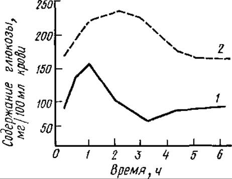
Fig. 6.11. Dynamics of human blood glucose levels following sugar ingestion:
1 — normal, 2 — in diabetes
In the absence of sufficient insulin, Muscles and the liver are unable to utilize glucose even when its blood concentration is high. This phenomenon is termed "starvation amidst plenty." As hyperglycemia progresses, Brain and myocardial tissues increase their glucose uptake. In this rather limited context, hyperglycemia can be viewed as a beneficial adaptive response. Insufficient insulin secretion underlies the onset of Diabetes Mellitus. Many consequences of diabetes mimic the effects of carbohydrate deprivation, despite the fact that blood glucose levels are sharply elevated in this pathology.
Glucagon, a hormone produced by the a-cells of the Pancreas, activates hepatic Glycogenolysis, resulting in the release of glucose into the bloodstream.
Adrenaline is a hormone produced by The adrenal medulla. Acting in concert with glucagon, it activates liver and muscle Glycogen phosphorylase, thereby increasing blood glucose levels. The enzyme exists in two forms: active (phosphorylase a) and inactive (phosphorylase b). The Molecular Weight of phosphorylase a is 380,000. Phosphorylase a consists of four subunits, each containing a phosphoserine residue and a coenzyme molecule—Pyridoxal phosphate—covalently bound to a Lysine residue. Phosphorylase phosphatase catalyses the hydrolytic Cleavage of the phosphate group bond from the Serine residue, leading to the formation of dimers.
Inactive glycogen phosphorylase b, composed of two subunits, is phosphorylated into active glycogen phosphorylase a by phosphorylase kinase. The latter is activated by cyclic AMP, which is generated by adenylate cyclase. Adrenaline stimulates The production of cAMP via adenylate cyclase. Thus, adrenaline accelerates glycogen breakdown. Conversely, by stimulating adenylate cyclase, adrenaline inhibits glycogen synthesis from UDP-glucose through the accumulation of a glycogen (starch) synthase form that requires high concentrations of glucose-6-phosphate.
The mechanisms by which adrenaline and glucagon regulate blood glucose levels are analogous.
Glucocorticoids (Hormones of the adrenal cortex) enhance gluconeogenesis, the de novo synthesis of sugar from non-carbohydrate precursors. This results in elevated blood glucose and hepatic glycogen levels. The pituitary Growth Hormone (somatotropin) reduces glucose utilization by peripheral tissues, enhances gluconeogenesis, inhibits insulin production, and ultimately raises blood glucose levels. Thyroxine and triiodothyronine (THYROID HORMONES) in moderate doses stimulate intestinal monosaccharide absorption. An excess of these hormones inhibits Oxidative Phosphorylation AND contributes to an elevated blood glucose concentration.
Hormone levels in the blood are regulated by the Central Nervous system, which controls blood glucose levels via the Endocrine System. However, In addition to its indirect influence on blood glucose, the central nervous system (CNS) also exerts a direct effect. A drop in blood glucose concentration below 60–70 mg% triggers a reflex excitation of the corresponding centers located in the Hypothalamus. This excitation is transmitted from the CNS along spinal nerve pathways, passes into the Sympathetic trunk, and reaches the liver via the sympathetic nerve. As a result of this neural activation, a portion of hepatic glycogen breaks down to form glucose. This process is termed glucose mobilization. Once the glucose concentration returns to baseline levels, the impulses originating from the CNS diminish, and glycogenolysis is inhibited.
Thus, through the regulatory control of the CNS and the endocrine system, a constant blood glucose level is automatically maintained via self-regulation, which can operate at various levels. For instance, blood with an excess glucose content reaching the pancreas directly stimulates insulin secretion. Insulin, in turn, enhances glucose uptake by tissues and its utilization for glycogen synthesis, thereby reducing glucose concentration back to normal. As demonstrated by the research of K. M. Bykov, the Cerebral Cortex also exerts a definite influence on blood glucose levels. An example of this is emotional hyperglycemia, which is an elevated blood glucose concentration occurring during states of excitement (such as athletes prior to competition or students before an exam).
Last update: 06/08/2026
Editorial and Educational Adaptation: This material has been compiled based on the primary/original source text. The project team performed an editorial review, corrected technical inaccuracies, structured sections, and adapted the content for an educational format.
What was processed:
- elimination of formatting defects (OCR errors, structural breaks, corrupted characters);
- editorial organization of content;
- standardization of terminology in accordance with academic sources;
- verification of factual statements against the original source text.
All mentions of the author, publication year, and origin of the primary text have been preserved in accordance with the source.