PLANT BIOTECHNOLOGY AND BIOSAFETY - A. P. Yermishin - 2015
CHAPTER 5. IN VITRO CLONAL PLANT PROPAGATION. PRESERVATION OF IN VITRO GENETIC COLLECTIONS
The application of in vitro plant Cell, tissue, and organ culture Methods opens up new opportunities for vegetative propagation and the maintenance of genetic collections. Agricultural or ornamental plants are propagated vegetatively when they possess specialized structures for this type of reproduction (tubers, bulbs, corms, stolons) or have a prolonged juvenile phase, which delays harvest times and complicates the breeding evaluation of individual genotypes (such as tree and shrub crops).
Cultivars and strains of vegetatively propagated plants are typically complex heterozygotes and frequently polyploids. Replicating their genotype through seed propagation is virtually impossible. Vegetative propagation makes it possible to obtain A large number of individuals sharing a single genotype of value to breeding and agricultural production, which is its main advantage over seed propagation. Furthermore, vegetative propagation significantly accelerates the yield from slow-developing plants. For instance, by grafting a scion into the canopy of a mature apple tree, fruits of the desired variety can be harvested within just a year or two.
However, vegetative propagation has several drawbacks. The propagation coefficient in natural vegetative propagation is generally much lower than in seed propagation. For example, propagating seed potatoes yields only 4 to 10 tubers per plant suitable for planting, whereas a single wheat plant can produce over a hundred seeds. Seed tubers and bulbs are much bulkier than conventional seeds, requiring large storage facilities maintained at precise temperatures and humidity levels, which frequently entails significant energy costs. During the growing season, vegetatively propagated crops accumulate various infections that are passed on to subsequent generations. While bacterial and fungal infections can be eradicated with appropriate chemicals, plant diseases caused by Viruses AND Viroids are practically untreatable. Consequently, seed production in vegetatively propagated crops is far more complex and costly than in seed-propagated crops. Certain forms of human-assisted vegetative propagation (grafting, layering, etc.) are highly labor-intensive and markedly seasonal, requiring a massive volume of work to be completed within a very short timeframe.
Maintaining genetic collections of vegetatively propagated crops also involves substantial expenses, often necessitating annual field planting. This requires plowing large areas, applying fertilizers, executing a full range of agrotechnical crop care measures, organizing plantation security, harvesting, sorting, and storing the gathered tubers and bulbs. Special demands are placed on keeping the plants healthy and culling those infected with viral diseases. Significant challenges may arise in preserving certain accessions if they are unadapted to the growing conditions. For example, wild potato species of high breeding interest—serving as sources of valuable genes for resistance to diseases and pests—may fail to tuberize under the long-day conditions typical of temperate latitudes.
Executing vegetative propagation under in vitro conditions substantially increases its efficiency and overcomes many of the aforementioned drawbacks. Modern in vitro clonal propagation technologies ensure very high multiplication rates. Thus, a single in vitro potato plantlet can yield over one hundred thousand plants in a year. A major advantage of this technology is its ability to efficiently propagate plants that are difficult to reproduce via Traditional Methods. All operations can be conducted year-round without requiring large land areas. Crucially, in vitro micropropagation poses no risk of reinfection by viral and other pathogens (provided the initial stock plant is healthy). Overall, in vitro clonal propagation is economically far superior to traditional vegetative propagation techniques and yields higher-quality planting material.
5.1. Utilization of Apical Meristem Culture to Obtain Pathogen-Free Planting Material
The goal of vegetative propagation is to derive the maximum number of healthy plants from a stock plant in the shortest possible time. At the same time, it is essential that these new plants faithfully reproduce the genotype and phenotype of the stock plant, particularly its traits of practical significance. In the vast majority of cases, traditional vegetative propagation essentially relies on stem buds (axillary, dormant, or tuber "eyes"). This is no coincidence, as the specific properties of apical Meristems housed within stem buds ensure the preservation of the stock plant's genotype during propagation, along with The production of healthy plants.
Meristems are formed during The Development of the zygotic embryo. Some meristematic Cells (initials) remain in an undifferentiated state and, through division, ensure continuous plant growth. Other cells gradually differentiate to form permanent Tissues (dermal, vascular, mechanical, etc.). Unlike cells of specialized tissues, the initial cells of apical meristems exhibit high cytogenetic stability: throughout the plant's life cycle, they generally preserve the zygote's genotype.
Another vital property of meristematic tissues is the absence or extremely low concentration of Viral Particles, even in virus-infected plants. Furthermore, the viral titer in specialized tissue cells increases with distance from the apical meristems. Typically, the apical stem meristem consists of a growth dome and one or two leaf primordia (Fig. 5.1). Its distal portion has an average diameter of about 200 μm and a height ranging from 20 to 150 μm. In the deeper layers, differentiating meristematic cells form the procambium, which gives rise to the vascular bundles. This structural architecture of the apical meristem prevents the rapid influx of viral particles via The Vascular System, though slow intercellular propagation through plasmodesmata connecting meristem cells remains possible. Additionally, the High Metabolic Rate of intensively dividing meristem cells is believed to inhibit viral Replication. High endogenous auxin concentrations in meristematic cells may produce a similar effect.
Class="center">Fig. 5.1. Structure of a stem bud

In 1952, J. Morel and C. Martin developed a technique for eradicating viruses in dahlias using in vitro culture of apical meristems approximately 100 μm in size. Since then, this method has been applied with varying degrees of success to a wide range of vegetatively propagated crops. It has been established that meristem culture eliminates not only viruses but also bacterial and fungal infections, viroids, and Mycoplasmas. Later Electron Microscopy studies revealed the occasional presence of viral particles in meristem cells as well. Nevertheless, in many instances—particularly during early infection stages—meristematic tissues remain virus-free for extended periods.
Consequently, stem meristems represent the most suitable explant for introducing plants destined for vegetative propagation into in vitro culture, as they ensure maximum preservation of the stock plant's genotype and the production of healthy progeny.
The first phase of commercial propagation involves selecting the most productive stock plant clones that closely match the Description of the cultivar, tree/shrub strain, or stand out for a complex of positive traits (such as so-called plus trees). Prior to in vitro Introduction, visually healthy selected stock plants are additionally screened for major viruses using various assays: bioassays (inoculation of indicator plants highly sensitive to the pathogen), electron microscopy, and enzyme-linked immunosorbent assay (ELISA). Recently, Methods based on pathogen nucleic acid analysis have gained prominence, including Electrophoresis, in situ Hybridization combined with various labels (e.g., nucleic acid spot hybridization - NASH), and Polymerase Chain Reaction (PCR) variants (e.g., reverse METABOLISM/31.html">Transcription polymerase chain reaction - RT-PCR).
In vitro apical meristem cultures are established by aseptically excising the upper part of the stem apex (approx. 200 μm in size) under a binocular Microscope and placing it onto an Agar-solidified nutrient medium (see Fig. 5.1). In addition to Cytokinins and Auxins, a small amount of gibberellin is added to the medium. Cultivation is carried out under light. The more leaf primordia the explant possesses, the easier it establishes on the nutrient medium and the better SHOOT regeneration proceeds. However, if the objective is to recover virus-free regenerants from infected plants or if latent viral contamination is suspected, the explant size should be kept to a minimum. Eradication efficiency depends not only on explant size but also on the viral species. For instance, apical meristem culture of potato yielded 70% potato virus Y (PVY) free regenerants, but only 10% free from potato virus X (PVX).
To enhance the efficiency of viral eradication via meristem culture, stock plants undergo thermotherapy—prolonged exposure to elevated temperatures—prior to explant excision. Thermotherapy is conducted in climate chambers at 37 °C, 90% relative humidity, an illumination of approximately 5,000 lux, and a 16-hour photoperiod. During the first week, plants are gradually acclimated to high temperatures by daily 2 °C increments from 25 to 37 °C. The duration of thermotherapy varies depending on plant species and viruses. For instance, 10 to 12 weeks of heat Treatment are sufficient for carnation sanitation.
Combining thermotherapy with meristem culture has yielded over 70% virus-free hop regenerants, 90% strawberry plants, 25% black and red currant plants, 50% raspberry plants, and over 80% potato plants. The efficacy of thermotherapy in treating mycoplasma-induced diseases has also been demonstrated.
However, most plant species tolerate high temperatures and humidity poorly, frequently perishing long before the heat treatment concludes. Many viruses are thermotherapy-resistant (for example, among potato viruses, only potato leafroll virus - PLRV is heat-sensitive). Furthermore, the Procedure itself is quite expensive and demands specialized equipment.
Interestingly, in certain cases, cold treatment rather than heat therapy serves as a highly effective planting material sanitation method. For instance, eliminating potato spindle tuber viroid (pSTV) was achieved by storing infected tubers in a refrigerator for 6 months followed by meristem culture initiation.
Another approach used alongside meristem culture to eliminate plant viruses is Chemotherapy. It involves supplementing the nutrient medium used for apical meristems with various antiviral compounds. These most frequently include synthetic purine and pyrimidine base analogues: cyanoguanosine, 2-thiouracil, D-ribofuranosyl-1,2,4-triazole-carboxamide (commercial name - virazole), as well as interferon, ridostin (sodium ribonucleate), binase (microbial Ribonuclease), and phenolic acids such as salicylic, gallic, syringic, caffeic, ferulic, and p-coumaric acids. Positive chemotherapeutic effects have been documented for several tree and shrub crops (sweet cherry, plum, raspberry, currant) and certain ornamental species. Chemotherapy increases the efficiency of meristem-mediated viral eradication from 40% up to 80–100%.
Applying thermo- or chemotherapy allows for an increased meristem explant size without compromising sanitation efficacy, thereby improving explant survival on the nutrient medium and ultimately yielding a greater number of healthy regenerants.
5.2. In Vitro Clonal Plant Propagation
Vegetative plant propagation utilizing in vitro cell, tissue, and organ culture techniques is termed in vitro clonal propagation. The term "clone" (derived from the Greek for twig, shoot, or offshoot) originally denoted a group of plants—primarily fruit trees—derived from a single parent (stock) plant via vegetative propagation methods. Later, the term came to refer not only to the entire group but also to its individual members (excluding the stock plant), with The process of obtaining them termed cloning. Currently, the biological definition of "clone" is broader; earlier in this manual, for instance, THE CONCEPT OF a "cell clone" was introduced, denoting progeny derived from the mitotic division of a single cell. Higher animals (Dolly the sheep), DNA molecules (molecular cloning), and even individual genes are routinely cloned. To designate in vitro plant cloning, terms such as "microclonal propagation", "clonal micropropagation", or simply "micropropagation" are also employed. Evidently, the prefix "micro-" originates from the microscopic dimensions of the explants used in apical meristems culture—the foundational step of in vitro clonal plant propagation.
5.2.1. Methods of In Vitro Clonal Plant Propagation
In practice, the following micropropagation methods for plant organisms are employed:
✵ activation of the development of existing meristems (stem apex, axillary, and dormant stem buds);
✵ induction of adventitious buds in an explant or callus culture;
✵ somatic Embryogenesis.
The activation of the development of meristems already present in a plant is based on the removal of apical dominance. Apical dominance is the suppression of the growth of lateral plant shoots in the presence of the apical bud. This can be overcome in in vitro plants in two ways: by removing the apical bud or by adding cytokinins to the nutrient medium.
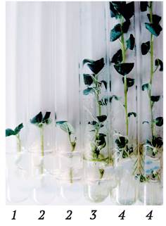
The first of these methods involves dividing in vitro plants into cuttings that typically contain a single axillary meristem. The cuttings are planted on a nutrient medium after completely or partially removing the leaf blade. After some time, the axillary meristem is activated, and one or more shoots develop. Simultaneously, rooting of the cutting occurs. As a result, within a relatively short period (3–4 weeks in the case of potato propagation), an in vitro plant is obtained, which can be cut and used to start a new cloning cycle (Fig. 5.2). One of the cuttings in this type of propagation contains the terminal bud instead of an axillary one. It is also activated when the cutting is planted on the nutrient medium and forms an in vitro plant within the same timeframe.
Fig. 5.2. Clonal propagation of potatoes via cutting of in vitro plants (through the activation of axillary and terminal buds): 1 - cutting containing an axillary bud after planting on a nutrient medium; 2 - activation of the axillary or terminal bud, onset of rooting; 3 - rooted in vitro plant three weeks after planting the cutting on the nutrient medium, ready to be divided into 4 cuttings to start the next cloning cycle; 4 - plant one month after cutting, potential propagation coefficient of 8
In the second method, The Use of cytokinins to lift apical dominance enables the development of axillary buds without removing the apical bud. In this process, a cluster of shoots (axillary branching) is formed from the explant buds and the shoots that have grown out from the apical and axillary buds of the explant (Fig. 5.3). Once they reach a certain length (1–2 cm), they are separated from the explant and transferred to a fresh nutrient medium containing cytokinins for the next propagation cycle, or to a medium with auxins (in some cases without growth regulators) for rooting and subsequent planting in soil. For the propagation of certain crops—primarily trees and shrubs—the grown shoots are rooted ex vitro using specialized substrates. This helps to reduce production costs.
Fig. 5.3. In vitro clonal Propagation of the medicinal plant Picrorhiza kurroa through the induction of axillary branching on a nutrient medium supplemented with kinetin. A cluster of shoots is formed, which can be separated for subsequent in vitro propagation or for rooting and planting in soil: a–d - various options for the composition and consistency of the nutrient medium (after H. Sood, R. Chauhan, 2009)

Micropropagation via meristem activation is the most efficient method of plant propagation. Compared to other techniques, it ensures the greatest preservation of the genotype of the original stock plant, since the resulting clones originate directly from meristematic cells, which are characterized by high cytogenetic stability.
Based on this method, propagation technologies have been developed and applied in practice for vegetable and industrial crops (potato, tomato, cucumber, pepper, pumpkin, asparagus, sugar beet, hops, tobacco, Jerusalem artichoke, Chinese artichoke, etc.), fruit and berry crops (apple, plum, cherry, pear, grapevine, raspberry, currant, gooseberry, etc.), woody plants (poplar, willow, alder, birch, rowan, sequoia, thuja, juniper, etc.), commercial floriculture crops (carnation, chrysanthemum, rose, gerbera, orchid), and tropical and subtropical plants (rhododendron, azalea, camellia, tea, etc.).
The method of in vitro clonal plant propagation through the induction of adventitious buds is based on the property of plant cell totipotency. Its essence lies in the fact that explants from various plant parts and Organs lacking stem buds are capable of forming them in vitro in the presence of factors that induce stem Organogenesis. Explants may include segments of leaf blades and petioles, young inflorescences, bulb scales and basal plates, corms, ROOT cuttings, lateral and intercalary meristems, and other organs and tissues. Stem buds arise de novo in both the epidermal and subepidermal layers of the explant. Because they appear in an "unusual" Location for the plant, they are referred to as adventitious.
There are A number of plants for which The formation of adventitious buds is a natural mode of vegetative propagation. For example, raspberries and blackberries propagate via stem buds originating on the roots. Begonias, Usambara violets (saintpaulias), and certain other ornamental plants are propagated primarily by leaf cuttings. In in vitro culture, through the use of appropriate growth regulators (see Chapter 3), this process can be significantly intensified; moreover, mass formation of adventitious buds can be achieved in plants that practically do not form them under in vivo conditions.
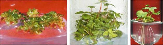
The induction of adventitious buds from explant tissues is the most widespread method of micropropagation for bulbous flower crops (lilies, daffodils, hyacinths, tulips) using bulb scales, segments of the basal plate, and leaf explants; for garden strawberries, using the base of shoots regenerated in meristem culture (Fig. 5.4); for Representatives of the genus Brassica (cauliflower, HEAD cabbage, Brussels sprouts, broccoli) using hypocotyl, cotyledon, and leaf segments; for chicory, using leaf blade segments; for petunias, using root segments; and for gloxinias, saintpaulias, streptocarpus, and echinapsis, using leaf blade segments.
Fig. 5.4. Microclonal propagation of the strawberry Fragaria × ananassa Duch. via the induction of adventitious buds from the base of shoots regenerated in meristem culture (after M. Biswas et al., 2009)
Plantlets regenerated using this method do not always fully reproduce the genotype of the original stock plant, as the cytogenetic stability of cells from specialized tissues is not as high as that of meristematic tissue cells. Plants propagated vegetatively over long periods may accumulate somatic Mutations, resulting in chimerism. Some ornamental variegated plants are genetic chimeras. The formation of adventitious buds from altered cells leads to the production of genetically distinct plantlets. Nevertheless, experience shows that when using this propagation method, the variation among regenerated plants in most cases does not exceed natural levels.
Adventitious stem buds can also form in in vitro callus culture (stem organogenesis). Fast-growing callus cultures that retain a high capacity for organogenesis provide additional opportunities to accelerate micropropagation. However, the genetic instability of callus cells can lead to the production of regenerated plants with significant genetic differences from the original stock plant (somaclonal variation) (Fig. 5.5). Such plants may be of breeding interest, but for commercial plant propagation, this Variability is a very significant drawback. Nevertheless, this method is justified in specific cases where other methods are ineffective, or for crops with low somaclonal variation (amaryllis, episcias, dracaenas, tomatoes, asparagus, and certain tree species).
Fig. 5.5. Propagation of orchids of the genus Phalaenopsis (a–c) in in vitro culture via regeneration from adventitious buds in callus culture (after A. Hussain et al., 2012); an example of somaclonal variability in the orchid Doritaenopsis during regeneration from adventitious buds (d–f) (after S. Y. Park et al., 2009): a - callus with a mass of developing adventitious buds; b - mass shoot regeneration; c - root formation on a hormone-free medium; d - flower of the original Doritaenopsis clone from which the explant was taken (normal flower); e and f - somaclonal variants with altered flower shape and size obtained via regeneration from adventitious buds

All of the above also applies to the method of in vitro clonal plant propagation based on the induction of somatic embryogenesis from explant tissues or callus cells in suspension culture. At the same time, certain positive characteristics of this method place it among the most promising. Technologies for the mass production of somatic embryos and their adaptation to ex vitro conditions have been developed for a number of important crops. These technologies include, among other things, the possibility of using bioreactors (see Chapter 4) and mechanized production of artificial seeds via embryo encapsulation, which makes it possible to substantially reduce labor costs (both during propagation and when transferring plants to ex vitro conditions) and production costs.
The concept of encapsulating somatic embryos to produce artificial seeds was proposed by T. Murashige as early as the 1970s. It was implemented in practice in the early 1980s using desiccated somatic embryos of carrot (S. Kitto, J. Janick, 1982) and hydrated (non-desiccated) embryos of alfalfa (K. Redenbaugh et al., 1984). The second of these technologies has become the most widely adopted; it involves encapsulating somatic embryos in various gel-like substances based on agar, alginate, carboxymethylcellulose, carrageenan, gellan gum, sodium pectate, etc. The best results have been achieved using alginate.
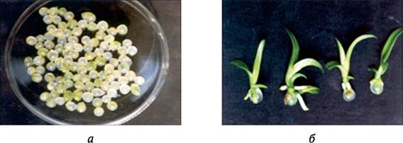
Alginic acid is a natural polysaccharide derived from seaweed fibers, consisting of two monomers: mannuronic and hyaluronic acids (in varying proportions). The long chains of these acids can cross-link to form three-dimensional networks, binding divalent Metal Ions via their carboxyl groups. The sodium salt of alginic acid is readily soluble in Water, forming viscous solutions that make it widely useful in the food industry as a thickener and stabilizer. To produce artificial seeds, sodium alginate is dissolved in the nutrient medium used to grow somatic embryos. Using specialized pipette-equipped devices, individual embryos are placed into separate drops of this solution. When immersed in a solution of calcium nitrate or calcium chloride, the alginate droplets containing the embryos form solid, spherical capsules as Na+ ions are replaced by Ca2+ ions. Capsule hardness depends on the extent of Ion Exchange (complexation is typically carried out using a 1-5% sodium alginate solution in 100 mM calcium nitrate for 20-30 minutes, after which the capsules are rinsed with water or nutrient medium) (Fig. 5.6).
Fig. 5.6. Artificial orchid seeds (somatic embryos encapsulated in alginate gel) (a) and seedlings derived from them (b) (adapted from G. Saiprasad, 2001)
Unlike zygotic embryos, somatic embryos lack an endosperm. To compensate for this deficiency, all essential nutrients are incorporated directly into the capsule matrix. This ensures embryo viability for a relatively long period (6 months or more) when stored at 4 °C, as well as The ability to germinate when the capsules are sown in soil. In addition, fungicides, pesticides, and Antibiotics are added to the matrix to improve capsule preservation, along with activated charcoal to enhance embryo Respiration. In some cases (such as the encapsulation of certain orchid seeds), mycorrhizal Fungi are included, which are essential for successful seed germination and seedling development.
To improve the quality of artificial seeds, embryo development is synchronized (via filtration through sieves of various pore sizes, centrifugation, or automated sorting), and a specialized pre-encapsulation conditioning regime is applied. This involves growing the embryos in a liquid nutrient medium supplemented with an osmotic agent (10% mannitol) under light (approx. 300 lux, 16-hour photoperiod). During this period, their size increases 3- to 4-fold, and chlorophyll content rises. Next, they are dried on filter paper under light for one week (reducing water content to 80-90%), and then cultivated for two weeks on a nutrient medium containing 2% sorbitol, 0.01 mg/L 6-BA, and 0.01 mg/L GA3 under an elevated carbon dioxide atmosphere (2%), which transitions the embryos to autotrophic Nutrition. Encapsulated embryos prepared in this manner exhibit high germination rates for artificial seeds—around 50%.
Overall, the low germination rate of artificial seeds remains the primary obstacle to their widespread application. A major drawback of this method is the potential for somaclonal variation in the resulting plants. Nevertheless, this technology is successfully employed for propagating several valuable crops: orchids (Dendrobium, Cymbidium, Phalaenopsis, Spathoglottis), the renowned Indian medicinal tree neem (Azadirachta indica), sandalwood (Santalum album), alfalfa, and cotton. For oil palm (Elaeis guineensis) propagation, the induction of somatic embryogenesis is the only viable method of vegetative propagation.
5.2.2. Transferring micropropagated plants from in vitro to ex vitro conditions
Plants obtained via in vitro clonal propagation develop under conditions vastly different from those in standard soil culture: high levels of inorganic and organic nutrients, sucrose as a carbohydrate source, exogenous growth regulators in the medium, high humidity, low light intensity, and restricted gas exchange. While these microclimatic conditions are highly favorable for rapid growth and multiplication, they induce structural and physiological traits in the plants that severely hinder their survival under ex vitro conditions.
Leaves formed under high humidity and heterotrophic nutrition exhibit poor anatomical differentiation: the palisade tissue is absent or poorly developed, the mesophyll consists mainly of spongy parenchyma cells with large intercellular air spaces, and Chloroplasts feature unorganized grana alongside low chlorophyll and protein contents. Photosynthetic Enzymes show reduced activity, and the leaf vascular system is underdeveloped. Numerous Stomata remain permanently open and fail to respond to environmental triggers (such as darkness, CO2, or Abscisic acid) that normally induce closure. A protective cuticular wax layer is virtually absent, and the wax that does form differs significantly in chemical composition from that of naturally grown plants (exhibiting lower hydrophobic properties). Stems of in vitro-grown plants are insufficiently lignified, containing significantly less collenchyma and sclerenchyma than normal plants. Their cells possess thin walls, enlarged intercellular spaces, and underdeveloped vascular systems. Furthermore, many plants fail to form root hairs under in vitro conditions.
These unique Anatomical and physiological features of in vitro plants create significant challenges during their environmental adaptation upon transfer to ex vitro conditions. It is common to observe growth arrest, leaf abscission, and plant mortality after transplanting into soil, primarily due to water balance disruptions. To minimize losses, various methodological techniques have been developed to improve plant survival rates, some applied in vitro and others following transplanting into soil.
The final subculture prior to transplanting is recommended to be carried out on a mineral salt-depleted nutrient medium, such as White's medium, with the sucrose concentration reduced to 1%. Cytokinins are completely omitted from the medium, leaving only auxins to stimulate root formation (such as IBA). It is also suggested to increase light intensity (up to 8-10 thousand lux) and slightly lower the cultivation Temperature compared to standard conditions. Effective treatments include The addition of growth retardants and adaptogens—such as succinic acid, kresacin, or Mival-Agro—to the nutrient medium; these compounds inhibit excessive stem elongation, promote root thickening, reduce leaf surface area, increase chlorophyll content, and stimulate cuticular wax deposition. A similar effect is achieved by lowering the relative humidity within culture vessels through desiccants, cooling the bottom of the vessels, or simply removing the stoppers from test tubes 7-10 days before soil transfer. For a number of woody species, mycorrhization of in vitro plants yields highly favorable results.
Transplanting in vitro plants into soil is typically performed in spring or early summer. Greenhouses or growth rooms with controlled lighting, temperature, and humidity are utilized. To mitigate dehydration-induced mortality, high air humidity is maintained post-transplant using misting systems, and plantings are covered with non-woven agricultural fabrics. An interesting approach to reducing leaf Transpiration, proposed by Indian researchers, involves spraying plants during the acclimatization period with a 50% aqueous glycerin solution or a 1:1 mixture of paraffin and diethyl ether.
Suitable substrates for transplanting in vitro plants include neutralized high-moor peat enriched with mineral salts, peat-sand mixtures (3:1), and perlite. Excellent results have been achieved with ion-exchange resin substrates developed at the Institute of Physical and Organic Chemistry of the National Academy of Sciences of Belarus; however, such substrates are quite expensive, which limits their large-scale use. Once rooted plants in the soil resume active growth, they are fed with complex fertilizers and treated for pests and diseases. Following the adaptation period (20-30 days), they are transplanted to their permanent location in a greenhouse or field (Fig. 5.7).
Fig. 5.7. Techniques applied for the acclimatization of micropropagated plants during their transfer from in vitro to ex vitro conditions

The challenges of transferring micropropagated plants to soil are related not only to environmental adaptation, but also to the complexity and labor intensity of the procedure itself, which resists mechanization. Industrial production requires planting vast numbers of in vitro plants (up to 100,000 or more) within short timeframes. At the same time, to avoid plant damage, they must be extracted from culture vessels and planted with extreme care. Great promise in solving these problems lies in utilizing pre-formed in vitro storage organs: tubers, bulbs, and corms (often referred to in literature as micro-tubers, micro-bulbs, and micro-corms), as well as artificial seeds (encapsulated somatic embryos). These propagules are much easier to store, transport, and plant directly into soil without requiring complex acclimatization.
Micro-bulbs are formed by inducing adventitious stem buds from scale segments or the basal plate of bulbous plants. For example, dividing a lily bulb scale into small segments can yield up to 100 micro-bulbs within 35-40 days.
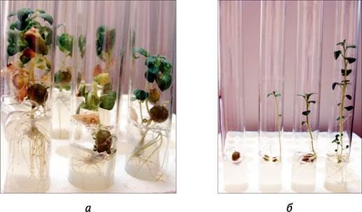
Under specific cultivation conditions, potato micropropagated plants are capable of forming micro-tubers (Fig. 5.8, a). Spontaneous micro-tuberization occurs if potato cultures are left on the nutrient medium until it dries out. To accelerate this process, media supplemented with cytokinins (3-5 mg/L 6-BA) and high sucrose concentrations (6-10%) are used. The addition of growth retardants (50-100 mg/L chlormequat chloride) to the medium can also be effective. Lower temperatures (15-18 °C), a shortened photoperiod (down to 10 hours), or transferring cultures to total darkness likewise promote tuberization. Both micro-tubers and micro-bulbs can be produced in liquid medium using bioreactors, significantly reducing production costs. Different potato varieties form in vitro micro-tubers with varying efficiency. This technology is unsuitable for certain cultivars, as only a small percentage of their micropropagated plants will form tubers even under optimal conditions. Therefore, micro-tuberization capacity must be evaluated for each specific cultivar and applied only to those yielding high productivity.
Fig. 5.8. Micro-tuber formation in potato micropropagated plants (a). Regeneration of plants from micro-tubers after cold storage (b) (from left to right): a micro-tuber with a sprouting bud; various stages of shoot growth, separated from the tuber for cutting or rooting; a rooted micropropagated plant separated from the micro-tuber
In vitro-derived micro-tubers are largely physiologically similar to tubers harvested from field-grown plants. In particular, they exhibit a prolonged dormancy period (3-4 months, depending on the cultivar). While this is a highly beneficial trait for maintaining potato germplasm collections, it presents certain inconveniences when applied to clonal propagation, as it shortens the micropropagation window: micro-tubers must be harvested three months prior to planting to allow completion of dormancy. Consequently, an optimal strategy combines two potato micropropagation methods: during the initial phase, plants are propagated via stem cuttings (via axillary meristem induction), after which a portion of the resulting micropropagated plants is transferred to micro-tuber induction conditions, while the remainder continues to be propagated through cuttings. As a result, both traditional micropropagated plants and dormant micro-tubers are planted in the soil, reducing overall labor costs.
The Institute of Genetics and Cytology of the National Academy of Sciences of Belarus has developed a potato micro-tuber production protocol that ensures workflow rhythmicity and enables the efficient utilization of equipment and personnel (A. Ermishin, 1994). The scheme comprises three distinct production units (nurseries) of specific capacities: Nursery 1—stock plants (for cuttings); Nursery 2—pre-growth nursery (where cuttings develop into plants before being transferred to optimal micro-tuber induction conditions); Nursery 3—tuberization nursery. A crucial feature ensuring technological rhythmicity is the schedule of stock plant propagation and nursery restocking. The daily volume of cuttings processed must precisely match two criteria: maintaining the operational schedule for restocking Nursery 2, while fully replenishing Nursery 1 to maintain a constant stock volume.
The dynamics of nursery restocking are illustrated in the table below.
Dynamics of nursery restocking during potato micro-tuber production (asterisks denote daily production volumes)
Time (weeks) |
Nursery 1 |
Nursery 2 |
Nursery 3 |
Storage |
0 |
***** |
|||
3 |
***** |
***** |
||
6 |
***** |
***** |
***** |
|
9 |
***** |
***** |
***** ***** |
|
12 |
***** |
***** ***** ***** |
||
15 |
***** ***** ***** |
***** |
The production cycle begins by having a predetermined number of in vitro plantlets in nursery 1. The formula for calculating its volume is as follows:
А = Rt/W(b-1),
where A is the volume of nursery 1; R is the number of microtubercles to be produced (target production plan); W is the total number of planned working days allocated for cutting propagation; b is the number of cuttings obtained from a single stock plant; t is the number of working days required to grow a plant suitable for cutting (i.e., capable of producing b cuttings). The values of B AND T are determined empirically for each specific cultivar, the cultivation conditions of stock plants, and the Organization OF THE production process.
To determine the number of lighting units and the production floor space required to fulfill the production plan, information regarding the maximum volume of nursery 2 is necessary. It is calculated using the formula
F = Re/W,
where F is the volume of nursery 2; e is the number of working days during which the in vitro plantlets are kept in nursery 2; W is the total number of planned working days.
Thus, if the objective is to produce R = 30 thousand microtubercles over W = 50 days, given that each stock plant yields b = 4 cuttings, and the time required to grow a plantlet ready for cutting takes t = 15 working days, then at THE START OF the production cycle, nursery 1 must house 3 thousand stock plants. At maximum capacity, the volume of nursery 2 will reach 9 thousand plants.
Producing 30 thousand microtubercles via traditional technology (first obtaining the required number of plants through cutting propagation and then transferring them to conditions optimal for microtubercle formation) would require a facility equipped with lighting units capable of holding 30 thousand test tubes (yielding 1 microtubercle per in vitro plantlet). In contrast, our example requires facilities for only 12 thousand test tubes (3 thousand in nursery 1 and 9 thousand in nursery 2), which is 2.5 times fewer.
At the beginning of the production cycle using traditional methods, cutting volumes are relatively small. However, towards the end of the cycle, a massive batch of 7.5 thousand plants (30 thousand divided by 4) must be cut within a very short timeframe. This is extremely demanding, considering that on average, a single employee handles 150–200 cuttings per working day. The proposed technology completely solves this problem: the workload is distributed evenly throughout the entire cycle, and daily output remains stable and relatively low.
5.2.3. Practical Applications OF In Vitro Plant Clonal Propagation
The advent of in vitro plant clonal propagation and the subsequent rapid integration of this technology into the industrial production of seed and planting material were pioneered by J. Morel (1960) using orchids. The success and rapid progress in this field were largely due to the fortunate choice of the research subject. Orchids are a high-value and immensely popular ornamental crop in the West. However, traditional vegetative propagation methods are largely ineffective, and seed propagation of orchids is equally problematic. Due to the high heterozygosity of orchid cultivars, their sexual progeny do not retain the traits of the parent forms. Orchid seeds are extremely minute and lack an endosperm. Upon germination, the embryo develops into a protocorm—a spherical, tuber-like structure that subsequently differentiates into a seedling. In many cases, this requires Symbiosis with specific fungi. Orchid seedlings develop extremely slowly, taking at least around 5 years for the plants to reach the flowering stage. J. Morel discovered that in apical stem meristem cultures of Cymbidium, protocorms are formed that closely resemble those resulting from embryo germination. Under specific cultivation conditions, these protocorms are capable of rapidly giving rise to several new protocorms, which are then separated from the original protocorm and transferred to a fresh nutrient medium for further propagation or seedling development (Fig. 5.9). Furthermore, the use of in vitro culture methods has significantly increased the efficiency of orchid seed propagation. Germinating orchid seeds on nutrient media under in vitro conditions considerably accelerates seedling development by shortening the juvenile phase without the involvement of mycorrhiza.
Fig. 5.9. Propagation of the orchid Phalaenopsis gigantea via in vitro culture of protocorms: a - 150-day-old protocorm planted on a nutrient medium; b - initiation of new protocorms; c - maturation of protocorms within 4 weeks after explant placement on the medium; 2 - developed orchid shoots after 10 months of explant cultivation (based on R. Murdad et al., 2006)
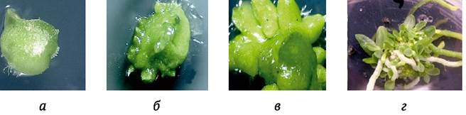
Naturally, these micropropagation technologies were rapidly adopted by orchid propagators worldwide. Today, industrial micropropagation technologies have been developed and are widely used for a vast number of orchid species, as well as other valuable floral and ornamental crops. For instance, in the Netherlands, tens of millions of plants are produced annually via micropropagation.
The second major Practical Application of in vitro plant clonal propagation technology is the production of high-quality seed potatoes. For this crop, the quality of planting material is paramount to achieving high yields. Traditional potato seed production relies on selecting healthy, productive plants of a specific cultivar that closely match its description, followed by their propagation using practices designed to maintain plant health (at least three phytosanitary roguing passes per season, insecticide treatments, early haulm destruction, etc.). The production cycle begins with establishing a preliminary testing nursery and selecting initial clones. Because the potato propagation rate is relatively low (4–6), this nursery must be quite large to yield a sufficient quantity of elite seeds over the subsequent 4–5 years of clonal multiplication. The health status of plants in this nursery is assessed primarily visually, as the sheer volume makes it practically impossible to evaluate every plant using ELISA or other virus-detection methods. Consequently, visually healthy plants harboring latent viral infections may remain in the nursery. As a result, despite all efforts to maintain plant health during propagation, pathogen accumulation occurs, ultimately diminishing the quality of the planting material.
The introduction of potato micropropagation technology makes it possible to establish an initial clone nursery using plants with a high probability of being virus-free. This is achieved as follows. The production cycle begins with the Selection of the most productive, visually healthy plants of a specific cultivar that conform to its description. The selected plants are then tested for the presence of major viruses and viroids in their tissues using sensitive immunoassays or pathogen nucleic acid testing. Virus-free plants serve as the explant source for establishing meristem cultures. To ensure the reliability of obtaining completely virus-free meristem clones, thermo- and/or chemotherapy are recommended. In vitro plantlets regenerated from meristems are further tested for viruses and viroids. If they test negative (pathogen-free), micropropagation of these plants begins via cuttings and microtubercle production (the production cycle in Belarus starts in September). In the spring of the following year, the produced plantlets and post-dormancy microtubers are planted in greenhouses, where they form so-called minitubers over the summer season. It is preferable to grow plants in greenhouses covered with gauze or non-woven fabrics, which prevent plant overheating in hot weather while protecting them from aphids—the vectors of viral infections. Necessary crop management is also implemented, including regular Fertilization and chemical treatments against plant pathogens and pests. The resulting minitubers are planted in the field the following year. Subsequently, the plants are propagated using traditional technology, carrying out the full range of measures to maintain their health.
The application of potato micropropagation technology enables the rapid production of large quantities of healthy initial clones. This shortens the production timeline for elite seed stock and consequently enhances its quality by minimizing the duration of viral accumulation. For example, in the Netherlands and other leading potato seed-producing nations, a three-year cycle is practiced: minitubers — super-super-elite — super-elite — elite.
Micropropagation technologies have also found widespread application in woody and shrub crops, encompassing not only various fruit and ornamental species but also plants used in urban landscaping and reforestation. This is largely because these technologies yield large quantities of healthy planting material of a defined genotype with predictable traits (cultivars, strains, rootstocks, elite trees). For instance, reforestation is predominantly carried out using open-pollinated seeds harvested from high-yielding elite or plus-trees. However, due to genetic segregation, the resulting progeny will not possess the full suite of positive traits of the parent forms. Micropropagation makes it possible to replicate the genotype of such trees across numerous clones. Its application is particularly crucial for crops that are nearly impossible to propagate via traditional vegetative methods, such as oil palms. Furthermore, it holds immense promise for propagating crops that do not produce fertile seeds (e.g., the triploid edible banana Musa x paradisiaca) or for dioecious plants (when growing papaya from seeds, the sex of individual plants is only revealed upon flowering, whereas female forms hold practical value).
Plant micropropagation methods are vital not only for the industrial production of planting material across various crops. The use of specialized micropropagation techniques and in vitro culture of plant cells, tissues, and organs enables the efficient maintenance of extensive genetic collections of source material—which is indispensable for success in modern breeding—as well as the conservation and propagation of rare and endangered plant species, and the exchange and transportation of collection accessions.
5.3. Conservation of Valuable Gene Pools In Vitro
The most effective method for maintaining genetic collections is seed storage at low temperatures. Standard technology used in gene banks involves drying seeds to a moisture content of 5–8% and subsequently storing them at -18 °C, allowing them to retain viability for many years without significant loss of germination capacity.
However, a large group of plants exists for which this technology is unsuitable: cultivars and strains of vegetatively propagated crops, sterile forms, and plants that produce seeds which rapidly lose viability or cannot withstand drying and low temperatures. Traditional vegetative propagation methods used to maintain genetic collections of such crops are costly and inefficient. To enhance the maintenance efficiency of genetic collections for these crops, the following methodological approaches are currently employed:
✵ maintaining collections as slow-growing in vitro plantlet cultures;
✵ cryopreservation of tissues or cell cultures from specific genotypes.
5.3.1. Conservation of genetic Collections as Slow-Growing In Vitro Plantlet Cultures
The development of micropropagation methods has successfully addressed most of the challenges associated with maintaining plant genetic collections. In many cases, this is achieved through regular subculturing of in vitro-propagated plantlets obtained via meristem culture. However, plant propagation and in vitro collection maintenance serve different purposes. While the primary goal of the former is to produce as many clones of a specific genotype as possible in the shortest possible time, the latter focuses simply on preserving the viability of in vitro plantlets at the lowest possible cost. To reduce the costs of maintaining in vitro collections, various growth-retardation techniques are employed to extend the time intervals between subcultures (Fig. 5.10). These techniques include:
✵ lowered cultivation temperatures;
✵ reduced atmospheric pressure or lowered oxygen concentration in the cultivation atmosphere;
✵ addition of osmotic agents or growth regulators to the nutrient medium to suppress plant growth; and
✵ production and storage at low temperatures of plant storage organs (microtubers, microbulbs, microcorms) or encapsulated somatic embryos.
Fig. 5.10. Main techniques for enhancing the efficiency of maintaining plant genetic collections in in vitro culture

The subculture interval for in vitro cultures can be extended to 6–24 months or even longer by cultivating the plantlets under low positive temperatures (1–10 °C). In this approach, light intensity is reduced (to 10–50 lux) while maintaining a standard 16-hour daily photoperiod, or cultivation is carried out in total darkness. The optimal temperature is determined empirically, with the objective of achieving maximum growth retardation without significantly compromising culture viability. As a rule, lower temperatures (1–4 °C) are used for cold-hardy cultures whose normal cloning temperature is 20–25 °C. For thermophilic cultures (cloned at approximately 30 °C), growth suppression is achieved by cultivating the plants at 8–16 °C. Using this method, R. H. Mullin and D. E. Schlegel (1976) maintained a collection of in vitro-propagated garden strawberry (Fragaria × ananassa Duch.) plantlets for 6 years without subculturing, simply by adding a few drops of fresh nutrient medium every three months.
Growth suppression in in vitro plant cultures can also be achieved by lowering the atmospheric pressure (down to 0.5 mm Hg) and/or modifying the gaseous environment by reducing the oxygen concentration (techniques adapted from the storage practices for vegetables, fruits, and cut flowers). Hypoxic conditions are created using a gas mixture of an inert gas (nitrogen) and 1–10% oxygen. When lowering atmospheric pressure, it is essential to maintain high humidity to prevent rapid desiccation of the nutrient medium. This method is particularly promising for preserving collections of thermophilic plants that are unsuited for low-temperature storage.
An effective way to reduce the growth rate of in vitro plants is to incorporate osmotic agents and retardants into the nutrient medium. To induce osmotic stress that slows down culture growth, elevated sucrose concentrations (6–10%) or 3–6% mannitol or sorbitol are added to the medium. Plant growth retardants such as chlormequat chloride, maleic hydrazide, daminozide (B-995), and Phosfon D are also utilized.
For the preservation of a range of crop collections, promising approaches include obtaining microtubers, microbulbs, or microcorms, as well as encapsulating somatic embryos or plant fragments in alginate beads for subsequent low-temperature storage. For instance, potato microtubers can be stored in sterile test tubes in a refrigerator for 6–12 months. After breaking dormancy, they sprout, producing long etiolated shoots. Once storage is complete, these shoots can be removed, the tuber cut in half, and placed on a nutrient medium where, under light, normal in vitro plantlets regenerate from dormant buds, ready for propagation or further in vitro storage (see Fig. 5.8, b).
Encapsulation in alginate beads is an effective method for preserving plant material (somatic embryos, axillary buds, stem segments of in vitro plantlets, root cultures) at low temperatures (1–4 °C). Encapsulation significantly slows down growth and transpiration, thereby ensuring the long-term viability of the cultures. When combined with dehydration, encapsulation has a highly beneficial effect on the post-storage survival of cultures during cryopreservation.
5.3.2. Cryopreservation of Plant Tissues and Cell Cultures
Cryopreservation is the Storage of Biological objects in a frozen state at ultralow temperatures (most commonly at -196 °C in liquid nitrogen) with the potential to fully restore their Functions upon thawing. In principle, this method allows for indefinite storage, as ultralow temperatures virtually halt all biochemical processes within cells. Cryopreservation is widely used for preserving microorganisms and certain animal specimens. Indeed, modern animal breeding is unimaginable without the use of animal semen stored in liquid nitrogen for Artificial Insemination. Methods for the cryopreservation of plant Cells and Tissues have also been developed. Initially, they were applied to store cell lines producing secondary metabolites. Later, techniques were established for preserving apical meristems, young embryos, and somatic embryos, making it possible to maintain genetic collections with high efficiency.
Low-temperature exposure is lethal to living organisms because it causes irreversible cellular damage. First, ice crystals forming during rapid temperature drops damage cell Organelles and the cells themselves. Second, cooling can cause cells to lose 80–90% of their water content, leading to a toxic increase in the concentration of intracellular solutes, the destruction of hydrated macromolecule complexes, and the loss of biopolymer activity due to alterations in their Tertiary and Quaternary structures.
Several methodological approaches have been developed to ensure cell survival during freezing. First and foremost, the choice of the biological object for cryopreservation is critical. Small, Cytoplasm-rich plant meristematic cells tolerate freezing much better than large, highly vacuolated cells. Therefore, prior to cryopreservation, cell Suspensions of secondary metabolite producers must be subcultured frequently, and cells for freezing should be harvested in the late lag phase or early exponential growth phase.
The use of cryoprotectants—substances that protect cells from freezing-induced injury—is crucial for successful cryopreservation. Although many substances possess cryoprotective properties, only a few are used in practice. Cryoprotectants are divided into two types: penetrating (intracellular) and non-penetrating (extracellular). Penetrating protectants (glycerol, propylene glycol, Ethylene glycol, dimethyl sulfoxide [DMSO], and Amino acids such as Proline and asparagine) inhibit ice crystal formation by forming Hydrogen Bonds with water molecules. Non-penetrating cryoprotectants (sucrose, trehalose, Ficoll, albumin, polyvinylpyrrolidone) are believed to shield cells from the osmotic shocks associated with freezing and to reduce the growth rate of ice crystals. Non-penetrating cryoprotectants are typically used as supplementary components in solutions containing penetrating cryoprotectants.
The efficiency of cryopreservation can be enhanced by preculturing the specimens under specific conditions designed to reduce cellular water content. For instance, beneficial results are obtained by preculturing in vitro plantlets—whose apical meristems are destined for cryopreservation—for several days on nutrient media supplemented with 2–6% mannitol or sorbitol, or 2.5–10% DMSO, at reduced temperatures (4–6 °C).
A drastic reduction of intracellular water to a minimum is achieved by drying the biological material in a laminar air flow cabinet using silica gel. This technique is applied to the cryopreservation of somatic embryos, stem meristems, and hairy root cultures. It is especially effective when combined with the encapsulation/dehydration technique. Meristems or embryos enclosed in calcium alginate beads are incubated in a nutrient medium containing 0.85 M sucrose for 4–16 hours, desiccated in a laminar airflow for 3–4 hours, and then plunged into liquid nitrogen. As we can see, sucrose serves as the sole cryoprotectant—it is one of the least toxic substances with cryoprotective properties.
The cryopreservation process for PLANT CELLS AND tissues comprises the following stages: freezing, storage, thawing, and reculturing (Fig. 5.11).
Fig. 5.11. The process of in vitro plant cryopreservation

Materials prepared for freezing (via preculturing, desiccation, or encapsulation/dehydration) are treated with cryoprotectants if necessary, packed into sterile 1–2 ml screw-cap polypropylene vials, and subjected to freezing. Although direct immersion of material into liquid nitrogen is possible in some cases, programmed freezing is typically employed. The temperature is lowered slowly (0.5–1 °C per min) down to -35 °C, held at that temperature for 15–30 minutes, and only then is the material immersed in liquid nitrogen. Programmed cooling is carried out using specialized commercially manufactured apparatus.
Material is stored in liquid nitrogen containers or refrigerators equipped with specialized racks to arrange the vials methodically, enabling rapid location and retrieval of any required sample. The vials must remain constantly immersed in liquid nitrogen. Due to natural evaporation, regular topping up with liquid nitrogen (typically once a week) is required.
Unlike the slow freezing process, thawing after storage is performed rapidly: vials retrieved from liquid nitrogen are immediately placed in a water bath heated to +37–40 °C. Rapid thawing prevents the formation of intracellular ice crystals, which can occur during gradual warming. A 1-ml vial thaws within 0.5–1 minute. Afterward, the cultures are washed free of cryoprotectants (many of which are toxic) using a nutrient medium or a 3–10% sucrose solution, and transferred to a fresh nutrient medium to resume growth. Following cryopreservation, cell suspensions are plated onto an agar-solidified nutrient medium, and the resulting callus is subsequently resuspended in a liquid nutrient medium. Embryos, somatic embryos, and apical meristems are placed on an agar-solidified medium supplemented with low concentrations of cytokinins and gibberellin, which promotes the regeneration of plantlets suitable for subsequent cloning. Encapsulated embryos and somatic embryos are handled similarly.
Below is an example protocol for the cryopreservation of a maize cell suspension culture according to L. Withers and P. King (1979):
1. Actively growing cell suspension cultures are subcultured onto a nutrient medium supplemented with 10% proline.
2. After 3–4 days, the cells are rinsed in a proline-free medium and subsequently resuspended in fresh nutrient medium.
3. The suspension is cooled in an ice bath, and an equal volume of chilled nutrient medium containing 20% proline is added (gradually, in 4 steps over 1 hour).
4. Aliquots of 1 mL of the suspension are transferred into sterile 2 mL polypropylene screw-cap cryovials.
5. The vials are placed in a programmable controlled-rate freezer and cooled to -30 °C at a rate of 1 °C per minute.
6. The vials are plunged into liquid nitrogen for long-term storage of the cultures.
7. Thawing is performed in a water bath at 40 °C for 1–2 minutes.
8. The thawed suspension is plated onto an agar-solidified nutrient medium to initiate culture recovery and growth.
A similar procedure is employed for the cryopreservation of shoot apices, except that DMSO (approximately 10%) is used instead of proline during pre-culture.
Modern in vitro plant clonal propagation technologies provide exceptionally high multiplication rates. A major advantage is their ability to efficiently propagate plant species that are otherwise difficult or recalcitrant to propagate using conventional methods. All operations can be conducted year-round without requiring large land areas. Crucially, micropropagated plants are free from reinfection by viral and other pathogens, provided that the initial stock plant is healthy. Overall, in vitro clonal propagation is economically more advantageous than traditional vegetative propagation techniques and yields higher-quality planting material.
Shoot meristems are the most suitable explant for introducing plants intended for vegetative propagation into in vitro culture, as they best preserve the genotype of the stock plant and ensure the production of pathogen-free plants. Unlike cells of specialized tissues, initial cells of apical meristems exhibit high cytogenetic stability, retaining the zygotic genotype throughout the plant's life cycle. Another essential property of meristematic tissues is the absence or extremely low concentration of viral particles, even in virally infected plants.
In 1952, G. Morel and C. Martin developed a technique for eliminating viruses from dahlia plants using in vitro culture of apical meristems approximately 100 µm in size (G. Morel, C. Martin, 1952). Since then, this method has been applied with varying degrees of success to a wide range of vegetatively propagated crops. Meristem culture has been shown to eradicate not only viruses but also bacterial and fungal infections, viroids, and mycoplasmas.
To enhance the efficiency of virus elimination via meristem culture, stock plants are subjected to thermotherapy prior to explant excision—a prolonged treatment (5–15 weeks) at elevated temperatures (37 °C, 90% relative humidity, light intensity of approximately 5,000 lux, and a 16-hour photoperiod)—or chemotherapy, which involves supplementing the nutrient medium for apical meristems with various antiviral compounds. Most commonly, chemotherapy utilizes synthetic analogues of purine and pyrimidine bases (such as cyanoguanosine, 2-thiouracil, and D-ribofuranosyl-1,2,4-triazole-carboxamide, commercially known as virazole), as well as interferon, ridostin (sodium ribonucleate), binase (microbial ribonuclease), and phenolic acids including salicylic, gallic, syringic, caffeic, ferulic, and coumaric acids.
In practice, the following in vitro clonal propagation methods are employed:
✵ activation of the development of pre-existing meristems (shoot apex, axillary, and dormant stem buds);
✵ induction of adventitious buds on explants or within callus cultures;
✵ somatic embryogenesis.
The activation of pre-existing meristems relies on breaking apical dominance in in vitro cultures by excising the apical bud or supplementing the nutrient medium with cytokinins. Compared to other micropropagation techniques, this method best preserves the genotype of the original stock plant because the resulting clones originate directly from meristematic cells, which are characterized by high cytogenetic stability.
In vitro clonal propagation via the induction of adventitious buds is based on the property of plant cell totipotency. This approach involves explants from various plant parts and organs lacking pre-existing shoot buds acquiring the capacity to form them in vitro when exposed to stimuli that induce shoot organogenesis, primarily appropriate growth regulators. Suitable explants include segments of leaf blades and petioles, young inflorescences, bulb scales, basal plates, corms, root cuttings, and lateral or intercalary meristems, among other organs and tissues.
Adventitious shoot buds can also form within in vitro callus cultures (shoot organogenesis). Fast-growing callus cultures that retain a high capacity for organogenesis provide additional opportunities to accelerate micropropagation. However, the genetic instability of callus cells can lead to regenerant plants with significant genetic deviations from the source stock plant (somaclonal variation).
All of the above also applies to in vitro clonal propagation methods based on inducing somatic embryogenesis from explant tissues or callus cells in suspension culture. Nevertheless, several positive attributes position this method among the most promising approaches. Technologies for the mass production of somatic embryos and their ex vitro acclimation have been developed for a number of important crops. These technologies include the potential use of bioreactors and mechanized production of synthetic seeds via embryo encapsulation, which significantly reduces labor costs (during both propagation and transfer to ex vitro conditions) and overall production expenses.
The microclimatic conditions during the cultivation of in vitro plants often induce structural and physiological traits that severely impede their survival under ex vitro conditions. Post-transplant setbacks such as growth arrest, defoliation, and plant mortality are frequently observed, primarily due to water balance disturbances. A range of methodological approaches has been developed to improve plant establishment rates. Pre-conditioning micropropagated plants prior to soil transfer involves using mineral-deficient nutrient media with lowered sucrose levels and no cytokinins. Supplementing the medium with growth retardants or adaptogens has also proven effective. Additional measures include increasing light intensity (up to 8,000–10,000 lux), lowering cultivation temperatures, reducing relative humidity within culture vessels using desiccants or bottom cooling, and simply removing culture closures 7–10 days prior to transplanting. For several woody species, mycorrhization yields beneficial effects. Following transfer, maintaining optimal temperatures, high atmospheric humidity, and specialized rooting substrates further AIDS establishment.
The challenges associated with transplanting in vitro plants stem not only from acclimation to novel growing environments but also from the complexity and labor-intensive Nature of the procedure itself, which hinders mechanization. Significant progress in overcoming these hurdles is linked to the use of pre-formed in vitro storage organs—such as tubers, bulbs, and corms (often referred to in literature as micro-tubers, micro-bulbs, and micro-corms)—as well as synthetic seeds (encapsulated somatic embryos). These structures are not only easier to plant directly into soil without prior acclimation, but they are also much easier to store and transport.
Industrial technologies for micropropagation of numerous valuable floral and ornamental species have been developed and are now widely used. They have found extensive application in the production of high-quality seed potatoes, as well as planting material for trees and shrubs (fruit-bearing, ornamental, and those used in urban landscaping and afforestation).
Plant micropropagation methods are of great importance not only for the commercial production of planting material, but also for maintaining genetic collections of source material through in vitro plant cultures and the cryopreservation of tissues or cell cultures from specific genotypes.
To reduce costs when maintaining in vitro collections, various growth-retardation methods are employed, which help extend the intervals between subcultures. These include:
✵ reduced cultivation temperatures;
✵ lowered atmospheric pressure or reduced oxygen concentration in the cultivation atmosphere;
✵ the addition of osmotica or growth regulators to the nutrient medium to slow plant growth;
✵ the production and low-temperature storage of plant storage organs (microtubers, microbulbs, microcorms) or encapsulated somatic embryos.
Storing biological specimens in a frozen state at -196 °C in liquid nitrogen allows genetic collections to be preserved indefinitely, as ultra-low temperatures virtually halt biochemical processes within cells. A number of methodological approaches have been developed to ensure cell viability is maintained during freezing. The efficiency of cryopreservation can be enhanced by pre-culturing the materials under specific conditions that help reduce the intracellular water
content: growing them on nutrient media containing 2–6% mannitol or sorbitol at low temperatures (4–6 °C), or drying in a laminar airflow combined with alginate encapsulation. A key factor in successful cryopreservation is the use of cryoprotectants—substances that protect cells from freezing damage—such as glycerol, propylene glycol, ethylene glycol, dimethyl sulfoxide, the amino acids proline or asparagine, trehalose, Ficoll, albumin, polyvinylpyrrolidone, and elevated sucrose concentrations.
Typically, slow freezing (0.5–1 °C per min) down to -35 °C is carried out using specialized commercial equipment, after which the material is plunged into liquid nitrogen. In contrast to freezing, thawing after storage is performed rapidly by placing the tubes retrieved from liquid nitrogen into a water bath heated to +37–40 °C for 1–2 min. Afterward, the cultures are washed free of cryoprotectants (many of which are toxic) and transferred to a fresh nutrient medium to resume growth.
1. What are the advantages of plant clonal micropropagation in vitro compared to traditional vegetative propagation?
2. What properties of shoot apical meristems make them the most suitable explants for establishing in vitro plant cultures for micropropagation?
3. What methodological approaches improve the efficiency of using meristem culture to produce virus-free in vitro plants?
4. List the substances with antiviral activity used in chemotherapy.
5. What methods are used for in vitro plant clonal propagation?
6. Describe the procedure for producing artificial seeds using alginate. For what purposes are artificial seeds used?
7. What are the reasons for the poor survival rate of in vitro plants when transplanted into soil?
8. What techniques help improve the ex vitro adaptation of in vitro-derived plants?
9. Provide Examples of the effective practical APPLICATION OF PLANT micropropagation technologies.
10. What techniques are used to slow the growth of in vitro plants when maintaining genetic collections?
11. How is the viability of biological samples maintained during freezing in liquid nitrogen?
12. Name the cryoprotectants used to preserve plant biological material in liquid nitrogen.
Last update: 11/08/2026
Editorial and Educational Adaptation: This material has been compiled based on the primary/original source text. The project team performed an editorial review, corrected technical inaccuracies, structured sections, and adapted the content for an educational format.
What was processed:
- elimination of formatting defects (OCR errors, structural breaks, corrupted characters);
- editorial organization of content;
- standardization of terminology in accordance with academic sources;
- verification of factual statements against the original source text.
All mentions of the author, publication year, and origin of the primary text have been preserved in accordance with the source.