Medical Genetics - V. M. Zaporozhan 2005
Congenital Malformations
Malformations associated with the action of teratogenic factors
Teratogens are defined as any factors whose influence on the embryo induces Congenital Malformations.
The teratogenic effect depends on the following factors:
1. The type of teratogen, which determines the Specificity of developmental defects.
2. The stage of Embryonic and Fetal development at which the exposure to the teratogen occurs.
As already noted, the embryo is most sensitive During the first 15 days of development (particularly the end of the first and the beginning of the second week) and during weeks 3–8 (the First and Second critical periods, respectively). Malformations also form during the fetal period. Overall, the first 10 weeks following Fertilization (the first 12 weeks of Pregnancy) represent the most hazardous period regarding the potential formation of malformations. Exposure to teratogenic factors during these periods may lead to the death of the embryo or The formation of congenital malformations. Phenotypically, the majority of hereditary and multifactorial malformations also manifest during this period. There are no periods when embryos are equally sensitive to different agents, nor are there stages when the embryo is resistant to all damaging influences.
3. The dose of the teratogen. In most cases, There is a threshold dose below which the teratogen has no effect.
4. The genotype of the mother and the fetus. This determines The activity of Enzymes involved in the METABOLISM of teratogens. For example, malformations developed in the fetus in only 20% of pregnant women who took thalidomide at the same gestational age. Among mothers who took hydantoin, 11% gave birth to children with fetal hydantoin syndrome.
5. The combination of teratogens with other adverse conditions.
Classification OF TERATOGENIC FACTORS (G. I. Lazyuk, 1991)
A. Endogenous causes:
1. Maternal endocrine diseases and Metabolic Disorders.
2. "Overripening of Germ Cells".
B. Exogenous causes:
1. Physical factors:
a) radiation;
б) mechanical influences;
в) hyperthermia;
г) non-ionizing radiation.
2. Chemical factors:
a) medicinal substances;
б) chemicals used in daily life, industry, and agriculture;
в) Hypoxia;
г) inadequate Nutrition.
3. Biological factors (TORCH infections):
a) Viruses;
b) Mycoplasmas;
c) Bacteria;
d) Protozoa.
Endocrine Disorders
and Maternal Metabolic Disturbances
Various hormonal imbalances and metabolic disorders in pregnant women frequently lead to spontaneous abortions or birth defects. A teratogenic effect is observed in cases of Diabetes Mellitus, hypothyroidism, virilizing tumors, phenylketonuria, galactosemia, and histidinemia in the mother. The most significant impact is caused by conditions such as diabetes mellitus and phenylketonuria.
In pregnant women with Insulin-dependent diabetes mellitus, the incidence of congenital malformations is 2 to 3 times higher than in the general population. Diabetic embryopathies and fetopathies have been described. Diabetic embryopathy manifests as a complex of musculoskeletal (Fig. 9.16), cardiovascular, and Central Nervous system defects. Caudal Dysplasia is the most characteristic feature (absence or hypoplasia of the sacrum and coccyx, and occasionally the lumbar vertebrae and femurs). Ventricular septal defects predominate among cardiac anomalies, whereas micro- and Hydrocephalus, microphthalmia, and colobomas are most common among central nervous system and sensory organ defects. These malformations are combined with intrauterine growth restriction.
Diabetic fetopathy is characterized by macrosomia, pancreatic hypertrophy, hepatic steatosis, and microangiopathy. Later in life, these children frequently experience developmental delays.
The Development of congenital malformations in diabetes may be caused by hypoinsulinemia, hypoxia, placental vascular disorders, and lipid or Amino acid metabolism abnormalities.
Phenylalanine embryofetopathy develops in the offspring of women with phenylketonuria who fail to follow a strict diet during pregnancy. Fetal damage occurs when maternal Blood phenylalanine levels exceed 30 mg/L. It manifests as spontaneous abortions or (if the pregnancy is carried to term) microcephaly, Congenital Heart defects, and prenatal hypoplasia. Subsequently, such children develop intellectual disability.
Class="center">
Fig. 9.16. Diabetic embryopathy (cleft lip, absence of upper extremities, pelvic, sacral, and Femoral hypoplasia)
"Gamete overripening." This term refers to the degenerative changes occurring in Gametes from the time of their release until the formation of a zygote. The longer this interval, the lower the fertilization capacity and the higher the incidence of abnormal embryos and fetuses.
At ovulation, an oocyte is released at the metaphase II stage of meiotic division. Meiosis is completed only after fertilization. Following ovulation, the oocyte retains its fertilizing capacity for 12–24 hours. Spermatozoa become capable of fertilization 1 hour after entering the female reproductive tract and remain viable for 24–72 hours, maintaining high fertility for 12–24 hours. The optimal time window for fertilization is 12 hours after ovulation for the oocyte and 12–24 hours for spermatozoa. If fertilization is delayed, metabolites accumulate in both the ovum and the spermatozoon, which can induce somatic Mutations in the embryo. It is generally believed that chromosome nondisjunction is the primary consequence of overripening.
Overripening may be caused by:
— intrafollicular factors (hormonal disorders, such as those during the premenopausal period, and delayed ovulation);
— extrafollicular factors (poor fallopian tube patency leading to delayed sperm transit, insufficient sperm motility, etc.);
— desynchronization between intercourse and ovulation;
— delayed ovulation during the winter-spring and autumn periods.

Fig. 9.17. Amputation of fingers and toes due to amniotic bands

Fig. 9.18. Lower limb amputation due to an amniotic band
Physical Factors
1. Radiation. The outcome of embryonic exposure depends on the type of ionizing radiation (X-rays and gamma rays produce the most adverse effects), the total dose (doses below 5 cGy do not induce malformations), the timing and duration of exposure, individual sensitivity, and other factors. A total ionizing radiation dose of 10 cGy received during blastogenesis leads to developmental arrest. The same dose administered during Organogenesis induces birth defects (microcephaly, eye anomalies, intellectual disability), whereas exposure during the fetal period results in prenatal hypoplasia and functional impairment (predominantly of the central nervous system). Ionizing radiation also exhibits mutagenic and carcinogenic effects.
Radioactive isotopes can cross the Placenta and accumulate in fetal tissues. For example, the fetal Thyroid Gland actively absorbs iodine isotopes 131I and 125I, which can lead to thyroid hypoplasia and hypothyroidism.
2. Mechanical factors: The most significant are amniotic bands, oligohydramnios, and uterine fibromyomas, among others. Amniotic bands (Streeter bands) are fibrous strands and threads formed As a result of mechanical trauma, inflammation, early amnion rupture, or endometritis. They constrict the limbs and other PARTS OF THE fetus, impairing their development. Amniotic bands can cause strangulation grooves on the Skin of a limb, and occasionally lead to complete amputation of fingers or limbs (Figs. 9.17, 9.18). The amputated parts are found in the Amniotic Fluid. The combination of amniotic limb defects with defects of the face, Skull, and Brain (exencephaly, encephalocele), and the abdominal wall (Evisceration of abdominal Organs) is known as ADAM complex or amniotic band syndrome (Fig. 9.19). It occurs with a frequency ranging from 1:5000 to 1:10,000.
In oligohydramnios, the fetus experiences pressure from the Uterus, and the incidence of Clubfoot increases.
Pronounced oligohydramnios is observed in fetal renal agenesis. This leads to a complex of secondary birth defects known as Potter sequence (pulmonary hypoplasia, anal atresia, esophageal and duodenal atresia, a characteristic facies with a flattened Nose, hypertelorism, epicanthus, narrow palpebral fissures, folds beneath the lower eyelids, micrognathia, etc.) (Fig. 9.20).
Uterine myomas are frequently associated with unilateral amelia and caudal dysplasia.
3. Hyperthermia caused by maternal illness, as well as overheating in hot tubs, baths, and saunas, can lead to microcephaly, microphthalmia, neural tube defects, and impaired differentiation of Nervous Tissue. It is recommended to avoid overheating during the first trimester of pregnancy.
4. Non-ionizing radiation: Teratogenic effects are characteristic of high-frequency ultrasound. Low-frequency ultrasound used for diagnostic purposes is considered safe.
Chemical Factors
1. Teratogenic effects of medications (Table 9.7). The teratogenicity of drugs depends on their chemical Structure, ability to cross the placental barrier, dosage, route of administration, and other factors.
The teratogenic effect of tranquilizers (thalidomide and diazepam) has been proven.
Thalidomide was the first drug whose teratogenic effect was described. It was widely used in Western Europe and Japan from 1958 to 1962 as a sedative for treating toxicosis in pregnant women. In 1961, W. Lenz first established that taking thalidomide between the 20th and 35th day after fertilization (34th–50th day of pregnancy) leads to thalidomide embryopathy. It is characterized by phocomelia—aplasia or hypoplasia of the long BONES OF THE limbs, causing the limb to resemble a seal flipper (Figs. 9.21, b). Other limb defects (Fig. 9.21, a), as well as defects of the ear, eyes, Cardiovascular system, Kidneys, and gastrointestinal tract, are also observed. Between 1958 and 1962, more than 10,000 children with thalidomide embryopathy were born in these countries.
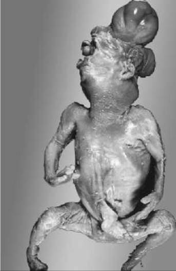
Fig. 9.19. Amniotic band syndrome

Fig. 9.20. Potter sequence
Today, the efficacy of thalidomide in treating HIV infection, leprosy, and tumors has been proven, so its teratogenic effect must be kept in mind.
The teratogenic effect of thalidomide exhibits species specificity—the consequences of exposure in laboratory animals and humans differ. For instance, in mice and rats, no toxic effects were observed even at doses exceeding 4000 mg/kg. In humans and monkeys, thalidomide caused teratogenic effects even when administered in single doses of 0.5–1.0 mg/kg.
According to Finnish and Norwegian researchers, diazepam increases the incidence of children born with cleft lip and palate.
Anticonvulsants (sodium valproate, trimethadione, phenytoin, phenobarbital, carbamazepine) are used to treat Epilepsy. They cause embryopathies such as cleft lip and palate, heart defects, and hypoplasia of the terminal Phalanges. Certain anticonvulsants lead to the formation of Spina bifida and delayed psychomotor development.
The teratogenic effect is caused by a decrease in Folic acid levels in the blood of pregnant women who took anticonvulsants. Folic acid plays an active role in the synthesis of Nucleic Acids and NUCLEOTIDES. Pregnant women taking anticonvulsants should be prescribed Folic Acid and monitored for potential developmental defects (ultrasound Diagnostics, AFP determination).
Anticoagulants: Teratogenic effects are characteristic of warfarin. When taken during the first trimester of pregnancy, it causes nasal hypoplasia, choanal stenosis, Optic nerve hypoplasia, focal chondrodysplasia, and developmental delay.
Antineoplastic alkylating agents (embichine, myelosan, mieleran, endoxan) interact with nucleic acids and enzymes, disrupt DNA reduplication and Protein Biosynthesis, and exert a cytostatic effect on tumors. They also suppress the proliferation of embryonic cells. When used in the first trimester of pregnancy, children may develop cleft palate, eye and Liver abnormalities, Kidney anomalies, inguinal hernias, and brain defects. These children experience delayed psychomotor development.
The teratogenic Effects of Other anticancer drugs—antimetabolites (aminopterin, methylaminopterin) and antimitotic agents (colchicine, actinomycin, etc.)—have also been described. They likewise exhibit mutagenic properties.
Antibiotics: Tetracycline accumulates in tooth tissue, alters coloration, and causes enamel hypoplasia. Both primary and permanent Teeth are affected. If tetracycline is used during the first trimester of pregnancy, the child may develop cataracts and upper limb defects.
Vitamins: A synthetic analogue of vitamin A (isotretinoin, or Accutane) is used in cosmetic creams to treat acne. Large doses of the drug lead to brain defects (hydrocephaly and microcephaly, hypoplasia or Aplasia of the cerebellar vermis), microphthalmia, microtia, anotia, atresia of the external auditory canal, and less commonly, cardiovascular defects. Vitamin A itself in large doses also produces a teratogenic effect.
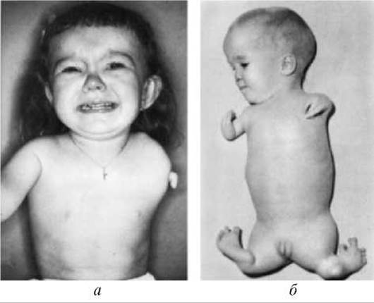
Fig. 9.21. Limb defects: a — amelia, b — phocomelia
Among hormonal drugs, the teratogenic effects of thyroid and antithyroid medications, as well as Sex Hormones, should be considered.
Thyroid and antithyroid drugs cause developmental delays. Affected children may exhibit either hypo- or hyperthyroidism.
Synthetic androgen analogs lead to the masculinization of female fetuses, ranging from clitoral hypertrophy to pseudohermaphroditism.
The estrogen diethylstilbestrol induces vaginal adenosis and cervical ectropion in female offspring, and external genital hypoplasia, Varicocele, etc., in males.
Sex Steroids are components of many oral contraceptives. Women may take them unaware of their pregnancy. This primarily concerns progestins (synthetic analogs of the corpus luteum) and their combinations with estrogens. It has been established that The Use of progestins, particularly medroxyprogesterone, and their combinations with estrogen during the first 4 months of pregnancy increases the incidence of cardiovascular anomalies in offspring by 6-fold.
Teratogenic effects have also been described for agents used in anesthesiology practice. Female anesthesia personnel experience higher rates of spontaneous abortions and a 1.5- to 2-fold higher incidence of congenital anomalies in their children (involving the cardiovascular and musculoskeletal systems, and the central nervous system) compared to female medical professionals in other specialties.
An elevated frequency of spontaneous abortions and birth defects has also been noted in women whose husbands worked in operating rooms (though to a lesser extent than among directly exposed workers). This suggests that the complex of occupational hazards in operating rooms exerts both teratogenic and mutagenic effects.
A comprehensive list of pharmacological agents exhibiting teratogenic activity is provided in Table 9.7.
2. Chemical substances used in daily life, industry, and agriculture. Alcohol consumption, drug abuse, and smoking are of paramount importance for pregnant women.
Alcohol. Fetal alcohol spectrum disorder develops in cases of chronic alcohol consumption during pregnancy. There is a correlation between The amount of alcohol consumed and the degree of fetal damage. However, no safe level of alcohol consumption exists during pregnancy. Fetal alcohol syndrome is characterized by congenital hypoplasia, postnatal growth and weight restriction, mild microcephaly, short and narrow palpebral fissures, epicanthic folds, a narrow sloped forehead, a long philtrum, mandibular hypoplasia (Fig. 9.22), among other features. Neurological impairments include hyperreflexia, tremors, and alterations in Muscle tone. Affected children lag behind in psychomotor development, while older individuals exhibit mental retardation and psychopathic behavioral reactions. Malformations of other Organ Systems have also been described, including cardiac and urogenital defects, and joint stiffness.
The development of these defects is associated with a depletion of folic acid in embryonic and fetal tissues, caused by incomplete ethanol metabolites such as acetaldehyde. Acetaldehyde easily crosses the placental barrier and circulates within the fetus for extended periods, as fetal activity of the enzyme acetaldehyde dehydrogenase is only 10% of adult levels.
Drugs of abuse. Maternal drug use leads to intrauterine growth restriction, preterm labor, breech presentation, and toxemia. In 65–75% of newborns, a withdrawal syndrome develops, which can be fatal.
Cocaine. The use of cocaine during pregnancy is associated with placental abruption and fetal cerebral vascular injury.
Organic Solvents (such as toluene) act as neurotoxins to the brain when inhaled.
Smoking. Smoking leads to prenatal hypoplasia. This is attributed to the direct vasoconstrictive effect of nicotine on uterine vessels, as well as elevated maternal carboxyhemoglobin concentrations. Obstetric complications, such as rupture of fetal membranes and premature placental abruption, occur more frequently.
Industrial and agricultural chemicals. Many industrial chemicals exhibit embryotoxic effects, including gasoline, benzene, phenols, dimethyldioxane, chloroprene, formaldehyde, nitrofuran compounds, nitrogen oxides, numerous pesticides, lead, mercury vapor, etc. Virtually all of these substances have been shown to induce congenital malformations in mammals experimentally. In humans, spontaneous abortions and intrauterine fetal demise are more frequently observed.
In 1977 in Japan, the birth of children with Minamata disease was described, manifesting as microcephaly, cerebral cortical atrophy, and/or hydrocephalus. The source of poisoning was traced to the consumption of fish and shellfish inhabiting bay waters contaminated with mercury salts.
3. Hypoxia is a major teratogenic factor. During the preimplantation period, it leads to the death of a portion of the embryos. Hypoxia in the Embryonic period proper inhibits placentation and embryonic development, and occasionally leads to Congenital malformations and fetal death. Chronic hypoxia results in delayed development, prenatal hypoplasia, and occasionally mild hydrocephalus.
Hypoxia may result from decompensated heart defects, anemia, uterine Hemorrhage, chronic respiratory disorders, and other maternal conditions.
4. Malnutrition — micronutrient deficiencies (zinc, copper, manganese) are of particular significance.
Zinc-deficiency embryofetopathy develops as a result of low dietary zinc intake (meat-free diets), the binding of zinc by salicylates, or impaired zinc absorption in chronic colitis. When maternal serum zinc levels drop below 1.3 µmol/L, zinc-deficiency embryopathy occurs in 13–18% of cases. It manifests as hydrocephalus, micro- and anophthalmia, cleft palate, spinal curvature, various types of hernias, and heart defects. In women, zinc deficiency manifests as acrodermatitis, impaired Olfaction and Hearing, diarrhea, preterm labor, uterine inertia, and atonic hemorrhage.
Table 9.7. Pharmacological agents with teratogenic properties (A. M. Serdiuk et al., 2003)
|
Pharmacological agents (trade name) |
Pathology |
|
Antihypertensives |
|
|
Reserpine (Adelphan, Adenosin, Sinepres) Captopril (Enalapril, Quinapril, Lisinopril, Ramipril) Diazoxide (Hyperstat) Losartan (Cozaar) Methyldopa (Aldomet, Dopegyt, Echybar) |
Microcephaly, Hydronephrosis, hydroureter, inguinal hernia Omphalocele, pulmonary hypoplasia Animal studies have shown skeletal and pancreatic anomalies Skull hypoplasia In some cases, esophageal atresia and cardiovascular malformations |
|
Cerebral Circulation modulators |
|
|
Cinnarizine (Verizin, Stugeron, Cinedil, Cinnabene) Nimodipine (Nimotop, Nemotan) |
Teratogenic effects identified in animal studies Teratogenic effects identified in animal studies |
|
Antiplatelet agents and anticoagulants |
|
|
Warfarin (Pelentan) Nadroparin (Fraxiparine) |
Microcephaly, microphthalmia, retinal atrophy, congenital cardiovascular anomalies, encephalocele, chondrodysplasia Increases the incidence of limb anomalies in animal studies |
|
ß-blockers |
|
|
Betaxolol (Lokren) |
Skeletal anomalies noted in animal studies |
|
Antiallergic agents |
|
|
Diphenhydramine (Dimedrol, Betadrin) |
Hypospadias, cleft palate, cardiovascular defects |
|
Hypoglycemic agents |
|
|
Chlorpropamide |
Limb and intestinal defects, microcephaly detected in animal studies |
|
Thyroid-regulating agents |
|
|
Propylthiouracil (Propicil 50) Thiamazole (Methizol, Mercazolil, Thyrozol) |
Syndactyly, hypospadias, anal atresia, aortic atresia Anal atresia, hypospadias, cranial bone aplasia, cataract |
|
Antigonadotropic agents |
|
|
Danazol (Danoval, Danol) Clomifene (Clomid, Serophene, Clostilbegyt) |
Spontaneous abortions Syndactyly, cardiovascular defects, microcephaly, hemangiomas, retinal aplasia |
|
Sex hormone preparations |
|
|
Medroxyprogesterone (Provera, Farlutal) Estrogens (Cliogest, Klimonorm, Ovestin, Microfolin, Femoden) Diethylstilbestrol (DES) |
Genital structural abnormalities, cardiovascular pathology Genital, auricular, and ocular structural abnormalities, cardiovascular pathology, increased incidence of Down syndrome Genital system anomalies |
|
Amphetamines |
|
|
Amphetamine (Phenamine) |
Cardiovascular and biliary tract defects, cleft lip and palate |
|
Anticonvulsants |
|
|
Sodium valproate (Acediprol, Depakine, Convulex, Everiden, Encorat) |
Cardiovascular neural tube defects, hydrocephaly, microcephaly, skeletal formation disorders |
|
Carbamazepine (Zeptol, Carbadac, Carbamen, Carbapin, Novocarbamoz, Stazepine, Tegretol, Finlepsin) |
Cardiovascular defects, Bone tissue developmental defects |
|
Trimethadione (Trimetin) Diphenylhydantoin (Phenytoin) |
Cleft palate, urogenital anomalies Facial defects, urogenital developmental defects, mental retardation |
|
Vitamins |
|
|
Isotretinoin (Roaccutane) Vitamin A in doses exceeding 8000 IU |
Hydrocephaly, cleft palate, cardiovascular defects Teratogen |
|
Antidepressants |
|
|
Amitriptyline (Amizol, Damilen Maleate, Novo-Tryptin, Tryptizol, Elivel) |
Skeletal anomalies, bilateral anophthalmia |
|
Imipramine (Depsopil, Imizin, Impramin, Melipramine) |
Skeletal anomalies, cleft palate |
|
Barbiturates |
|
|
Amobarbital (Estimal) Phenobarbital (Andipal, Belaspon, Valocordin, Corvalol, Pentalgin, Plivalgin, Sedalgin, Spasmoveralgin, Teofedrin N) |
Congenital heart defects, anencephaly, hydrocephaly, polydactyly Congenital heart defects, anencephaly, hydrocephaly, polydactyly |
|
Benzodiazepines |
|
|
Alprazolam (Alzolam, Zoldac, Casadon, Neurol) |
Down syndrome, hydrocephaly |
|
Diazepam (Aparin, Valium Roche, Dikam, Calmpose, Relanium, Seduxen, Sibazon, Faustan, Reladorm) |
Congenital heart defects, cleft lip and palate |
|
Temazepam (Signopam) Prazepam (Demetrin) Triazolam (Halcion) Clorazepate (Tranex, Tranxene) Chlordiazepoxide (Librium, Napoton, Chlozepid, Elenium, Amixid, Librax) |
Congenital heart defects Cleft lip and palate Hydrocephaly, cardiovascular defects, cleft lip and palate, renal ectopia Hydrocephaly, cardiovascular defects, cleft lip and palate Deafness, duodenal atresia |
|
Estazolam |
Hydrocephaly, cardiovascular defects, cleft lip and palate, renal ectopia |
|
Lithium |
|
|
Lithium (Quilonium, Contemnol, Lithium Carbonate, Litosan SR, Mikalit) |
Cardiovascular malformations |
|
Antipsychotics |
|
|
Haloperidol (Haloper, Senorm, Trankodol) |
Limb defects, cardiovascular malformations |
|
Trifluoperazine (Stelazine, Trazin, Trifutazin, Stelabid, Espazine Plus) |
May increase perinatal mortality; teratogenic in animals at high doses |
|
Fluphenazine (Lyogen, Liorodin Depot, Myrenil, Moditen, Prolinat) |
Cranial bone developmental disorders, cleft palate |
|
Sedatives |
|
|
Bromides |
Microcephaly, gastrointestinal malformations |
|
Meprobamate |
Cardiovascular defects, Down syndrome, limb deformity |
|
Glucocorticoids |
|
|
Cortisone |
Cataract, cardiovascular defects, hydrocephaly, limb defects |
|
Nonsteroidal anti-inflammatory drugs |
|
|
Acetylsalicylic acid (Aspirin, Acesal, Acetylin, Novandol, Plidol 300, Aspirin-C, Plidol C, Fortalgin C, Finrexin C, Alka-Seltzer) |
Cardiovascular defects |
|
Antibiotics |
|
|
Doxycycline (Vibramycin, Doxybene, Doxycycline, Medomycin, Monocline, Unidox) |
Skeletal System defects |
|
Rifampicin (Benemicin, Rimactane, Rifadin, Rifamor, Rifampin, Tubocin) |
Spina bifida and cleft palate detected in animal studies |
|
Kanamycin |
Affects hearing |
|
Antitubercular drugs |
|
|
Isoniazid |
Hypospadias, meningocele, impacts mental development |
|
Antifungals |
|
|
Griseofulvin (Fulcin) |
Increases the frequency of spontaneous abortions |
|
Miconazole (Daktarin) |
Cleft palate, cardiovascular defects |
|
Fluconazole (Diflucan) |
Skeletal system defects |
|
Antivirals |
|
|
Acyclovir (Virolex, Herpex, Zovirax) |
Polydactyly, hypospadias, central nervous system dysfunction |
|
Ribavirin (Virazole) |
Exhibits teratogenic properties in animal studies |
The teratogenic effect of zinc deficiency is linked to the suppression of zinc-dependent enzymes involved in DNA Synthesis (DNA polymerase, thymidine kinase), which act as Transcription factors. This disrupts NUCLEIC ACID METABOLISM and the Cell Cycle, and inhibits cellular Cleavage.
Biological teratogens include viruses, certain bacteria, mycoplasmas, and protozoa. Many infectious agents cross the placenta, causing its infection. The most significant are pathogens grouped under the general term TORCH infections. The acronym TORCH stands for the initial letters of the following words:
1. Toxoplasma (T) — affects fetal development, particularly the central nervous system.
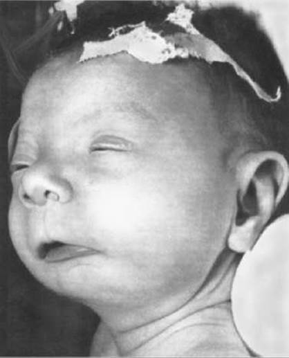
Fig. 9.22. Fetal alcohol syndrome (microcephaly, characteristic facies — epicanthal folds, short palpebral fissures, ptosis, long philtrum, thin upper lip, micrognathia, macrotia)
2. Other (O) — other pathogens (Influenza Viruses, Coxsackie, measles, HIV infection, hepatitis A and B, enteroviral infections, varicella-zoster, Treponema pallidum — the CAUSATIVE AGENT OF Syphilis, etc.). They disrupt the Development of the brain and Skeleton.
3. Rubella (R) — affects the central nervous system, organs of Vision and hearing, and The Heart.
4. Cytomegalovirus (C) — leads to microcephaly, intracerebral calcifications, microphthalmia, hearing impairment, and delayed psychomotor development.
5. Herpes (H) — Herpes simplex virus types I and II — causes central nervous system damage.
Infectious agents directly affect the fetus and can cause fetal demise, intrauterine growth restriction, congenital malformations, and mental retardation. Intrauterine infections account for approximately 49% of intellectual disability cases in children, leading to severe disability.
The severity of clinical manifestations of intrauterine infections largely depends on the gestational age at which the infection occurred. If infection happens within the first 8 weeks post-fertilization, severe and life-threatening fetal malformations frequently develop, resulting in Spontaneous Abortion. When infected after 8–12 weeks of gestation, the fetus most often survives; however, by birth, infection-related changes occur that may cause stillbirth, severe illness, or neonatal death. Inflammatory changes in organs can lead to structural malformations. If the infection occurs In the second half of pregnancy, the neonatal period may manifest signs of generalized infection or localized inflammatory organ involvement (hepatitis, myocarditis, meningitis, meningoencephalitis, chorioretinitis, etc.).
The most Typical symptoms of intrauterine infection appearing in the early neonatal period include:
1. Hyperplasia or hypotrophy of the newborn.
2. Petechiae or ecchymoses.
3. Hepatosplenomegaly.
4. Jaundice.
5. Chorioretinitis.
6. Anemia, thrombocytopenia.
7. Skeletal lesions.
This set of clinical symptoms occurs in intrauterine infections of various etiologies. In English-language literature, the term "TORCH syndrome" is used to describe the clinical manifestations of intrauterine infection.
In the fetus, the development of The Immune System begins in the first trimester of pregnancy. Any intrauterine infection causes an increase in IgM production. Determining its level in cord blood can serve as a valuable screening test. If the IgM level exceeds 20 mg/mL, an intrauterine infection can be suspected.
Last update: 11/08/2026
Editorial and Educational Adaptation: This material has been compiled based on the primary/original source text. The project team performed an editorial review, corrected technical inaccuracies, structured sections, and adapted the content for an educational format.
What was processed:
- elimination of formatting defects (OCR errors, structural breaks, corrupted characters);
- editorial organization of content;
- standardization of terminology in accordance with academic sources;
- verification of factual statements against the original source text.
All mentions of the author, publication year, and origin of the primary text have been preserved in accordance with the source.