Orthopedics - Oleksa A.P. 2006
Scoliosis
Idiopathic scoliosis
Among all types of Scoliosis, the idiopathic form is the most prevalent (Fig. 269), accounting for 90% of cases (Vovk M.M., 2001). Although numerous theories have been proposed and the most probable Pathogenesis of this condition has recently been elucidated, the exact Etiology of idiopathic scoliosis remains fully unresolved to this day.
Experimental studies (Kazmin A.I., 1963 and others) have established similarities between idiopathic and dysplastic scolioses, which corroborates the views of Bogdanov F.R. (1961) and other authors.
Movshovych I.A. (1964) posited that the progression of both idiopathic and dysplastic scoliosis requires at least three contributing factors:
1) a primary pathological factor disrupting normal spinal growth (located in the Spinal Cord, vertebrae, or discs, or resulting from dysplastic changes due to Gene or Chromosomal aberrations, or acquired factors);
2) a factor establishing a pathological systemic Background which, in progressive scoliosis, triggers the manifestation of the primary factor across an entire spinal segment (e.g., metabolic-hormonal disorders, Osteoporosis);
3) a factor involving static-dynamic disturbances, which play a crucial role during the Formation of Structural alterations in the vertebrae.
Class="center">
Fig. 269. Idiopathic scoliosis: a - with the torso in a vertical position, b - with the patient in a flexed position.
Currently, most orthopedists believe that the primary indicator determining the severity of spinal involvement is impaired metabolic processes within the Connective Tissue. This metabolic disruption weakens the connective tissue structures of the spine, creating conditions for disc epiphysiolysis and the Displacement of the Nucleus pulposus toward the convex side of the curvature (Fig. 270).
Analyzing surgical specimens, Fishchenko V.Ya. identified immature Cellular forms of The Nucleus pulposus with altered lysosomal apparatus and impaired lysosomal Enzymes (cathepsin B1), which regulate Collagen METABOLISM. This triggers connective tissue Metabolic Disorders and Changes in the fibrous rings of the discs. Based on biochemical studies, Magomedov S., Uleshchenko D.V. et al. (2001) indicate disruptions in Bone tissue collagen metabolism and mineral balance, which may be of significant importance in the onset of scoliosis.
The loosening of collagen fibers in the annulus fibrosus promotes early migration of the nucleus pulposus toward the future convex side of the spinal deformity, as confirmed by discography of the apex of the primary curvature.
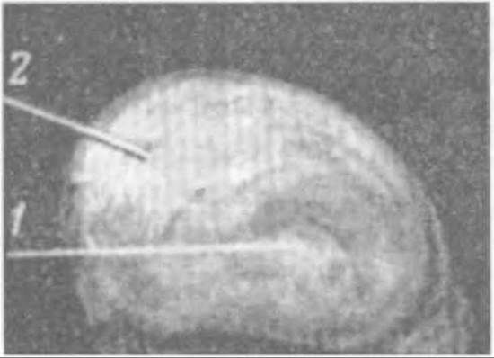
Fig. 270. Changes in the intervertebral disc (2) with displacement of the nucleus pulposus toward the convex side of the scoliosis (1).
This hypothesis is further supported by the fact that secondary compensatory spinal curves exhibit no dysplastic changes in the intervertebral discs or displacement of the nucleus pulposus, even in the presence of pronounced structural alterations in the vertebral bodies.
Studies by Kazmin A.I. and Abalmasova E.O. have established that the greatest amplitude of rotational movement in the spine occurs in the Regions of the ThVIII — ThIX and LI — LII vertebrae, which correspond to the locations of the apex of the primary spinal curvature.
Epiphysiolysis and pathological changes in the discs may merely serve as a triggering mechanism, after which spinal curvature progresses under The Influence of antigravity Muscle contraction, driving The formation of both primary and compensatory spinal curves. Muscle imbalance induces secondary structural changes, along with disruptions in bioelectrical activity and biochemical processes.
Yankovska A.S. performed simultaneous electromyography of two symmetrical areas on the right and left sides of the torso in scoliosis patients. She observed a decrease in electrical potentials, manifested as reduced electrical activity on the functionally weakened concave side of the curve. Conversely, potentials of increased magnitude were detected in the stretched Muscles on the convex side of the spinal curvature.
Spinal muscle bioelectrical Asymmetry, predominantly on the convex side, was also documented by Roaf R. (1960), with heightened activity additionally observed in the deep rotators of the spine (mm. rotatores semispinalis).
Spinal deformation occurs As a result of torsional processes in the vertebrae and discs during a child's growth period; consequently, scoliosis progression is most pronounced during this time. Progression ceases once the iliac apophyses fuse (Risser J.C., 1958; Prokhorova A.T., 1963 and others).
As the spinal curvature progresses, the articular processes on its concave side experience increasingly heavy loads (Movshovych I.A., 1963). In the region of the curvature, the vertebral body twists relative to the arch, a feature also visible in transitional vertebrae (Koveshnikova A.K., 1936). The body and arch on the convex side are taller than on the concave side, where growth is retarded.
As a result of torsion, the vertebral body deviates toward the convex side due to twisting between the body and the Base of the arch on the concave side. The displacement of the pulpal nucleus toward the convex side widens the curvature arc and expands the intervertebral spaces on that side.
Thus, in lateral spinal curvature, torsion (twisting) is what drives scoliosis.
As noted by Reinberg A.S. (1968), the onset of scoliosis is diagnosed significantly faster radiologically than through physical examinations by physicians during routine check-ups. This prompted the Introduction of spinal fluorography for schoolchildren. Radiologists describe the fluorograms and provide Conclusions that should not, however, be overestimated, although children are placed on dispensary observation registers to monitor the process clinically, with follow-up radiological examinations repeated after 6 months if necessary.
Clinical Diagnostics. During preventive examinations of children in organized groups, it is essential to identify all deviations from normal spinal alignment and subsequently refine the Diagnosis.
Pre-scoliotic posture is characterized by shoulder girdle asymmetry, a slight lagging of one scapula, and even lateral spinal curvature visible both clinically and on radiographs when the child is standing. However, this spinal curvature disappears when the child bends forward and during radiography in a prone/supine position. In cases of true scoliosis, the spinal curvature remains noticeable under these conditions.
Since orthopedists rarely participate in routine preventive check-ups, pediatricians, school physicians, and surgeons should be well-acquainted with the signs of scoliosis.
Initially, it is essential to take a thorough medical history and perform a comprehensive physical examination of the patient. The examination is conducted with the child in both standing and supine positions. In the sitting position, the physician evaluates the degree of lumbar lordosis, lateral deviation of the trunk and spine, and the spinal length from the seventh cervical vertebra to the first sacral vertebra.
In the "attention" (standing upright) position, visual inspection and Palpation of the spinous processes are used to determine the spinal axis, waist triangles and their asymmetry, the degree of lateral spinal curvature, as well as the alignment of the scapulae and shoulder girdles (Fig. 271). The lower limbs are measured to check for contractures or anatomical shortening of one of the limbs, which can also act as a cause of spinal curvature.
In the presence of scoliosis, one shoulder is elevated higher than the other, and the scapula on the concave side of the spine lies closer to the spinous processes than on the convex side. Asymmetry of the waist triangles is clearly visible. When the child bends forward, paraspinal asymmetry becomes apparent. The severity of these clinical signs depends on the degree of spinal curvature and its rotational component.
When torsion (rotation) is present, paraspinal asymmetry becomes pronounced, and a rib hump appears as early as the Second Stage of scoliosis; by the Third Stage, it is markedly pronounced, particularly upon forward bending. A number of scoliometers (designed by Schulthess, Ehler, Nedrygailova, Gamburtsev, Yatskevitch, and others) are available to determine the shape and magnitude of the rib hump. While all of them are practically straightforward to use, they lack precision because rib humps vary significantly: they may involve 5 or more Ribs, and can be either shallow or sharp. A photograph taken with the patient in a flexed position is more informative (Fig. 269, b).
Next, the distance between the spinous process of C7 and the upper medial angle of the scapulae is measured. The range of motion of the spine in all planes is evaluated, and any limitations in mobility are determined. Back muscle tone can also be assessed via palpation.
Examination of the child from the front reveals THE POSITION OF the Sternum, lateral displacement of the umbilicus from the midline, and the presence of a chest wall prominence on the side opposite the posterior rib hump (representing scoliotic deformation of the Thorax). The distance from the xiphoid process to both anterior superior iliac spines (spina iliaca anterior superior) is measured. Afterward, by gently tractioning the child upward by the HEAD, the distractibility of the spine is assessed. Normally, the spine elongates by 2%, and in cases of non-fixed scoliosis, it lengthens even further due to the flattening of the curvature arc.
In all cases of spinal pathology, follow-up radiographic evaluation is mandatory. For scoliosis, spinal radiographs are performed every 6 months, capturing the entire lumbar and thoracic spine on a single film.
To determine the degree of fixation of the curved spine, radiographs are taken with the child in both standing and supine positions. If the scoliosis is fixed, the degree of spinal curvature in the supine position (unloaded) does not decrease. The scoliosis stability index is more precisely determined using A.I. Kazmin's formula, which calculates The ratio of the scoliosis angles in the patient's standing and supine positions. Additionally, fixed spinal curvature can be identified radiologically through functional tests, where the curvature arc remains unchanged.
Radiographs obtained in the supine position allow for a more precise measurement of the spinal curvature angle in S-shaped scoliosis.
First, the CHARACTERISTICS OF THE vertebrae are determined, specifically the basal vertebrae—those that remain pathologically unaltered and form the foundation of the spinal Column. The upper basal vertebra adjacent to the deformed spine.
The apex (keystone) vertebrae, defined as those most distant from the imaginary central axis of the spine, represent the vertex of the primary and compensatory (secondary) curves.
Wedge-shaped (transitional) vertebrae originate immediately adjacent to the apex vertebrae and transition into neutral vertebrae, in which the intervertebral spaces are unaffected or minimally altered.
The unaltered (neutral) caudal and cranial vertebrae mark the BOUNDARIES OF THE scoliotic curves; their number can vary.
Building upon these vertebral landmarks, a number of Methods for measuring the degree of scoliosis have been proposed (Chaklin, Enchur, Abalmasova, Ferguson, Cobb, and their modifications such as Lekum's, etc.).
The simplest and relatively accurate methods for measuring spinal curvature angles are those described by Ferguson and Cobb (Fig. 271).
Ferguson's Method.
A line is drawn between the central points of the body of the lower neutral basal vertebra and the vertebra located at the apex of the primary curvature. A second line is drawn through the centers of the bodies of the neutral vertebrae located above the primary curve. The intersection of these two lines forms an angle that defines the scoliotic angle, expressed in degrees. Using a similar approach, the angle of the compensatory spinal curvature can be additionally determined, which typically equals or is slightly smaller than the primary angle.
Cobb's Method.
Two horizontal lines are drawn passing through the intervertebral spaces of the neutral (unaltered) basal vertebrae and the neutral vertebrae situated above the primary spinal curve. Outside the spinal column, two perpendicular lines are drawn to the aforementioned lines, and their intersection opposite the apex of the deformity forms an angle that indicates the Cobb angle of scoliosis.
It should be noted that in severe lateral and rotational spinal curvatures, determining the angle can be difficult because the shadows of the vertebrae overlap, the intervertebral spaces are poorly visualized, and drawing precise lines becomes practically impossible.
In such cases, F.R. Bogdanov utilized a modification of V. Lekum's measurement technique (1951). After identifying the boundary vertebrae of the curve, lines are drawn from the center of the shadow of the most rotated vertebra at the apex of the curvature to the centers of the shadows of the terminal vertebrae. The angle of curvature was defined as the difference between the angle formed by these lines and a straight line (180°).
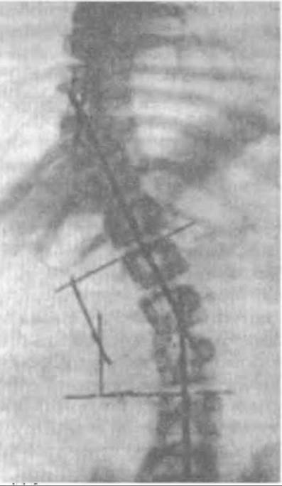
Fig. 271. Radiograph of a scoliotic spine and measurements of the curvature angles using Ferguson's and Cobb's methods.
Radiologically, it is possible to determine the onset and degree of vertebral torsion (rotation) using M.D. Cherfas' method (1962) or the technique employed by F.R. Bogdanov and V.S. Shargorodsky.
Patients undergo anteroposterior and lateral spinal radiographs. On the radiographs, the distance from the midpoint of the vertebral body to the tip of its spinous process is measured as distance h1, and the distance from the midpoint of the vertebral body to the base of the transverse process as h2. The ratio h1/h2 represents the tangent of the vertebral rotation angle (Fig. 272). Using trigonometric tables, the value of the angle in degrees is determined from its tangent.
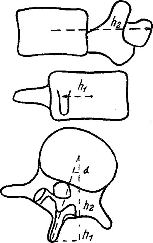
Fig. 272. Determination of vertebral torsion in scoliosis according to Bogdanov-Shargorodsky.
Bogdanov (1968) provides the following example. If the deviation of the spinous process from the midline of the vertebral body is 35 mm in the anteroposterior projection and 75 mm in the lateral projection, the tangent of the vertebral torsion angle will be 35:75, i.e., 0.47, and the torsion angle will be 28°.
In clinical practice, physicians routinely measure spinal curvature angles on radiographs but almost never calculate the angle of vertebral torsion.
Based on clinical signs and the radiographically determined angle of spinal curvature, scoliosis is classified into four grades.
Grade 1 scoliosis is characterized by asymmetry of the waist triangles, although spinal curvature is mild and disappears in the supine position. Initial Impairment of the spinal axis can only be detected when the patient bends forward, making the spinous processes prominent.
The deviation of the spinous processes indicates early spinal rotation, although it is absent on radiographs in the supine position and does not exceed 10° in the standing position. A rib hump is also absent.
Grade 2 scoliosis is clearly visible upon inspection. On the concave side of the spine, the shoulder is somewhat lowered; there is pronounced asymmetry of the waist triangles and a mild S-shaped lateral curvature of the spinal axis with a distinct primary curve, the onset of a compensatory curve (in its upper section), and a rib hump. During physiological growth, structural changes occur in the vertebrae. Radiography reveals deformation of the vertebral bodies at the apex of the curve, and the curvature angle varies, reaching up to 30° (25° according to Chaklin, 39° according to Bogdanov).
Grade 3 scoliosis is clinically manifested by pronounced spinal deformity. On radiographs, the curvature angle ranges from 30° to 50° (26–50° according to Chaklin, 40–60° according to Bogdanov). At the apex of the spinal curve, the vertebral bodies are wedge-deformed, spinous processes are rotationally displaced, and intervertebral spaces are narrowed on the concave side and widened on the convex side. As a result of spinal rotation, a well-defined rib hump appears on the convex side of the spine and anteriorly on the opposite side. The rib hump is particularly prominent when the patient is in a flexed position.
Grade 4 scoliosis is a severe, mostly fixed spinal deformity with a curvature angle exceeding 50°, accompanied by a sharply pronounced rib hump and chest deformity. Radiographic findings show signs of spondylarthrosis and calcification of the spinal ligamentous apparatus. Spinal mobility is severely restricted. The trunk is significantly shortened. Occasionally, pelvic deformity occurs during a child's growth due to uneven tension of the back and abdominal Muscles Attached to it.
The diagnosis should specify the type, level, shape, and grade of scoliosis. Clinical and radiological examinations of the patient are performed semi-annually to determine the disease course, The rate of scoliosis progression, and the degree of deformity correction during Treatment.
One of the pressing issues is detecting the progression of scoliosis, which determines the treatment strategy. The patient's age and medical history data are considered the most informative indicators of potential progression. The younger the age at the onset of idiopathic scoliosis, the higher the likelihood of its progression. Krys-Pugach A.P., Kinchaya-Polishchuk T.A., and Hayko O.H. (2000) point out that at the age of 6–7 years, scoliosis progresses in 30% of children, at 8–11 years in 41.5%, and at 12–15 years in 66.3%.
In clinical practice, the following dynamic radiological signs are utilized: Kohn's sign (based on the widening of the intervertebral space on the convex side), Movshovich's sign (based on relative osteoporosis of the lower-lateral region of the convex and rotated vertebrae), Met's sign (Determination of the difference in costovertebral angles), and Risser's sign (based on iliac apophyses; when the apophyseal growth Cartilage disappears, growth and scoliosis progression cease).
As Vovk M.M. (2001) points out, existing signs and the scoliosis prognostication system based on periodic examinations and monitoring only allow for assessing the course of the disease and do not answer whether scoliosis will progress.
Attempts to answer this question were made by Abalmasova E.A. (1972) and Alekseyev N.V. (1978), who evaluated the child's growth potential. Efforts to identify a primary prognostic sign that would allow for accurate forecasting have been unsuccessful because its prognostic significance varies across different age periods (Movshovich I.A., 1965; Kon I.I., 1989).
Vovk M.M. believes that the most reliable prognosis can be made based on the sum of the following prognostic indicators: the patient's sex and age, duration of the deformity, observation period, magnitude, type, and side of the spinal curvature, as well as the magnitude and dynamics of the central angle of primary curvature, torsion, and secondary curvature. To this end, he developed four prognostic index tables for potential outcomes: non-progression, slow, moderate, and rapid progression. Each sign is coded according to a reference table. A higher sum of prognostic indices corresponds to the most probable prognosis.
Scoliosis is accompanied by degenerative disk changes, osteophytes, osteochondrosis, spondylosis, and arthrosis of the intervertebral joints.
Simultaneously with the progression of scoliosis, torsional changes in the vertebrae and discs lead to a gradual increase in both spinal and thoracic deformity. This results in impaired cardiovascular and respiratory function, which constitutes a crucial pathogenetic link in the disease. Elevated Diaphragm position and reduced pleural cavity volume cause cardiac displacement, kinking of the great vessels, and The Development of Pulmonary Atelectasis and emphysema. All of this hampers Blood flow within the pulmonary artery system, leading to increased venous pressure, overload, hypertrophy, and subsequent dilation of the right Heart ("scoliotic heart"). A decrease in vital capacity, impaired external Respiration, and cardiac dysfunction cause hypoxemia, which in turn disrupts the acid-base balance of the blood.
There are contrasting views on this issue in literature. For instance, Dekhtyarova Z.Y. and Shumskaya T.N. (1982) argued that children and adolescents with scoliosis do not exhibit acid-base balance disorders because they lack overt signs of respiratory failure. Cardiorespiratory disorders reflecting respiratory failure appear in grades 3–4 scoliosis and in older patients.
Storozhenko L.L. and Donchenko L.I. (2001) analyzed blood samples prior to treatment in children with varying degrees of scoliosis. In grade 1 scoliosis, biochemical blood parameters were characterized by calcium deficiency (2.38 mmol/L), elevated Cholesterol (5.1 mmol/L) and triglyceride levels (1.2 mmol/L). High amylase (61.0 IU) and acid phosphatase (4.1 IU) activity, alongside low alkaline phosphatase activity (412.2 IU), were detected. The de Ritis ratio was 2.1 (norm: 1.3), which, according to some authors, indicates functional changes in The Cardiovascular system.
Features of metabolic processes in children with grade 2 scoliosis were characterized by impaired glucose and Protein metabolism, reflecting more pronounced destructive processes in The Musculoskeletal System.
In grade III–IV scoliosis, metabolic processes were characterized by the "normalization" of protein, lipid, and Carbohydrate Metabolism parameters, which should be regarded as the formation of a pathological adaptive syndrome.
These data should serve as the foundation for comprehensive Pathogenetic Treatment of scoliosis patients.
Treatment. It is essential to detect posture disorders and early spinal deformity in a timely manner in order to take appropriate measures to prevent the onset and progression of scoliosis. To this end, systematic annual medical examinations of children in organized groups (preschools, schools) are of paramount importance. Children with identified orthopedic pathology are placed on dispensary registration and referred to an orthopedist for treatment.
Shevchenko S.D. (2001) points out that the treatment plan for a patient with idiopathic scoliosis should be based on the following principles:
1. Realignment of the vertebrae that form the spinal curvature arc.
2. Reduction of the functional component, i.e., spinal instability. The presence of a spinal curvature is always accompanied by functional compensation and the development of counter-curves. The more mobile the spine, the more pronounced the curvatures become under load.
3. If decompensation is not eliminated, varying degrees of curvature magnitude will inevitably lead to an increase in the smaller curve and trunk deviation.
4. Development of a muscular corset to reduce spinal instability and decompensation.
5. Spinal offloading.
6. Medical and physiotherapeutic interventions targeting the body as a whole to normalize metabolic processes.
All therapeutic measures must be applied taking into account the patient's AGE AND SEX, the degree of spinal deformity, and the prognosis.
Patients with first- and second-degree scoliosis are treated conservatively. Treatment is carried out in outpatient clinics, specialized preschool institutions in large cities and interregional boarding schools, sanatoriums, and healthcare facilities.
For children with pre-scoliotic posture and early-stage scoliosis, along with a course of conservative therapy at school, remedial gymnastics classes in special groups are prescribed. Schoolchildren are seated in an appropriate row at an individually adjusted desk so that the child's posture helps correct the spinal curvature. Children are advised to swim in a pool twice a week.
If proper treatment conditions are lacking, especially in rural areas, children should be placed in a specialized boarding school for children with scoliosis or in children's sanatoriums. In such schools and sanatoriums, children receive treatment and develop better physically and mentally, as they do not feel disadvantaged among peers with similar conditions.
School classes are held in a single shift with an individual daily routine tailored to specific groups, taking into account age, sex, physical development, and the severity of scoliosis.
Conservative treatment of scoliosis encompasses: 1) spinal offloading; 2) therapeutic corrective gymnastics; 3) muscle massage; 4) physical and balneotherapy; 5) The Use of corrective corsets, specialized beds, etc.
Spinal offloading, especially in weakened children with progressive scoliosis, is achieved through traction (which not only straightens the spinal axis but also reduces vertebral torsion) and by periodically placing children during study hours on a Pokydanov desk-couch, whose special design allows them to read and lie down while writing. Children Sleep on beds with an inserted board beneath the mattress. When necessary, a plaster bed is made for gradual spinal correction.
Therapeutic corrective gymnastics is conducted for 25–30 minutes in the morning, between classes, and after afternoon nap in mirrored halls using expanders, wands, ribbons, dumbbells, and balls according to a special audio-recorded program.
Children are divided into groups based on right-sided and left-sided scoliosis, as well as The Nature and degree of deformity. Individual therapeutic gymnastics is provided when necessary. It is crucial that children constantly monitor their posture and see in the mirror reflection how correctly they are performing gymnastic exercises. All corrections are made by the instructor.
In cases of neurodysplastic scoliosis with severe pain or discomfort during exercise therapy (ET), gentle individual exercises are administered.
To strengthen muscles, correct the spine, and improve pulmonary function, children engage in therapeutic swimming in a pool twice a week.
Massage helps improve Blood supply and tone in weakened muscles. The massage primarily focuses on strengthening the erector spinae and scapular muscles on the convex side of the curved spine, as well as the oblique Abdominal muscles, which helps reduce spinal deformity.
Favorable effects are achieved through physical and balneotherapy courses involving quartz therapy (in winter), diathermy, amplipulse therapy, paraffin and ozokerite Applications, as well as pine, salt, or sea baths. Manual therapy, which is so frequently promoted, should be avoided as it leads to spinal segment instability and progression of scoliosis (Usleshchenko V. et al., 1998).
The daily routine includes an afternoon nap in the fresh air or in well-ventilated rooms. Children receive five balanced meals a day. In spring, they are prescribed pharmacotherapy (calcium supplements, multivitamins).
Our observations confirm the high efficacy of treating children with scoliosis at the specialized boarding school (Sokal, Lviv Oblast). This treatment is based on clinical examinations and the analysis of clinical and radiological data conducted every three and six months, respectively, with measurement of spinal curvature angles. Scoliosis did not progress in 80% of cases, with nearly a third of patients showing a reduction in the curvature angle in unrigid second-degree scoliosis. These findings are consistent with reports by Loskutov A. et al. (1998) and Usleshchenko V. et al. (1998), who analyzed the outcomes of treating children in specialized schools in Dnipro and Kyiv, respectively.
It should be noted that scoliosis progresses much more frequently when a child is treated on an outpatient basis, as proper regimens and systematic, comprehensive treatment are often not adhered to.
Our observations indicate that some children who study and receive treatment at the specialized school experience a slight regression of the spinal correction achieved at school after spending a two-month summer vacation in a home environment. It is generally accepted that scoliosis stops progressing only after growth cessation, leaving behind any uncorrected spinal curvature that persisted despite treatment.
However, Mikeltadze V.Z., Ovechkina A.V., and Shumskaya T.N. (1986), based on their observations, point out that progression of scoliosis is also possible in patients older than 17 years who exhibit Ossification of the iliac apophyses (Risser sign) and vertebrae.
If scoliosis progresses and parents refuse Surgical treatment for the child, conservative management is continued using unloading, corrective, and functional braces such as Vreden, Blount, Boston, Zuckerman and its modifications, as well as Ducroquet, Milwaukee, and telescoping devices.
The application of corrective braces and appliances helps to some extent restore the vertical alignment of the spine, correct lateral curvature in both the primary and compensatory curves, and achieve vertebral detorsion, particularly in grade I and II scoliosis.
Treatment with removable braces should be combined with a comprehensive conservative management program to be effective for many children.
Prolonged immobilization in a brace without adjunctive therapy leads to torso muscle atrophy, and upon brace removal, spinal curvature increases sharply. This is especially true for unfixated, progressive forms of scoliosis. Unloading and corrective braces are beneficial during periods of rapid growth in children and as part of their comprehensive conservative care.
Surgical treatment for scoliosis has a relatively long history and is constantly evolving. The indication for surgery is the failure of conservative management and the progression of grade II scoliosis.
The progressive course of scoliosis, in the absence of proper and consistent long-term treatment, is a pathogenetically predictable phenomenon.
Given that the most pronounced progression occurs during rapid growth spurts in children, it is crucial to employ all available therapeutic modalities during these periods.
In rapidly progressive idiopathic grade II scoliosis, when conservative treatment fails, surgery performed directly on the vertebral growth plates or discs is advisable to influence proper vertebral growth and prevent torsion. These are less invasive, relatively minor Procedures (such as nucleus pulposus enucleation, discectomy, or spondylolaminar epiphysiodesis) that prove quite effective.
However, parents do not always consent to surgery for such children, which is why orthopedists frequently have to operate on patients with grade III and IV scoliosis.
The primary goals of surgery are maximum correction of the deformity and spinal stabilization.
We will not dwell in detail on surgical procedures that are no longer used as standalone methods. However, we should mention the once-popular operation introduced by L.I. Shulutko (1961), which involved ligamentocapsulotomy on the concave side of the spinal curvature to eliminate the contracture component, followed by correction of the spinal axis and posterior spinal fusion. Nevertheless, nearly half of the patients who underwent this Procedure experienced continued progression of scoliosis.
An original and less invasive method for correcting spinal curvature was proposed in 1953 by A. Gruca. He believed that dynamic spinal correction using spring-loaded hooks was feasible in grade II scoliosis (with a curve up to 30°). The hooks of the spring were attached to the transverse processes of the terminal vertebrae of the primary curve on the convex side, allowing a force of 7-9 kg to maintain the corrected curve.
At the same time, the majority of orthopedists concluded that all forms of progressive scoliosis require radical correction and surgical spinal fixation, as this is the only way to prevent deformity progression and relapses in adolescents and adults (F.R. Bogdanov, 1968).
Surgical technique. The operation was performed following staged correction of the spinal curvature using traction on an orthopedic table with the application of a plaster cast, serial plaster casts, skeletal traction, or I.G. Herzen and V.F. Wenger's rod apparatus directly attached to the vertebral elements.
The surgery is performed under endotracheal anesthesia with blood transfusion and full anesthetic support due to its high invasiveness. A linear incision of the Skin, subcutaneous tissue, and fascia, up to 25-30 cm in length, is made alongside the spinous processes. The fascia is incised on both sides of the process tips, and the muscles are stripped away from them and the vertebral arches. Decortication is then performed to prepare a bed for the grafts.
The procedure is carried out swiftly with bleeding control achieved using Sponges soaked in warm saline or via electrocoagulation.
Steindler, Chaklin, Bogdanov, and others considered it necessary to perform resection of deformed articular processes of the vertebrae—commonly found in grade III and IV scoliosis—along with joint arthrodesis. Long allografts are placed into the prepared beds on both sides of the spinous processes, tightly adapting them to the decorticated surfaces. The gaps are packed with small autografts or cancellous allografts. To prevent displacement of the long grafts, orthopedists secure them to the spinous processes with wire.
It is essential that the length of the graft spans across the baseline neutral vertebrae, covers the entire curve of the spinal deformity, and encompasses two neutral vertebrae located above. As a rule, fixation of seven to ten vertebrae is required.
Considering the invasiveness of the procedure and blood loss in grade III and IV scoliosis, the surgery is performed in two stages. First, bone-plastic fixation of the primary curve is carried out, followed by the compensatory curve. The wound is closed in layers and drained for 24-48 hours. Postoperatively, the torso is immobilized in a plaster spica cast extending to the thighs.
The rib hump is eliminated through a separate procedure—subperiosteal resection of the convex ribs. The surgery is performed under endotracheal anesthesia to prevent complications in the event of an accidental pleural tear. By making a tissue incision and retracting the muscles laterally, the ribs forming the hump are exposed. Resection begins at the most prominent point of the hump; the periosteum is incised linearly along the length of the rib and separated using a rib elevator. The rib must be isolated carefully to avoid damaging the intercostal vessels running directly beneath it and to prevent injury to the parietal Pleura. The rib is transected at the lateral edge of the hump and removed along with its head. The remaining ribs are resected in a similar manner. Hemostasis is then checked, the periosteum of each rib is sutured, and the wound is closed in layers. The patient is placed in a plaster bed, and cotton-gauze pads are used to eliminate the soft tissue bulge.
At KITO in the 1960s, A.T. Sachenko achieved a 40° reduction in spinal curvature for scoliosis by applying a torso plaster cast with thigh extensions combined with serial corrections. Without removing the cast, she cut a window in it corresponding to the PROJECTION OF THE spinal curves and performed a similar bone-plastic operation. However, loss of correction was observed postoperatively, with only 19% of patients maintaining the initial correction. In 50% of patients, the loss of correction ranged from 10-20°.
The loss of correction can be attributed to inadequate pre-operative correction in grade IV scoliosis, insufficient graft length, and unfixated corrected compensatory curves.
For grade IV scoliosis, R.J. Roaf proposed in 1954 performing a wedge resection of the vertebral bodies at the apex of the curvature curve. Concurrently, during the procedure, he excised the laminae and articular processes on the convex side and corrected the spinal axis. Postoperative traction was used to achieve additional correction until the osteotomized vertebrae fused.
Today, wedge resection is considered a fully established procedure and is widely utilized by certain spine surgeons.
Surgical technique. The operation is performed under endotracheal anesthesia with comprehensive anesthetic support. On the convex side of the spine, a linear paravertebral incision is made to expose the rib hump and the long back muscles. The ribs forming the hump are resected over a length of 8-9 cm. The erector spinae muscles are detached from the transverse and articular processes and the vertebral arches. Next, the corresponding transverse processes are resected, and the heads of the resected ribs are removed. Vertebral resection begins with the removal of the laminae and articular processes, thereby opening the spinal canal along its posterior and lateral surfaces. This is performed cautiously to avoid injuring the vessels and intercostal nerves. Subsequently, a wedge is excised from the exposed side of the disc and the adjacent vertebral bodies, tapering toward the concave side of the spine. Hemostasis is verified, and the wound is closed in layers. The patient is placed in a plaster bed. Two weeks later, the spinal axis is corrected via traction on an orthopedic table, and a plaster jacket is applied. Bone-plastic spinal fixation is performed after 2-3 months, and the cast is replaced. The patient must remain in the brace for up to one year.
Wedge osteotomies of the vertebrae were performed by Gruca, who used springs postoperatively, and Kazmin, who, following resection in the thoracic region, corrected the primary spinal curvature using a distractor of his own design. After surgery, Kazmin placed the patient in a plaster bed, and ten days later additionally aligned the spinal axis using lateral traction. Once full correction was achieved, he performed posterior spinal fusion (Fig. 273, a).
Currently, wedge osteotomies at the Ukrainian Institute of Orthopaedics are performed by V.Ya. Hlushchenko (Fishchenko), whose clinic has performed nearly 4,400 surgeries on scoliosis patients.
It is worth noting that at one time Kazmin's distractor was widely promoted and used even in children with grade II scoliosis as an independent method without bone-grafting surgery. This procedure was typically based on eliminating the primary lumbar spinal curve.
Surgical technique. Following preoperative preparation via traction and maintaining traction on the operating table, the distractor was placed along the concave side of the spine. One incision was made alongside the spinous processes over the projection of the upper section of the curve, thereby exposing the articular and transverse processes of the transitional or neutral vertebra just above the curve. A second small tissue incision was made in the posterior region of the iliac crest, exposing it to serve as a support for the distractor.
Then, these two incisions were connected through a soft-tissue tunnel, through which a suitably sized distractor was advanced. The lower end of the distractor rested against the iliac crest, while the upper end rested against the base of the transverse process. By rotating the threaded tube of the distractor during surgery, even greater correction of the curvature was achieved, and the wounds were closed in layers. Once the wounds healed, patients were allowed to resume a normal lifestyle.
However, Kazmin's distractor, when implanted in growing children, gradually loosens, leading to a loss of spinal axis correction. We observed two cases where, upon completion of the girls' growth, the lower end of the distractor was located within the soft Tissues above the iliac crest, accompanied by recurrent spinal curvature. Therefore, using the distractor as a standalone method without bone grafting is impractical.
However, the bone-grafting procedure results in posterior spinal fusion, which permanently immobilizes the fixed segment of the spine. Therefore, in the search for new methods of scoliosis treatment that would ensure reliable fixation of the corrected spine regardless of the child's growth, as well as the restoration of movement in all planes of the spine after growth cessation, Bliskunov's fixator and the Rodnyansky-Gupalov endocorrector were proposed, both of which feature telescoping designs capable of longitudinal extension.
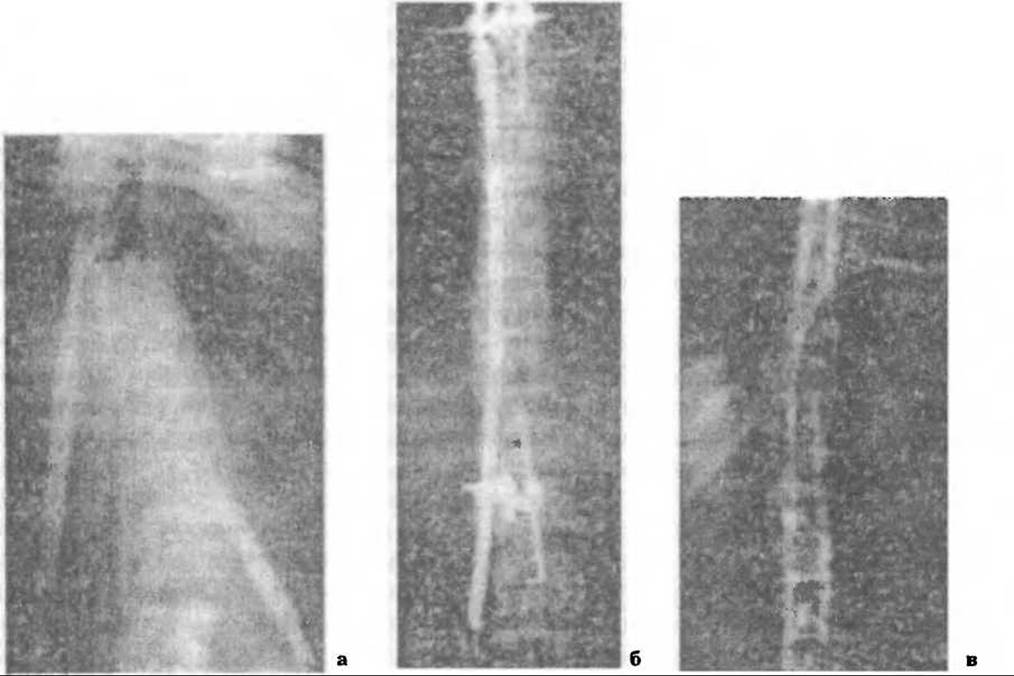
Fig. 273. Spondylofixation following correction of the spinal axis: a — using Kazmin's method (with distractors and a bone graft), b — using Rodnyansky's fixator-corrector, c — following vertebrotomy.
However, Bliskunov's fixator did not gain support among orthopaedic surgeons and is now practically obsolete. On the other hand, the Rodnyansky-Gupalov endocorrector is still used in Russia today, as the implantation procedure is straightforward and minimally invasive, with a blood loss of up to 200–400 ml. Applying this device across all grades of scoliosis, Rodnyansky encountered numerous postoperative complications resulting from excessive pressure in grade III and especially grade IV scoliosis (pressure ulcers, fistulas, etc.). We believe that this corrector should not be discredited by indiscriminate use, as it is undoubtedly most effective for progressive grade II scoliosis in growing children (Fig. 273, b).
Surgical technique. Preoperative traction is used to correct the spinal axis, which is then maintained by traction on the operating table. Guided by radiographs, the neutral baseline vertebrae and the vertebrae above the compensatory curve are identified. Tissue incisions up to 12–15 cm long are made along the spinous processes in these projections. First, the muscles are detached from the spinous processes of three to four upper thoracic vertebrae and, following hemostasis, a serrated clamp ("comb") is applied and secured to them. Through a second incision below the primary curve, 3–4 spinous processes are likewise exposed down to their base, and another serrated clamp is fixed in a similar manner.
The upper clamp is connected to the end of a long plate, which is advanced through a tunnel alongside the spinous processes from the previously convex side of the curve. The distal part of the plate is passed through a metal loop ("eyelet"), and the loop is secured to the lower serrated clamp. Thus, the strong, long plate rests against the spinous processes, supporting the spinal axis and, as the patient grows, sliding within the loop without shifting its position. The wound is closed in layers and drained for 48 hours in the area of the installed clamps. No additional spinal immobilization is used. The blood-soaked dressing is changed the following day.
Two weeks after surgery, following suture removal, the patient is allowed to walk. The corrector is removed after the patient has finished growing.
Our observations confirm the effectiveness of the Rodnyansky-Gupalov endocorrector in grade II scoliosis. It is crucial to administer antibiotic therapy before and after surgery, ensure proper drainage of the surgical wounds, and prevent suppuration, as it is difficult to treat and necessitates the removal of the metal hardware with all ensuing consequences (Fig. 273).
At one time, operations utilizing spinous processes of varying lengths to correct the spinal axis were quite widespread.
Building on our modern understanding of the pathogenesis of scoliotic disease, surgical interventions on the intervertebral discs have been developed. Procedures such as discectomy, disc enucleation, or open and even closed papainization are quite effective for early-stage grade I–II scoliosis, as they interrupt the pathogenetic process.
Cotrel and Dubousset (1988) developed a method in 1984 for correcting scoliotic spinal deformities using an endocorrector. The purpose of this device is to achieve three-dimensional correction of the spine by rotating the correction rod around its axis and ensuring more stable fixation in multisegmental deformities. Since spinal torsion is a leading factor in the progression of scoliosis, performing derotation makes it possible to eliminate spinal curvature in three planes—namely, torsional, sagittal, and frontal.
This surgical technique has been endorsed by orthopaedic surgeons and has been refined through improvements in the quality of metal implants. This particularly concerns the fixation of correction rods using wire loops around the vertebral arches, as well as hooks with supra- and infralaminar heads whose tips are inserted into the spinal canal—a design that carries the risk of damaging the spinal cord or dislodging during spinal derotation (Been H.D., Kalkman C.J., Traast H.S., 1994; Mezentsev A.A., 2001) (Fig. 274).
Due to the significant number of neurological complications associated with using wire loops and hooks for rod fixation, many orthopaedic surgeons have abandoned this method (Goli S.P., Balderston R.A., Stambough J.L. et al., 1988). A number of spine correction and fixation systems (TSRH, ISOLA, SSI, etc.) have been developed utilizing pedicle screws instead of hooks. This prevents neurological complications; these screws are used to connect special pedicular hooks (Ariet V., Marchesi D., Aebi M., 1998; Grossman B.S., Sarwark J.F., Lim R.D., 1999; Ariet V. et al., 1999; Mezentsev A.A., 2001) or derotational cables/rods (Dwyer; Fig. 275).
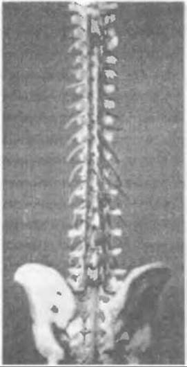
Fig. 274. Spine fixed in the corrected position using rods with hooks outside the arches and wire around the vertebral arches.
In Ukraine, three-dimensional spinal correction for idiopathic and dysplastic scoliosis has been performed at the M.I. Sytenko Institute of Spine and Joint Pathology in Kharkiv since 1998 (Mezentsev A.A., 2001). If children's evaluations show that the magnitude of the primary scoliotic curve on functional spondylograms is less than 40°, a single-stage posterior approach is performed using a multisegmental metal construction for correction, combined with posterior bone grafting using iliac bone autografts.
However, despite the effectiveness of this method for correcting spinal deformity, rigid kyphoscoliotic curves also require anterior mobilization to eliminate excessive thoracic Kyphosis (Newton P.Q., Wenger D.R., Mubarak S.J., Meuer R.S., 1997).
In cases where the curvature angle exceeds 40°, The First stage of surgery involves anterior mobilization at the apex of the spinal curve, followed 2–3 weeks later by the second stage—correction and stabilization of the spine using a multisegmental construct with posterior spinal fusion, although in some cases, according to Mezentsev, children are operated on in a single stage.
When determining the lower limit of spinal fusion, surgeons are guided by the level of the lumbar vertebrae where the wedging of the intervertebral discs and pathological vertebral rotation disappear.
If, during lateral bending of the spine toward the concavity of the main curvature, the upper thoracic compensatory curvature exceeds 20°, the upper limit of arthrodesis is moved 1–2 vertebrae above the upper neutral vertebra.
To determine the degree of lumbar lordosis and thoracic kyphosis according to Cobb, lateral radiographs of the spine were performed from the cervical vertebrae to the level of the femoral heads with the patient in a standing position.

Fig. 275. Spinal correction by torsional rod rotation. Stages of Dwyer spinal fusion.
If the scoliotic deformity is combined with pronounced kyphosis, anterior spinal mobilization is performed.
Surgical technique. Under endotracheal anesthesia with full anesthetic support, a transthoracic approach is performed on the convexity side of the primary curvature through the bed of the rib corresponding to the vertebra located one or two levels above the apical one. After retracting the lung, the pleura is incised and the segmental vessels are ligated. At the apex of the deformity on the concaved side of the curvature, the intervertebral discs along with the rib heads are removed. Depending on the specific case, four to seven discs are resected.
In children with incomplete skeletal growth (Risser sign 0–3), the epiphyseal plates of the vertebrae are disrupted. The resulting defects are filled with bone chips harvested from the resected rib during the approach.
For primary main curves in the lumbar spine, an oblique extraperitoneal approach to the anterolateral regions of the vertebrae is used, with a similar surgical procedure.
According to A. A. Mezentsev, the average duration of anterior spinal mobilization is 157 minutes with a blood loss of 245 ml.
Technique of posterior spinal correction and arthrodesis. Under endotracheal anesthesia with full anesthetic support, a posterior approach along the projection of the spinous processes exposes the posterior elements of the vertebrae by stripping muscles and connective tissue structures. The sites and levels for placing "open" hooks and screws are determined using the Cotrel-Dubousset method (Fig. 276). Pedicle hooks are used in the thoracic spine and are locked with screws after being placed under the vertebral pedicle. Transpedicular screws are used in the lumbar spine. Following mobilization of the posterior spinal segments, the correcting rod is bent 10° less than the existing spinal deformity, the open heads of the hooks and screws are positioned on the concave side and lightly secured with nuts. The rod is then rotated 90–120° around its axis and the nuts are firmly tightened (Fig. 277).

Fig. 276. Instruments for the Cotrel-Dubousset procedure.
If the rod cannot be rotated due to spinal rigidity, the correcting rods are contoured in situ or a construct scheme with a short apical rod is used. It is fixed in two hooks on the concave side at the apex of the curvature and pulled toward the main correcting rod using cross-links.
Next, a rod is placed on the convex side of the curvature, and cross-links are positioned between the long rods. Iliac crest autografts are then harvested and implanted into the decorticated areas of the spinous process bases and vertebral laminae. The wound is closed in layers and drained for 24 hours.
According to A. A. Mezentsev, the surgery lasts an average of 256 minutes with a blood loss of 637 ml. Blood transfusion is repeated in the intensive care unit. After suture removal, the patient is allowed to get out of bed and walk while wearing a plastic immobilization brace for six months postoperatively (Fig. 278).
The use of locking pedicle hooks with ends positioned outside the spinal canal during correction and stabilization of the thoracic spine eliminates the risk of spinal cord compression. As A. A. Mezentsev notes, securing the hooks with screws to the posterior elements of the spine significantly facilitates the placement of the correcting rod and prevents their dislocation during its rotation. Rod rotation is feasible when the magnitude of the main curvature on functional spondylograms (with the patient lying supine with a bolster beneath the apex of the curve) does not exceed 30°. In other cases, the rod must be contoured "in situ" or a metal construct with a short apical rod should be used, which is fixed in two hooks and pulled toward the main correcting rod with a cross-link. For rigid spinal curves, an open anterior mobilization must be performed first.
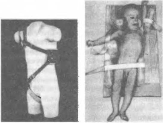
Fig. 277. Methods of spinal axis correction in congenital scoliosis.
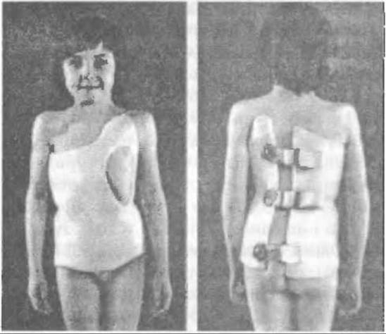
Fig. 278. Removable plastic brace.
The use of multisegmental metal implants in the Surgical Treatment of scoliosis makes it possible to correct all components of spinal deformity and restore or improve physiological curvatures.
Last update: 10/08/2026
Editorial and Educational Adaptation: This material has been compiled based on the primary/original source text. The project team performed an editorial review, corrected technical inaccuracies, structured sections, and adapted the content for an educational format.
What was processed:
- elimination of formatting defects (OCR errors, structural breaks, corrupted characters);
- editorial organization of content;
- standardization of terminology in accordance with academic sources;
- verification of factual statements against the original source text.
All mentions of the author, publication year, and origin of the primary text have been preserved in accordance with the source.