Chemistry and Biology of Proteins - F. Haurowitz 1953
Conjugated Proteins
Hemoglobin
The prosthetic group of Hemoglobin and other similar Proteins is heme, which is an iron-porphyrin complex. The intensive and thorough study of hemoglobin was prompted, on the one hand, by its biological role as an oxygen carrier, and on the other hand, by the fact that it can be readily obtained in crystalline form and has an intense color, making colorimetric determinations possible. Another crucial factor is that Changes in the Native State of hemoglobin can be easily detected by shifts in its color and absorption spectrum. It is beyond The Scope of this work to examine The Structure of heme and various Porphyrins; therefore, the subsequent Discussion will be devoted exclusively to questions concerning the Structure and properties of the protein moiety of hemoglobin.
To prepare hemoglobin solutions, whole Blood is centrifuged, the plasma is removed, and the Blood Cells are washed with an isotonic salt solution. They are then hemolyzed by adding Water, diethyl ether, or toluene [118—120] and centrifuged to precipitate the stroma. If ether or toluene was used to hemolyze THE RED BLOOD cells, a significant portion of the stroma remains suspended between the aqueous layer and the organic solvent layer. In such cases, the hemoglobin solution can be drawn off using a siphon. Hemoglobin is precipitated from its solutions by the careful addition of alcohol at a low Temperature. Some Hemoglobins are almost insoluble in salt-free water and precipitate in crystalline form upon dialysis; others can be precipitated by passing a stream of oxygen and carbon dioxide through their solutions (the carbon dioxide serves to maintain a slightly acidic reaction). The prosthetic group of hemoglobin, which is cleaved by the action of acids, is identical in all hemoglobins and myoglobins. It is called protoheme and is a compound of protoporphyrin and iron.
Class="center">
The protein component of hemoglobin is the protein globin. Although pure hemoglobin was isolated as early as 1867, native globin was not obtained until 1926. All previously prepared globin samples, as shown by Hill and Holden [121], were actually denatured globin.
Native globin is obtained by the careful addition of dilute Hydrochloric acid [121] or oxalic acid [122] to a hemoglobin solution. The cleaved hemin is extracted with diethyl ether; it is also possible to precipitate native globin in an excess of acetone [122—124].
Denatured globin, obtained by older Methods, combines with reduced protohemin to form hemochromogen; however, when native globin is mixed with protohemin and a reducing agent at pH 8–9, hemoglobin is re-formed [121]. Globin differs from most other proteins by its high Histidine content (see Table 1), which reaches 6–10% in various globin preparations compared to 2–3% in most other proteins. Due to this high histidine content, the isoelectric point of globin lies at pH 6.8–7.0; the isoelectric point of denatured globin lies higher, near pH 8.0. Native globin is soluble over a wide pH range, whereas denatured globin precipitates under mildly alkaline conditions, which can be exploited to separate denatured globin from native globin.
Although free heme is a very unstable compound and is immediately oxidized by atmospheric oxygen to form hemin, the combination of hemoglobin with oxygen does not entail The oxidation of iron, which remains ferrous in the resulting oxyhemoglobin. Consequently, the oxygen bound to hemoglobin is readily released in a vacuum. These data clearly indicate that globin protects the heme iron from passing from the ferrous to the ferric state [125], thereby enabling The formation of an HbO2-type compound. Since no other protein can replace globin in this regard, it must be assumed that globin likely contains a special "hemaffinity" group or possesses a specific spatial arrangement of molecular groups that bind to heme [126, 127].
"Synthetic" hemoglobin, prepared by combining native globin with heme, differs from natural hemoglobin by a greater sensitivity to heat [128] and to the action of sodium hydroxide solutions [129]. In other respects, particularly in its ability to bind oxygen, it does not differ from native hemoglobin.
The Molecular Weight of mammalian blood hemoglobin is 66,000–68,000. Since all hemoglobins contain 0.34% iron, The amount of iron per mole of hemoglobin is 230 g, which means there are 4 iron atoms per hemoglobin molecule. Since each heme molecule contains 1 iron atom, the hemoglobin molecule must incorporate 4 heme molecules as its prosthetic group [130, 131].
Upon the action of urea [132], dilute hydrochloric acid [133], or salts [134], the hemoglobin molecule dissociates to form particles with a molecular weight of 34,000. Disaggregation also occurs when native globin is prepared from hemoglobin by the action of hydrochloric acid [135]. All these findings provide sound justification for the Conclusion that the hemoglobin molecule consists of four smaller subunits, each containing 1 heme molecule as a prosthetic group. The reaction of these subunits with large ions, such as ferricyanide ions, indicates that heme is located on their surface [136].
The Study of the absorption spectra of hemoglobin crystals at various orientations of the optical axes led to the conclusion that all four heme groups in the hemoglobin molecule are arranged in parallel [137]. The hemoglobin molecule has the shape of a cylinder with a height of 33.5 Å and a diameter of 57 Å; the heme molecules appear to be located On the surface of the cylinder. Red blood cells contain about 34% hemoglobin, which is the maximum possible concentration at which the hydrated hemoglobin cylinders are still able to rotate freely around their three axes [139].
The structure of hemoglobin and its derivatives can be represented by the following formulas, in which the 4 nitrogen atoms represent the respective porphyrin rings:

The side chains of protoheme are not critical for the formation of hemoglobins, as the latter can also be obtained when globin is combined not with protoheme, but with mesoheme dimethyl ester, in which the unsaturated vinyl groups of protoheme are replaced by saturated groups, and the carboxyl groups by methyl groups [128]. Although this cannot yet be considered definitively established, most authors adhere to the view that in hemoglobin, the heme iron is linked to the imidazole group of histidine. This view is supported by the following evidence: 1) hemin readily forms compounds with imidazole groups; 2) histidine-cobalt compounds can reversibly bind oxygen; 3) the oxygen affinity of hemoglobin changes markedly when the pH is varied from 5 to 8, i.e., in the pH range where imidazole groups are titrated [116]. The ability of hemoglobin to bind bases within the specified pH limits also changes significantly upon oxygenation [116, 142]. Upon oxyhemoglobin formation, the pH values shift from 7.93 to 6.68 and from 5.25 to 5.75 [116]. This means that in neutral solutions, oxyhemoglobin can bind more bases than hemoglobin, whereas in acidic solutions the reverse relationship holds [143].
All of these facts, as mentioned above, support the hypothesis that the heme iron is bound to the imidazole group of histidine. However, it is known that the absorption spectra of hemoglobin and oxyhemoglobin, which are readily shifted by the action of dilute acids, are resistant to concentrated alkali solutions [144]. The latter findings argue rather in favor of the heme iron being linked to an acidic group, such as a carboxyl [126] or sulfhydryl group. Sulfhydryl groups are not detected in native globin at a neutral solution pH; however, they appear upon alkalinization of globin solutions [145] or upon its Denaturation [146].
The Nature of the bonds of iron atoms in the hemoglobin molecule was elucidated to a certain extent by determining their magnetic moments. A ferrous ion has 4 unpaired electrons, whereas a ferric ion has 5 such electrons. Each of these electrons possesses a spin, and consequently, we can measure the resulting magnetic moment. If the number of unpaired electrons in a molecule or ion is $n$, the magnitude of the magnetic moment, expressed in Bohr magnetons, will be equal to:
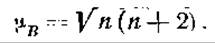
From the given equation, it is easy to determine that the magnetic moment of a ferrous ion is 4.9 magnetons, and that of a ferric ion is 5.9 magnetons. Determinations of the magnetic moments of hemin and heme yielded values of 6 and 4.7 magnetons, respectively [147, 148]. It follows that both in hemin and in heme, iron is present as the corresponding ions. The same holds true for hemoglobin [148]. Oxyhemoglobin and carboxyhemoglobin, however, are diamagnetic, i.e., they exhibit no magnetic moment [148]. This indicates that the electron orbitals of the iron ion undergo profound changes upon the binding of oxygen or carbon monoxide. It is plausible that all unpaired electrons of the hemoglobin iron take part in the formation of oxyhemoglobin or carboxyhemoglobin, which is why the latter lack magnetic properties. It must also be assumed that in this process not only do bonds form between iron and oxygen or carbon monoxide, but the bonds between iron and both globin and porphyrin also lose their ionic character and become true covalent bonds. Oxyhemoglobin and carboxyhemoglobin are similar in this respect to diamagnetic iron compounds, a typical representative of which is the ferricyanide complex.
As noted above, the calculated maximum magnetic moment of hemoglobin is 4.9 magnetons. Meanwhile, measurements have yielded a noticeably larger value of 5.43 magnetons [148]. The reason for this rather significant discrepancy remains unclear. It cannot be related to interactions between heme groups, since a similar discrepancy has also been found for Myoglobin, whose molecular weight is 17,000 [149] and whose molecule therefore contains only a single protoheme [149].
All changes in the state of the iron atom and its bonds with globin are accompanied by alterations in the absorption spectra of hemoglobins. As is well known, diamagnetic compounds—oxyhemoglobin, carboxyhemoglobin, and hemochromogen—are characterized by two absorption bands in the green region of the visible spectrum [150].
Upon partial Hydrolysis of hemoglobin by Trypsin, a compound is obtained in which all the hemin originally present in the hemoglobin molecule remains firmly bound to the globin Cleavage product [151]. Partial hydrolysis of globin with 70% sulfuric acid at 37° yields a polypeptide with basic properties containing a significant amount of histidine [152]. This indicates that histidine residues constitute the most stable part of the globin molecule.
When hemoglobin combines with oxygen, not only The properties of the prosthetic group change, but also the physical and Chemical properties of the molecule as a whole. It was pointed out earlier that the ability of hemoglobin to bind bases increases upon its conversion to oxyhemoglobin. As a consequence, arterial and venous blood have nearly identical acid-base reactions. The higher carbonic acid content in venous blood is compensated for by the higher acidity of arterial oxyhemoglobin. The curve of oxyhemoglobin formation as a function of oxygen pressure [153] is characterized by a specific, unusual for such processes, sigmoidal shape (Fig. 41). Since myoglobin, whose molecule contains only a single iron atom, does not yield such a curve, it is reasonable to think that its sigmoidal shape is due to the interaction of the four heme groups in the hemoglobin molecule. Oxygen always binds to all 4 Hemes of the hemoglobin molecule. The formation of intermediate compounds containing 1, 2, or 3 oxygen molecules per hemoglobin molecule is described by a hyperbolic curve [155]. When the first oxygen molecule binds to one of the hemes, the oxygen affinity of the remaining hemes increases, causing them to become saturated with oxygen faster than the hemes of another hemoglobin molecule.
The Thermodynamics and kinetics of these highly complex reactions were studied by Roughton, who proposed the following equation [156]:

In this equation, $y$ is the percentage of oxygen saturation, $p$ is the oxygen pressure, and $K_1$ is the Equilibrium Constant of the primary reaction
Нb4 + O2 = Нb4O2,
and λ is approximately equal to K13(K4/4), where K4 is the equilibrium constant of the final reaction
Нb4(O2)3 + O2 = Нb4(O2)4.
It was also assumed that the equilibrium constants K1, K2, and K3 decrease in the specified order, while K4 is significantly larger than K1 [156]. The high value of K4 indicates interaction between all heme groups within each hemoglobin molecule. This hypothesis is quite plausible, as it is difficult to conceive that four heme groups attached to the same globin molecule would not interact with one another in some manner [157]. Heme groups are likely arranged in pairs, meaning that the hemes of one pair can react with each other more readily than hemes belonging to different pairs [116].

Fig. 41. Equilibrium state between hemoglobin and oxygen [116]. Solid line — sheep hemoglobin [153]. Data points on the solid line — human hemoglobin [154]. Dashed line — curve calculated using Pauling's equation for an interaction constant of 12.
Differences in the crystal shapes of hemoglobin and oxyhemoglobin also indicate that oxygenation alters not only the Properties of the heme, but also the properties of the hemoglobin molecule as a whole [158] (Fig. 42).
Fig. 42, at the top of the microphotograph, shows the transformation of hexagonal hemoglobin plate crystals into long oxyhemoglobin needles under METABOLISM/18.html">The Influence of diffusing oxygen. The dramatic change in crystal Morphology upon the transition from hemoglobin to oxyhemoglobin suggests that the shape and surface of hemoglobin and oxyhemoglobin molecules differ substantially. This difference evidently plays a role in the formation of sickled erythrocytes. Such erythrocytes are known to occur in the blood of patients suffering from this specific form of anemia, with sickle cells appearing in the blood when oxygen tension drops [159].
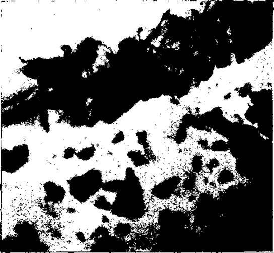
Fig. 42. Crystals of horse hemoglobin and oxyhemoglobin [158].
The equilibrium between hemoglobin and oxygen is usually expressed by the following equation:

Taking into account, however, that hemoglobin is a hydrated compound, it has been proposed to write this equation as follows [160]:
Нb(Н2О) + О2 ⇄ HbO2 + H2O.
When hemoglobin is thoroughly dried in a vacuum, the broad absorption band of hydrated hemoglobin is replaced by two narrow bands of anhydrous hemoglobin, which resemble the absorption bands of hemochromogen. Hydration of anhydrous hemoglobin restores the original absorption band [160, 161].
The oxygen affinity of hemoglobin decreases in the presence of salts [162]. However, this affinity increases in concentrated urea solutions, wherein hemoglobin molecules undergo disaggregation [116, 163]. Acidification of hemoglobin solutions with carbon dioxide or other acids reduces the oxygen affinity of hemoglobin. This phenomenon is the reverse of the Bohr effect described above—namely, the increase in acidity (base-binding capacity) upon The conversion of hemoglobin to oxyhemoglobin [116]. These phenomena are attributed to the presence in globin of an ionic group situated adjacent to the iron-binding group, which influences the iron's affinity for oxygen.
It is well established that carbon monoxide displaces oxygen from its combination with hemoglobin. The absorption spectrum, crystal shape, and certain Other properties of carboxyhemoglobin resemble those of oxyhemoglobin. The main difference between these two compounds is that carboxyhemoglobin is a much more stable compound than oxyhemoglobin, and its dissociation into hemoglobin and carbon monoxide proceeds considerably more slowly [164]. Furthermore, carboxyhemoglobin is split into its components by light [165], with each carboxyhemoglobin molecule absorbing 1 quantum [166]. Unlike hemoglobin and oxyhemoglobin, carboxyhemoglobin lacks an absorption band in the near-infrared region (λ = 900—1 000 mμ) [167]. Carboxyhemoglobin is easily distinguished from oxyhemoglobin by the bright red color of its solutions, which persists even after Treatment with copper sulfate, sodium hydroxide, or tannin. Upon such treatment, oxyhemoglobin is converted into a brownish compound. The resistance of carboxyhemoglobin to the action of the aforementioned substances also attests to the greater Stability of the carboxyhemoglobin molecule compared to the oxyhemoglobin molecule, which, when cleaved under these conditions, yields brown hemin derivatives.
While oxygen and carbon monoxide bind to the iron of heme, carbon dioxide attaches to the globin. A significant portion of blood carbon dioxide normally exists in combination with hemoglobin as carbaminohemoglobin [168]. The nature of this compound is not yet entirely clear. It is known that protein-bound Carbon dioxide is not precipitated as barium carbonate. On this basis, it has been suggested that carbaminohemoglobin is a carbamic compound in which carbon dioxide is bound as follows [169]:

It should be noted, however, that carbamic compounds are formed only in an alkaline reaction and are unstable at the pH of normal blood [116, 162, 170]. It is possible that bicarbonate ions are so tightly bound to the  groups of hemoglobin that they become inactivated—much in the way calcium or phosphate ions are inactivated when combined with casein—and that precisely for this reason they fail to precipitate with barium baryta (see Ch. V).
Under the action of oxidizing agents, the ferrous iron of hemoglobin is converted into ferric iron, forming methemoglobin. The properties of methemoglobin resemble those of an indicator, as it imparts a brown color in acidic solutions and a red color in alkaline solutions [171]. Both forms of methemoglobin correspond to two forms of iron in equilibrium: ferric hydroxide and its cationic form [172]:

Methemoglobin combines with cyanides, fluorides, sulfides, and peroxides to form complex compounds possessing characteristic absorption spectra, which have been isolated in crystalline form [173, 174]. Normal human blood contains about 0.1% methemoglobin [175].
When hemoglobin solutions are exposed simultaneously to hydrogen sulfide and oxygen or hydrogen peroxide, sulfhemoglobin is formed—a green-colored compound [176, 177]. Sulfhemoglobin can also be generated during the putrefaction of hemoglobin.
Hemoglobins from different animals exhibit species Specificity. The specificity of individual hemoglobins can be established not only immunologically [178] (as is done for many proteins), but also through differences in crystal morphology [179, 180], solubility, and Amino Acid Composition (Table 14) [181 — 183]. The Terminal Groups of the peptide chains in various types of hemoglobins also differ. In human hemoglobin, these terminal groups consist of 5 valine molecules; in horse hemoglobin, 6 valine molecules; and in bovine, sheep, and goat hemoglobin, 2 valine molecules and 2 Methionine molecules [184].
Table 14. Amino acid composition of hemoglobin in various animals
|
Hemoglobin |
Number of amino acid molecules per 1 protein molecule (mol. wt. 66,700) |
||
|
histidine |
isoleucine |
||
|
Human |
35 |
16 |
0 |
|
Horse |
33 |
14 |
— |
|
Sheep |
32 |
15 |
— |
|
Dog |
— |
— |
6 |
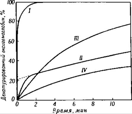
Fig. 43. Denaturation of various hemoglobins by 0.25 N sodium hydroxide solution [193].
I — mother; II — newborn infant; III — rabbit; IV — horse.
Hemoglobins from different animal species undergo denaturation by 0.25 N sodium hydroxide at varying rates. Human hemoglobin is 90% denatured within 1 minute, whereas bovine hemoglobin requires 24 hours to undergo the same degree of denaturation (Fig. 43) [185]. These differences made it possible to detect a specific hemoglobin in human embryonic blood [186]; the denaturation time of this hemoglobin is 60 minutes [186]. Hemoglobin crystals from adults [187] and newborns differ in shape (Figs. 44 and 45) [186]. The N-terminal amino groups of human embryonic hemoglobin consist of valine, with a ratio of 2.6 groups per every 4 iron atoms [184]. The ratio of 2.6 : 4 indicates that this hemoglobin is not a homogeneous compound.
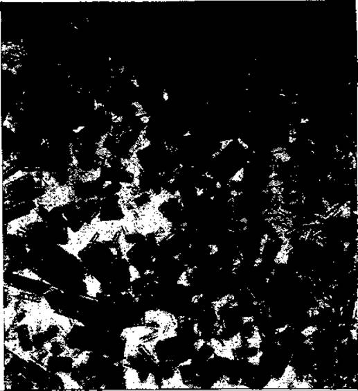
Fig. 44. Adult human hemoglobin [189].
Indeed, it has recently been demonstrated that both adult and embryonic hemoglobins are mixtures of at least 3 to 5 different hemoglobins [188]. Adult and embryonic hemoglobins differ not only in crystal morphology but also in their X-Ray Diffraction identity periods [189]. However, the molecular weight of both hemoglobins is identical [190]. A third type of hemoglobin, distinguished from normal adult hemoglobin by its resistance to sodium hydroxide, has been found in the blood of children over 3 years of age [191].
Although the absorption spectrum and oxygen affinity are Functions of the prosthetic group, both properties depend to a certain extent on the globin moiety as well. For example, the distance between the absorption maxima of oxyhemoglobin and carboxyhemoglobin varies from 53 to 62 Å among different animal species. Similar differences have been observed in oxygen affinity [192]. The oxygen affinity of embryonic erythrocytes is higher than that of adult erythrocytes. Following hemolysis, the oxygen affinity of embryonic hemoglobin remains unchanged, whereas that of adult hemoglobin increases [193, 194]. These findings clearly indicate that hemoglobin's oxygen affinity is determined not only by its prosthetic group and globin, but also by certain factors present within the red blood cells. Substantial differences between embryonic and maternal hemoglobin are found only in humans; less pronounced variations, particularly regarding oxygen affinity, have also been detected in other animal species, such as cows and goats [195].

Fig. 45. Oxyhemoglobin of a newborn infant [189].
It is interesting to note that in patients with pernicious anemia, where hemoglobin regeneration is enhanced, the adult type of hemoglobin is synthesized rather than the embryonic type [171]. In contrast, in anemia characterized by the formation of sickle-shaped erythrocytes1, a different type of hemoglobin is found that differs electrophoretically from normal adult hemoglobin. The hemoglobin comprising sickle cells also differs from normal adult hemoglobin in its net charge: at pH 6.9, this charge is positive, whereas at the same pH, normal adult hemoglobin is negatively charged [159].
As already mentioned, hemoglobins from various animals exhibit species specificity. However, it is difficult to assert that they represent homogeneous compounds. For instance, electrophoretic and spectrometric studies of bovine and equine hemoglobins have demonstrated that these hemoglobins consist of several distinct molecular species.
Aside from animal blood, hemoglobin has also been detected in the ROOT nodules of leguminous plants [199–201]. At present, it remains unclear whether it plays any role in nitrogen assimilation there.
Myoglobin, the pigment of red Muscle, contains the same protoheme prosthetic group as hemoglobin [202]; however, its protein component differs from globin.
Crystalline myoglobin has been obtained by dialyzing muscle extract against a concentrated ammonium sulfate solution [203]. It can also be resynthesized from its constituent parts [204]. The oxygen affinity of myoglobin is higher than that of hemoglobin [205]. It also differs from hemoglobin in its absorption spectrum, amino acid composition, and higher resistance to sodium hydroxide [206]. The molecular weight of myoglobin is 17,000, and its molecule contains a single iron atom [203]. The N-terminal α-amino group of myoglobin is Glycine [184]. Myoglobin has also been detected in the urine of individuals suffering from severe muscle crushing injuries (Crush syndrome) [207]. Of great interest is the fact that, phylogenetically, myoglobin corresponds to the hemoglobin of invertebrates [208].
The Quantitative determination of hemoglobin and its derivatives is performed largely colorimetrically, particularly in clinical laboratories. Since neither hemoglobin nor oxyhemoglobin are stable compounds, they cannot serve as primary standards. A brown-colored acid hematin solution is frequently used as a standard instead. In this method, the blood sample is first mixed with diluted hydrochloric acid. It must be noted, however, that this method often yields erroneous results due to solution turbidity caused by the gradual flocculation of the pigment. This turbidity means that the light incident on the solution is not only absorbed but also scattered [209]. Solution turbidity may likewise result from blood Lipids [210] or the flocculation of Plasma Proteins [211]. More reliable results are obtained by colorimetry of alkaline solutions, while the optimal method is the colorimetric or photometric determination of cyanmethemoglobin [212], formed by adding hydrochloric acid and potassium cyanide to blood [213]. This method has been evaluated across various laboratories and has yielded excellent results [214]. A major advantage of this Procedure is The ability to use cyanhematin as a standard, which shares the identical color and absorption spectrum as cyanmethemoglobin. The Van Slyke manometric gas method also yields good results in hemoglobin determination. Using these methods, the hemoglobin content in the blood of healthy individuals has been found to be 15.7–16.1% [215]. Methemoglobin in the presence of hemoglobin can be quantified by saturating the blood with oxygen or carbon monoxide before and after reduction with dithionite (Na2S2O4) [216]. This salt is one of the few reducing agents suitable for converting oxyhemoglobin or methemoglobin to hemoglobin, as most other reducing agents concurrently cause the irreversible denaturation of globin. A minor drawback of this method, however, is that trace amounts of "inactive pigment" incapable of binding oxygen are also converted to hemoglobin by the action of Na2S2O4 [217]. Very small quantities of oxygen and oxyhemoglobin can be determined polarographically [218]. Carboxyhemoglobin and methemoglobin can also be measured via infrared spectrophotometry [219]. Spectrophotometric techniques are similarly employed when it is necessary to quantify a specific hemoglobin derivative present in a mixture with other derivatives [171, 220].
Under pathological conditions involving The breakdown of hemoglobin and the release of significant amounts of hematin, a fraction of the hematin binds to serum albumin. This complex is designated as methemalbumin1 [221].
Proteins containing the heme moiety also include catalase, peroxidase, dithiochromes, and cytochrome oxidase. The properties of these compounds will be examined in the following chapter.
The blood of certain invertebrates contains true hemoglobin (Sabella spallanzanii, Daphnia magna), whereas others contain hemoglobin-like substances [222]. One such substance is erythrocruorin, which exhibits the same red color as hemoglobin. Another pigment, chlorocruorin, is green. Erythrocruorin contains the same protoheme moiety as hemoglobin; however, its protein component differs significantly from globin. It contains less histidine, and its isoelectric point lies near pH 5.0 [223]. Erythrocruorin has been found both in the erythrocytes and the plasma of invertebrates. The intracellular erythrocruorins of Dasybranchus, Glycera, Thione briareus, Arca pescata, and Petromyzon fluviatilis have molecular weights ranging from 16,700 to 56,500 [224, 225]. Extracellular erythrocruorins dissolved in the Blood Plasma of Arenicola, Planorbis, Lumbricus, and other invertebrates possess molecular weights ranging from 350,000 to 2,800,000 [225, 226].
Chlorocruorin was discovered by Fox [227] in the blood of Spirographis and Serpula. This crystalline protein contains 1.2% iron [228]. Its molecular weight is very high, and its isoelectric point lies at pH 4.3 [229]. Each iron atom of erythrocruorin (as well as chlorocruorin) binds to 1 molecule of oxygen.
1 Work by A. M. Charny and L. A. Blumenfeld (DAN SSSR, 73, 1001, 1950) demonstrated that in methemalbuminemia, a transfer ("shuttling") of heme from hemoglobin to plasma albumin can occur, indicating a weakening of the heme-protein bond in hemoglobin. — Ed. Note.
Last update: 06/08/2026
Editorial and Educational Adaptation: This material has been compiled based on the primary/original source text. The project team performed an editorial review, corrected technical inaccuracies, structured sections, and adapted the content for an educational format.
What was processed:
- elimination of formatting defects (OCR errors, structural breaks, corrupted characters);
- editorial organization of content;
- standardization of terminology in accordance with academic sources;
- verification of factual statements against the original source text.
All mentions of the author, publication year, and origin of the primary text have been preserved in accordance with the source.