Molecular Biology of the Cell - Volume 1 - Alberts B., Bray D., Lewis J., Raff M., Roberts K., Watson J. 1994
Molecular Organization of Cells
Energy Conversion: Mitochondria and Chloroplasts
Chloroplasts and Photosynthesis
All animals and most microorganisms need a constant supply of large quantities of Organic compounds from their environment. These substances provide the carbon skeletons for Cell/9.html">Biosynthesis and the metabolic energy required for all cellular processes. It is believed that the earliest organisms on primordial Земля had an Abundance of organic compounds produced by geochemical processes (see Section 1.1.1). However, most of these resources were depleted billions of years ago. Since then, nearly all organic Materials essential for living Cells have been produced by photosynthetic organisms, including various types of photosynthetic Bacteria. The evolutionarily most advanced among these bacteria—the cyanobacteria—have minimal nutritional requirements. To convert atmospheric carbon dioxide (СО2) into organic compounds, they utilize sunlight as an energy source and Water as a source of electrons. Furthermore, during the splitting of water [via the reaction nН2О + nСО2→ n(СН2О)n + nО2], they release into the atmosphere the oxygen required for Oxidative Phosphorylation. As we will explain later, it was likely the evolution of cyanobacteria from more primitive photosynthetic bacteria that enabled The Development of the first aerobic life forms.
In plants, which appeared later, Photosynthesis takes place within specialized intracellular Organelles known as METABOLISM/14.html">Chloroplasts. However, chloroplasts supply metabolic energy only during daylight hours; at night, they cease the synthesis of high-energy metabolites, and during this time, plants obtain ATP through The activity of Mitochondria, which are remarkably similar to those found in animal cells.
Based largely on biochemical evidence, it is believed that chloroplasts are the descendants of cyanobacteria that were engulfed by eukaryotes via endocytosis and established a symbiotic relationship with them. THE ORIGIN OF mitochondria is explained in a similar fashion. According to this theory, the numerous differences between chloroplasts and mitochondria stem partly from their derivation from different bacterial ancestors and partly from subsequent evolutionary divergence. Nevertheless, the fundamental mechanisms of ATP Synthesis in chloroplasts and mitochondria are remarkably similar, although in the former it is driven by light energy, and in the latter by energy derived from Respiration.
Class="center">
Fig. 7-38. Chloroplasts contain three membranes—outer, inner, and thylakoid—which divide the organelle into three internal compartments: the intermembrane space, the stroma, and the thylakoid space. The thylakoid membrane houses all the energy-transducing systems of the chloroplast. In electron micrographs, these membranes appear fragmented into discrete flattened vesicular structures (see Fig. 7-39), but within the intact chloroplast, they are likely continuous, forming a single highly folded membrane. As shown in the figure, individual thylakoids are interconnected into stacked structures known as grana.
7.3.1. Chloroplasts Resemble Mitochondria but Contain One Additional Compartment
Like mitochondria, chloroplasts utilize a chemiosmotic mechanism for energy conversion, and both organelles share the same fundamental organizational principles (Figs. 7-38 and 7-39). Chloroplasts also feature a highly permeable outer membrane and a much less permeable inner membrane embedded with specialized transport Proteins, with a narrow intermembrane space separating the two. The inner membrane encloses a large central region called the stroma, which is the functional equivalent of the mitochondrial matrix and contains various Enzymes, Ribosomes, RNA, and DNA.
However, There is a fundamental difference. The inner membrane of chloroplasts does not form cristae and lacks an Electron Transport Chain. The photosynthetic light-harvesting system, The electron transport chain, and ATP synthase are instead located in a third membrane that forms a set of flattened, disclike sacs called thylakoids (Fig. 7-38). The internal cavities of the thylakoids are believed to be interconnected, forming a third, internal chloroplast compartment known as the thylakoid space. This space is separated from the stroma by the thylakoid membrane.
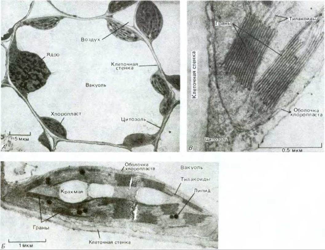
Fig. 7-39. Cytology/cytology/93.html">ELECTRON MICROGRAPHS OF chloroplasts. A. A cell from a wheat leaf, showing a thin layer of Cytoplasm containing chloroplasts surrounding a large vacuole. B. A thin section of a single chloroplast, revealing starch grains and lipid droplets accumulating in the stroma as products of biosynthesis. C. Grana at high magnification, showing the stacked thylakoid membrane. (Courtesy of C. Plaskitt.)

Fig. 7-40. Comparison of Mitochondrial and Chloroplast Structure. Typically, a chloroplast is much larger and contains a thylakoid membrane and thylakoid space. The inner membrane of mitochondria forms cristae.
Figure 7-40 illustrates the structural Similarities and differences between Mitochondria and chloroplasts. Broadly speaking, a chloroplast can be envisioned as a greatly enlarged mitochondrion whose cristae have developed into chains of interconnected submitochondrial particles within the matrix. In chloroplasts, the spherical HEAD of the ATP synthase—where ATP is generated—projects from the thylakoid membrane into the stroma, just as it projects from the inner membrane into the matrix in mitochondria (see Fig. 7-51).
7.3.2. Chloroplasts Perform Two Unique Reactions: Light-Driven Generation of ATP and NADPH, and The conversion of СО2 into CARBOHYDRATES [26]
The diverse reactions of photosynthesis can be divided into two major categories.
1. Photosynthetic Electron transfer reactions (sometimes referred to as light reactions) use radiant energy to excite an electron in a chlorophyll molecule, enabling its passage along an oxidation-reduction chain in the thylakoid membrane, analogous to the electron transport chain in The inner mitochondrial membrane. This electron transport drives the pumping of protons across the thylakoid membrane, generating a proton-motive force that provides the energy for ATP synthesis in the stroma. Simultaneously, the high-energy electrons produced by the oxidation chain reduce NADP+ to NADPH. The electrons for this process are derived from The oxidation of water, which releases О2.
2. Carbon fixation reactions (sometimes called dark reactions) convert СО2 into carbohydrates, utilizing the ATP and NADPH synthesized during the photosynthetic electron transfer reactions as sources of energy and reducing power, respectively. Initiated in the chloroplast stroma and completed in the Cytosol, these reactions produce sucrose in the leaves, which is then transported to other PARTS OF THE plant to supply energy for growth and serve as a building block for organic molecules.
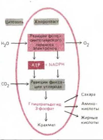
Fig. 7-41. Photosynthetic reactions in chloroplasts can be subdivided into electron-transport reactions and carbon-fixation reactions. The first group of reactions oxidizes water and releases О2, whereas the second group assimilates СО2 and produces organic molecules.
Thus, the evolution of molecular oxygen (which requires the direct input of radiant energy) and the conversion of СО2 into carbohydrates (which does not directly require light) are two distinct processes (Fig. 7-41). However, as we will see later, these two processes are linked by a delicate feedback mechanism essential for Metabolic Regulation. For example, The production of ATP and NADPH in the thylakoid membranes fluctuates According to the cellular demand for these molecules, while certain chloroplast enzymes required for carbon fixation are inactivated in the dark and reactivated by light-stimulated Electron transport processes.
7.3.3. Carbon Fixation Is Catalyzed by Ribulose Bisphosphate Carboxylase [28]
Earlier in this chapter, we explored how cells harness the vast amount of energy released during the oxidation of carbohydrates to СО2 and Н2О to synthesize ATP. It should be clear from this that the reverse process—the FORMATION OF CARBOHYDRATES from СО2 and Н2О—requires a substantial energy input and can occur only when coupled to energy-yielding reactions.
Figure 7-42 illustrates the central reaction that converts inorganic carbon into organic form: atmospheric СО2 reacts with water and the five-carbon compound ribulose 1,5-bisphosphate to yield two molecules of the three-carbon compound 3-phosphoglycerate. Discovered in 1948, this reaction is catalyzed within the chloroplast stroma by a large enzyme (molecular mass ~500,000) known as ribulose bisphosphate carboxylase. Because this enzyme operates very slowly (catalyzing the turnover of only about 3 substrate molecules per second, compared to ~1,000 molecules per second for typical enzymes), a massive amount of ribulose bisphosphate carboxylase is required. The enzyme often accounts for more than 50% of the total chloroplast protein and is considered to be the most abundant protein on Earth by total mass.
7.3.4. The Carbon-Fixation Cycle Consumes Three Molecules of ATP and Two Molecules of NADPH per Fixed СО2 Molecule [29]
Although the carbon-fixation reaction itself does not require light energy, it depends on a continuous supply of the high-energy compound ribulose 1,5-bisphosphate to trap СО2 (Fig. 7-42). Research into the complex pathway of ribulose 1,5-bisphosphate regeneration represents one of the earliest and most successful Applications of radioisotopes in biochemistry. As shown in Fig. 7-43, the fixation of three molecules of СО2 by ribulose bisphosphate carboxylase yields six molecules of 3-phosphoglycerate, containing a total of 6 × 3 = 18 carbon atoms: 3 from СО2 and 15 from ribulose 1,5-bisphosphate. These 18 carbon atoms then pass through a cycle of reactions that regenerate the 3 molecules of ribulose 1,5-bisphosphate (containing 3 × 5 = 15 carbon atoms) consumed at THE START OF the cycle, yielding a net gain of one molecule of glyceraldehyde 3-phosphate (3 carbon atoms). In this carbon-fixation cycle (the Calvin-Benson cycle), the incorporation of a single molecule of СО2 consumes three molecules of ATP and two molecules of NADPH. The overall stoichiometric equation for the cycle is
3СО2 + 9АТР + 6NADPH + Water → Glyceraldehyde-3-phosphate + 8Pi + 9ADP + 6NADP+.
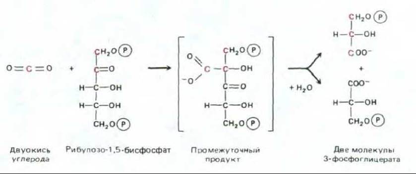
Fig. 7-42. The initial reaction in which Carbon dioxide is converted into organic carbon. This reaction is catalyzed in the chloroplast stroma by ribulose bisphosphate carboxylase, which is present there in very large amounts, resulting in The formation of 3-phosphoglycerate, which is also a crucial intermediate of Glycolysis (see Fig. 2-20). If the same enzyme attaches oxygen instead of СО2 (see Section 7.3.5), the two carbon atoms highlighted in color are used to form phosphoglycolate.
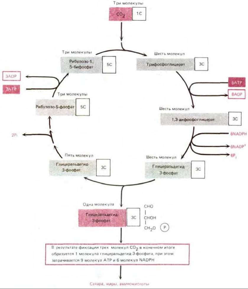
Fig. 7-43. The carbon-fixation cycle, in which organic molecules are produced from СО2 and Н2О. For simplicity, many intermediates on the pathway from glyceraldehyde-3-phosphate to ribulose-5-phosphate are omitted. The participation of water in the cycle is also not shown.
Thus, the synthesis of organic molecules from СО2 and Н2О requires the energy of phosphate bonds (in the form of ATP) and reducing power (in the form of NADPH). We will return to this point later.
Glyceraldehyde-3-phosphate, produced in the chloroplasts during the carbon-fixation cycle, is a three-carbon sugar and a key intermediate in glycolysis (Section 2.3.2). Most of the glyceraldehyde-3-phosphate enters the cytosol, where it is rapidly converted into fructose-6-phosphate and glucose-1-phosphate via the reversal of certain glycolytic reactions (Section 2.5.3). Glucose-1-phosphate is then converted into a nucleotide sugar derivative, UDP-glucose, which reacts with fructose-6-phosphate to form sucrose phosphate, the direct precursor of the disaccharide sucrose. In plants, sucrose plays the same role that glucose does in animals: it is the primary sugar form transported from one cell to another. As needed, sucrose moves from the leaves to the rest of the plant through the vascular bundles (see Fig. 7-45), much like glucose is carried by the bloodstream in an animal.
In the stroma, most of the remaining glyceraldehyde-3-phosphate retained in the chloroplasts is converted into starch. This high-molecular-weight glucose polymer serves as a reserve carbohydrate, much like Glycogen in animal cells. Starch is synthesized in the chloroplast stroma during periods of high photosynthetic activity and is stored there as large grains (see Fig. 7-39, B). Starch synthesis occurs via the reversal of stromal glycolytic reactions: glyceraldehyde-3-phosphate is converted into glucose-1-phosphate, which then forms ADP-glucose, the direct precursor of starch. At night, starch is degraded to meet the metabolic needs of the plant.
7.3.5. To facilitate the growth of certain tropical plants under low СО2 concentrations, carbon fixation in their leaves is compartmentalized [30]
Although ribulose bisphosphate carboxylase predominantly adds СО2 to ribulose-1,5-bisphosphate, at low carbon dioxide concentrations it will also add O2. This is an apparently wasteful pathway that yields one molecule of 3-phosphoglycerate and one molecule of the two-carbon compound phosphoglycolate, rather than two molecules of 3-phosphoglycerate (see Fig. 7-42). Phosphoglycolate is converted into glycolate and transported to Peroxisomes, where two molecules of glycolate are synthesized into one molecule of 3-phosphoglycerate (three carbon atoms) and one molecule of СО2. Because this process consumes O2 and releases СО2, it is termed Photorespiration. In many plants, about one-third of the fixed carbon is re-released as СО2 via photorespiration. It remains unclear whether photorespiration serves any beneficial function in plants or is merely a mechanism for recovering a portion of carbon that was diverted into phosphoglycolate due to the undesirable interaction of oxygen with ribulose-1,5-bisphosphate.
Photorespiration can become a significant drawback in hot, dry conditions where plants are forced to close their Stomata (Pores in the leaves responsible for gas exchange) to prevent excessive water loss. As a result, the internal СО2 concentration in the leaves drops sharply, leading to an increase in photorespiration. However, the leaves of many plants adapted to hot, dry climates, such as corn and sugarcane, possess a specific adaptive mechanism. In these plants, the Reactions of the carbon-fixation cycle shown in Fig. 7-43 occur exclusively in the chloroplasts of specialized bundle-sheath cells, which contain all of the plant's ribulose bisphosphate carboxylase. These cells are shielded from the air and surrounded by a layer of mesophyll cells that "pump" СО2 into the bundle-sheath cells, thereby maintaining a high СО2 concentration around ribulose bisphosphate carboxylase and strongly suppressing photorespiration.
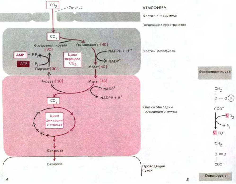
Fig. 7-44. The СО2-transport cycle in plants such as maize. A. Carbohydrate synthesis occurs exclusively in the bundle-sheath cells, which harbor all the ribulose bisphosphate carboxylase. The СО2-trafficking cycle begins in the mesophyll cells; the cycle involves the four- and three-carbon compounds indicated in the scheme. Variants of this cycle are also found in other СО2-transporting plants. B. The reaction of phosphoenolpyruvate with СО2 in mesophyll cells.
The СО2-"pumping" mechanism is driven by a cycle of reactions that begins with the fixation of СО2 in the cytosol of mesophyll cells by an enzyme with a high affinity for carbon dioxide (in the form of bicarbonate). The resulting four-carbon compound is transported to the bundle-sheath cell, where it is cleaved into one molecule of СО2 and one three-carbon molecule. The latter returns to the mesophyll cells, where, through an ATP-driven reaction, it is converted into the active form capable of capturing the next СО2 molecule and repeating the СО2-transport cycle (Fig. 7-44).
By pulse-labeling a plant equipped with such a СО2 pump with radioactive 14СО2, it was established that the first labeled organic substance appearing in the mesophyll as a result of 14СО2 assimilation contains four carbon atoms, whereas in other plants it is three-carbon (see Fig. 7-43). For this reason, СО2-transporting species are referred to as C4 plants, while all others are called C3 plants (Fig. 7-45).
Like any directed transport process, The transfer of СО2 into bundle-sheath cells requires an Energy Expenditure. In hot, dry conditions, this cost is often far less than the losses caused by photorespiration in C3 plants, giving C4 plants a distinct advantage. Furthermore, because C4 plants can carry out photosynthesis at low internal leaf СО2 concentrations, they keep their stomata open less and are thus able to fix roughly twice as much carbon per unit of water lost compared to C3 plants.
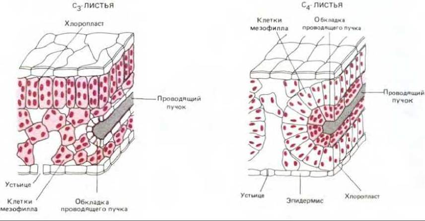
Fig. 7-45. Comparison of leaf anatomy between C3 and C4 plants. In both cases, cells whose chloroplasts carry out the normal carbon-fixation cycle are highlighted in color. In C4 plants, mesophyll cells are specialized for The Active Transport of СО2 rather than carbon fixation; it is these cells that generate a high СО2:O2 ratio within the bundle-sheath cells. The carbon-fixation cycle in these plants takes place exclusively in the bundle-sheath cells (see Fig. 7-44). Through the vascular bundles, the sucrose produced in the leaf is distributed to all other Tissues of the plant.
7.3.6. Photosynthesis is governed by the Photochemistry of the chlorophyll molecule [31]
Having examined the carbon-assimilation reactions, we now return to the question of how photosynthetic electron transport in the chloroplast generates the ATP and NADPH required to synthesize carbohydrates from СО2 and Н2О (see Fig. 7-41). The necessary energy is harvested from sunlight absorbed by chlorophyll molecules (Fig. 7-46). Energy conversion begins when a chlorophyll molecule is excited by a quantum of light (photon), which promotes an electron to a higher energy level. Such an excited molecule is unstable and tends to return to its ground state in one of three ways: (1) by converting the excess energy into heat (molecular motion), or into heat and longer-wavelength light (fluorescence), when radiant energy is absorbed by an isolated chlorophyll molecule in solution; (2) by transferring the energy (rather than an electron) directly to a neighboring chlorophyll molecule via a process known as Resonance energy transfer; or (3) by transferring a high-energy electron to a nearby molecule (an electron acceptor) and returning to its ground state by accepting a low-energy electron from another molecule (an electron donor, Fig. 7-47). The latter two mechanisms play a crucial role in photosynthesis.
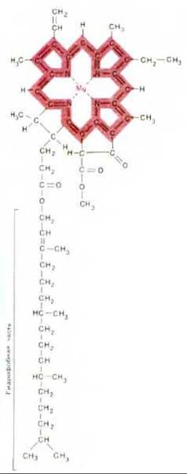
Fig. 7-46. Structure of chlorophyll. A magnesium atom is coordinated within a porphyrin ring, which is structurally similar to the iron-coordinating porphyrin ring in heme (compare with Fig. 7-27). The system of conjugated double bonds is highlighted in color.
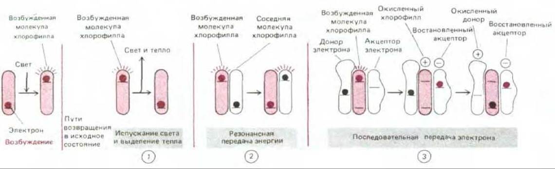
Fig. 7-47. Three possible pathways by which an activated chlorophyll molecule (containing a high-energy electron) returns to its unexcited ground state. In the first pathway (1), radiant energy absorbed by an isolated chlorophyll molecule is entirely released as light and heat. By contrast, during photosynthesis, chlorophyll either transfers its energy to another molecule within the antenna complex (2) or ejects an excited electron at a reaction center (3), as described in more detail in the text.
7.3.7. The photosystem consists of a reaction center and an antenna complex [32]
Photosystems are multi-Protein Complexes that catalyze the conversion of light energy—via the energy of excited chlorophyll molecules—into biologically useful forms. A photosystem comprises two closely linked components: a photochemical reaction center and an antenna complex (Fig. 7-48).
The antenna complex is essential for capturing light. In chloroplasts, it consists of an aggregate of several hundred chlorophyll molecules bound to proteins that firmly anchor these molecules within the thylakoid membrane. Depending on the plant species, each complex also contains accessory pigments, such as carotenoids, which can absorb light of different wavelengths. Upon excitation of a chlorophyll molecule in the antenna complex, energy is rapidly transferred from one molecule to another via resonance energy transfer until it reaches two special chlorophyll molecules in the photochemical reaction center. Thus, each antenna complex acts as a "funnel" that collects energy and directs it to specific sites where it can be utilized most efficiently (Fig. 7-48).
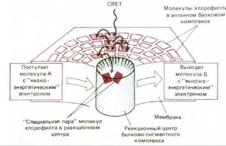
Fig. 7-48. A photosystem consists of a reaction center and an antenna complex. The reaction center is a transmembrane protein complex that holds a "special pair" of chlorophyll molecules in a fixed orientation relative to other electron carriers (see Fig. 7-49). The reaction center catalyzes the third process shown in Fig. 7-47. Viewing this center as an enzyme, its substrates are a weak electron donor (molecule A) and a weak electron acceptor (molecule B), and its reaction products are a strong electron acceptor (oxidized molecule A) and a strong electron donor (reduced molecule B). The antenna complex contains the majority of the chlorophyll in the thylakoid membrane and acts as a funnel, directing the energy of the excited electron toward the reaction center. Many of these energy-transfer events occur between identical chlorophyll molecules, transmitting excitation randomly (process 2 in Fig. 7-47). However, the average time interval between the absorption of an exciting quantum and the transfer of excitation to the reaction center is only 10-10-10-9 s, so that a very small fraction of the absorbed energy is lost through the wasteful process 1
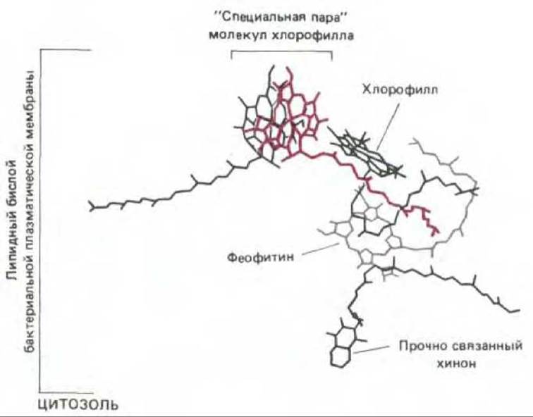
Fig. 7-49. The arrangement of electron carriers in the bacterial photochemical reaction center, determined by X-ray crystallography. The depicted pigment molecules are embedded within the transmembrane protein and surrounded by a lipid bilayer. Excitation from the antenna chlorophyll is transferred to an electron of the special pair of chlorophyll molecules via a resonance mechanism (process 2 in Fig. 7-47), after which the excited electron is transferred from the special pair to a quinone (through a series of intermediate steps, see Fig. 7-50).
The photochemical reaction center is a transmembrane protein-pigment complex that forms the very "Heart" of photosynthesis. It is believed that this complex first appeared in primitive photosynthetic bacteria more than 3 billion years ago. The special pair of chlorophyll molecules in the reaction center acts as an efficient trap for excitation energy because the reactive electrons of these molecules are directly transferred to a chain of acceptors located in close proximity to the chlorophyll within the same protein complex (Fig. 7-49). By rapidly removing the high-energy electron from chlorophyll, the reaction center transfers it to nearby molecules where the electron can reside in a much more stable state. This makes the electron available for subsequent photochemical reactions that require time to take place. As we will see, the net result of these slower reactions is that a low-energy electron within a weak electron donor (such as water) is converted into a high-energy electron within a strong donor (such as quinone).
7.3.8. Radiant energy absorbed by the reaction center chlorophyll is used to replace a weak electron donor with a strong one [33]
Electron-transfer processes in the photochemical reactions just described have been intensively studied using rapid spectroscopic Methods, particularly in the Photosystem of purple bacteria, which is simpler than the evolutionarily related chloroplast photosystem. Bacterial reaction centers can be solubilized and isolated in active form using a detergent. These are large protein-pigment complexes, and in 1985, X-ray crystallography made it possible to determine their complete three-dimensional structure (see Figs. 6-72 and 7-49). This structure, combined with kinetic data, provides the best insight into the electron-transfer reactions underlying photosynthesis.
Figure 7-50 schematically illustrates The sequence of these reactions in the purple bacterium reaction center. An electron excited by Light absorption is rapidly transferred from the special pair of chlorophyll molecules through a series of other pigments (Fig. 7-49) to a tightly bound quinone electron acceptor, designated QA. As a result of this electron transfer—which occurs in less than 10-9 s and is virtually irreversible—a positively charged "hole" with a very high electron affinity is created in the chlorophyll. This hole is filled by capturing an electron from a nearby cytochrome (normally a weak electron donor). Subsequently, the high-energy electron held by QA is transferred to a second quinone, QB, after which it leaves the reaction center and passes to a mobile quinone molecule (Q) in the photosynthetic membrane. Once reduced, this quinone serves as a strong electron donor whose reducing power can be used to drive proton translocation.
The fundamental principle of the described process is that the photosystem makes it possible to use light energy to transfer an electron from a weak electron donor—that is, a molecule with a high affinity for electrons (in this case, a cytochrome)—to a molecule such as quinone, which in its reduced form acts as a strong electron donor. Thus, excitation energy that would otherwise be dissipated as heat and/or fluorescence is harnessed to raise the energy of the electron and generate a strong electron donor. As we will see, in higher plant chloroplasts the initial electron donor is not a cytochrome, but water, which explains the oxygen evolution observed in plant photosynthesis. Before examining the processes occurring in the more complex chloroplast photosystem that ultimately provide the energy for ATP and NADPH synthesis, let us see how these end products are formed in purple bacteria via a less complex but analogous mechanism.
7.3.9. During Bacterial photosynthesis, an electrochemical proton gradient is generated across The Plasma Membrane, and its energy is used to synthesize both ATP and NADPH [34]
The energy of the electrons carried by reduced quinone is utilized in the plasma membrane of purple bacteria in two different ways for two purposes: the synthesis of ATP and the generation of NADPH. ATP is synthesized via a proton-pumping mechanism similar to the one we encountered when discussing mitochondria (Section 7.1.8): protons are transported across the bacterial plasma membrane as a result of high-energy electron transfer by quinone to a b-c1 complex embedded in this membrane. The b-c1 complex then passes its electrons to a soluble cytochrome, from which the electrons (now of low energy), after passing through another tightly bound cytochrome, return to the reaction center, thereby completing a cyclic pathway (Fig. 7-51, A).
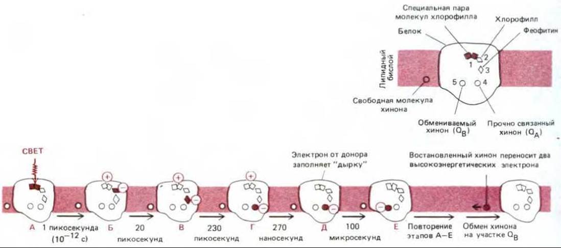
Fig. 7-50. Electron transfer occurring in the photochemical reaction center of purple bacteria. A similar reaction sequence is believed to operate in the evolutionarily related Photosystem II of plants. Top right: a schematic representation of the electron-carrying molecules depicted in Fig. 7-49, along with the exchangeable quinone (QB) and the mobile quinone Q dissolved in The Lipid Bilayer. Electron carriers 1–5 are specifically associated with a transmembrane protein consisting of 596 amino acid residues folded into two distinct subunits (see Fig. 6-32). Following excitation by a light photon, an energy-rich electron moves from one pigment molecule to another, leading to charge Separation as shown at the bottom of the figure (stages B–D; pigment molecules carrying high-energy electrons are highlighted in color). Upon entering the lipid bilayer, the fully reduced quinone picks up two protons and loses its net charge (see Fig. 7-30).
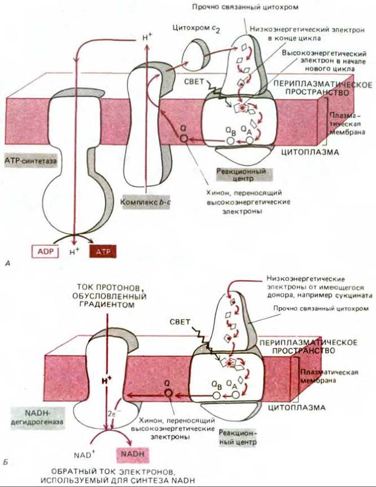
Fig. 7-51. Two electron-transfer pathways in the photosynthetic system of purple bacteria. These reactions take place in the plasma membrane, and cytochrome c2 is located in soluble form within the periplasmic space beneath the outer membrane (see Fig. 6-54). A. Cyclic electron flow generates an electrochemical proton gradient across the plasma membrane. The energy of this gradient is utilized by ATP synthase to synthesize ATP in the bacterial plasma membrane. B. Reverse electron flow through NADH dehydrogenase, driven by the energy of the same proton gradient, is used to synthesize NADH.
NADPH is produced during a second electron-transport process in which high-energy electrons are transferred from quinone not to the b-c1 complex, but to NAD. The resulting NADH is subsequently converted into NADPH by a transhydrogenase. Because the high-energy electrons carried by quinone are at a lower energy level than the electrons in NADH (recall that in mitochondria, electrons are transferred from NADH to quinone rather than the reverse—see Fig. 7-34), the formation of NADH from NAD requires an input of energy. In purple photosynthetic bacteria, the electrochemical proton gradient established across the plasma membrane forces protons to flow back into The Cell through the NADH dehydrogenase complex, supplying this complex with the energy required for the reverse transfer of electrons from quinone to NAD (Fig. 7-51, B).
Thus, in the plasma membrane of purple bacteria, reaction centers are used to build up a large pool of reduced molecules. A fraction of these molecules drives the establishment of a substantial electrochemical proton gradient across the plasma membrane. The energy of this gradient fuels two processes: (1) the synthesis of ATP via ATP synthase, and (2) the generation of a reverse electron flow from the remaining pool of reduced quinone to NAD, thereby generating the reducing power required for the synthesis of organic molecules.
7-24;
7-25;
7-26
7.3.10. In plants and cyanobacteria, both NADPH and ATP are produced via noncyclic Photophosphorylation [31, 35]
Photosynthesis is most complex in plants and cyanobacteria. Here, both ATP and NADPH are synthesized simultaneously in a two-step process known as noncyclic photophosphorylation. Because Two Photosystems sequentially excite an electron, the electron is able to travel the entire path from water to NADPH. As these high-energy electrons pass through the coupled photosystems, a portion of the energy contained within the electrons is used to generate NADPH, while another portion is diverted to synthesize ATP.
In the first of the two photosystems, historically named photosystem II, the oxygen of two water molecules is bound by a cluster of magnesium atoms with the assistance of a poorly characterized water-splitting enzyme. The extracted electrons fill the electron "holes" in the reaction center chlorophyll generated by light one by one. Once four electrons have been extracted (requiring four light quanta), the enzyme releases O2; thus, photosystem II catalyzes the reaction 2H2O → 4H+ + 4e- + O2.
The "core" of the reaction center in photosystem II is homologous to the bacterial reaction center just described and likewise generates strong electron Donors in the form of reduced quinone molecules in the membrane. These molecules transfer electrons to the b6 - f complex, which is similar to the bacterial b-c complex and the b - c1 complex in the mitochondrial Respiratory Chain. As in mitochondria, the b6 - f complex pumps protons across the thylakoid membrane into the thylakoid space (in chloroplasts) or from the cytosol through invaginations of the plasma membrane (in cyanobacteria), and the resulting electrochemical gradient provides energy for ATP synthesis by ATP synthase (Figs. 7-52 and 7-53). The final acceptor in this electron-transport chain is the second photosystem (Photosystem I), which accepts electrons into the "holes" created by light in the chlorophyll of its reaction center. While the electrons activated by photosystem II have too little energy to be transferred to NADP+, each electron leaving photosystem I is at a very high energy level due to sequential activation by two light quanta. As a result, these electrons can be transferred to the iron-sulfur center of ferredoxin and reduce NADP+ to NADPH (Fig. 7-53), with one proton being extracted from the medium.
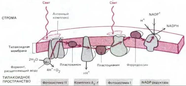
Fig. 7-52. Electron transport during photosynthesis in the thylakoid membrane. The mobile electron carriers in this chain are plastoquinone (very similar to mitochondrial ubiquinone), plastocyanin (a small copper-containing protein), and ferredoxin (a small protein containing an iron-sulfur center). The bb-f complex is very similar to the mitochondrial b-c1 complex and the bacterial b-c complex (see Fig. 7-63): all three complexes accept electrons from Quinones and pump protons. Note that the protons released during water oxidation and the protons consumed during NADPH formation also contribute to the electrochemical proton gradient that provides energy for ATP synthesis.
The zigzag scheme of photosynthesis shown in Fig. 7-53 is called the Z-scheme. As a result of two separate excitation steps, each catalyzed by its own photosystem, an electron is transferred from water, which normally holds its electrons tightly (redox potential +820 mV), to NADPH, which has a rather weak affinity for electrons (redox potential -320 mV). A single quantum of visible light cannot provide an electron with enough energy to travel the entire path from the start of photosystem II to the end of photosystem I; apparently, this requires as much energy as is needed to transfer an electron from water to NADP+. In addition, The Use of two separate, consecutive photosystems allows them to be linked by an electron-transport chain in which the energy of the electrons is sufficient to move H+ across the thylakoid membrane (or the plasma membrane of cyanobacteria), thereby directing some of the light-excited electrons toward ATP synthesis.
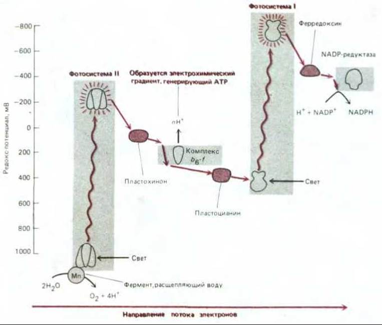
Fig. 7-53. Changes in redox potential during electron transport in photosynthesis, leading to the formation of NADPH and ATP in plants and cyanobacteria. Photosystem II is very similar to the reaction center of purple bacteria (see Fig. 7-50), to which it is evolutionarily related. Photosystem I differs from these two systems: it is believed to be evolutionarily related to the photosystems of another group of prokaryotes, the green bacteria. In photosystem I, electrons from excited chlorophyll pass through a series of tightly bound iron-sulfur centers. Two photosystems connected in series provide a net flow of electrons from water to NADP+, forming NADPH. In addition, ATP is generated by ATP synthase (not shown) using the energy of the electrochemical proton gradient created by the electron-transport chain linking photosystem II to photosystem I. This Z-scheme of ATP production is called noncyclic photophosphorylation, in contrast to the cyclic scheme shown in Fig. 7-54 (see also Fig. 7-52).
7.3.11. In The process of cyclic photophosphorylation, chloroplasts can synthesize ATP without producing NADPH [31, 36]
In the noncyclic photophosphorylation discussed above, high-energy electrons leaving photosystem II drive ATP synthesis, whereas the energy of electrons leaving photosystem I is used to form NADPH. In this process, slightly more than one ATP molecule is synthesized for every pair of electrons transferred from H2O to NADP+ to form an NADPH molecule. However, carbon fixation requires significantly more ATP than NADPH (see Fig. 7-43). To generate additional ATP, chloroplasts can switch photosystem I to a cyclic mode of operation, in which the system's energy is directed toward ATP production rather than NADPH synthesis. This process, called cyclic photophosphorylation, involves an electron flow very similar to that used by photosynthetic bacteria to generate ATP (see Fig. 7-51, A). In this case, high-energy electrons activated by photosystem I do not pass to NADP+ but instead return to the b6-f complex, thereby driving proton translocation across the thylakoid membrane. The resulting electrochemical gradient provides the energy for ATP synthesis (Fig. 7-54).
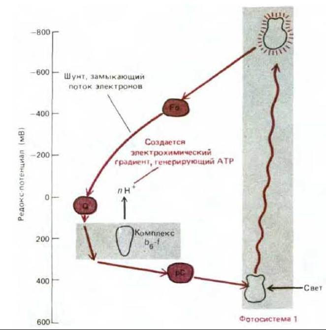
Fig. 7-54. The electron transport pathway in cyclic photophosphorylation. This pathway allows for the synthesis of ATP only, without the formation of NADPH and O2. Whether electron flow is noncyclic or cyclic depends on where ferredoxin transfers its reactive electrons—either to NADP+, as shown in Fig. 7-53, or to components leading back to the b6-f complex. Whenever NADPH accumulates and the level of NADP+ drops accordingly, favorable conditions are created for photosynthesis to proceed via the cyclic pathway. Other, less direct regulatory mechanisms also ensure that ATP and NADPH are produced in the proper proportion during photosynthesis. F — ferredoxin; PQ - plastoquinone; PC - plastocyanin.
Thus, the process of noncyclic photophosphorylation, which involves the reduction of NADP+ using water as an electron donor, is carried out by the joint action of photosystems I and II, resulting in the formation of NADPH, ATP, and O2. In contrast, cyclic photophosphorylation, which involves only photosystem I, synthesizes ATP alone—no NADPH or O2 is formed. Consequently, the relative intensity of cyclic and noncyclic electron transport determines what fraction of light energy is used to generate reducing power (NADPH) and what fraction is converted into the energy of phosphate bonds (ATP). This balance is regulated according to the demand for NADPH. Whether electron flow is cyclic or not depends on where ferredoxin transfers its reactive electrons—either to NADP+ or to components leading back to the b6-f complex (compare Figs. 7-53 and 7-54). At low concentrations of NADP+, caused by the accumulation of NADPH, the cyclic process will predominate, leading to ATP synthesis.
The Effect of the NADPH level on cyclic photophosphorylation is only part of an extensive regulatory network controlling the activity of photosystems I and II. For example, excessive activity of photosystem II leads to an increase in The ratio of reduced to oxidized quinone in the thylakoid membrane, whereas excessive activity of photosystem I has the opposite effect (see Fig. 7-53). However, whenever the ratio of reduced to oxidized quinone exceeds a certain threshold, a protein kinase is activated that phosphorylates the major light-harvesting pigment-protein in the antenna complex. This promotes the detachment of the antenna complex from the photosystem and even its migration from the region of photosystem II localization (grana) into the thylakoid membrane where the components of photosystem I are concentrated. As a result, photosystem I receives a larger share of radiant energy until the quinone level returns to normal.
7-27
7.3.12. The geometry of proton movement in mitochondria and chloroplasts is similar [37]
At first glance, the presence of a third internal compartment in chloroplasts—the thylakoid space—makes them look very different from mitochondria. However, the geometry of proton movement in these two organelles is very similar. As shown in Fig. 7-55, in chloroplasts, Protons are pumped from the stroma (pH 8) into the thylakoid space (pH of about 5), creating a gradient of 3 to 3.5 pH units. This generates a proton-motive force of about 200 mV across the thylakoid membrane (almost entirely due to the pH gradient rather than the Membrane Potential), which drives ATP synthesis by the membrane ATP synthase.
In the mitochondrial matrix, just as in the chloroplast stroma, the pH is close to 8, but this is achieved by pumping protons out of the organelle into the cytosol (pH of about 7) rather than into an internal compartment. Therefore, the pH gradient is relatively small, and the proton-motive force across the inner mitochondrial membrane, which is similar in magnitude to that across the thylakoid membrane of the chloroplast, is generated primarily by the membrane potential (see Section 7.1.7). However, in both mitochondria and chloroplasts, the catalytic site of ATP synthase is located in the large compartment of the organelle (the matrix and stroma, respectively), which has a pH of about 8.0 and is filled with soluble enzymes. Therefore, this is where all of the organelle's ATP is synthesized (Fig. 7-55).
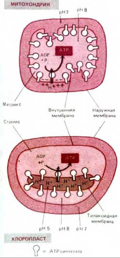
Fig. 7-55. Comparison of proton flow and ATP synthase orientation in mitochondria and chloroplasts. Compartments with similar pH are colored similarly. The proton-motive force across the thylakoid membrane is almost entirely due to the pH gradient; the high permeability of this membrane to Mg2+ and Cl- ions allows the flow of these ions to dissipate most of the membrane potential. It is believed that mitochondria could not withstand the degree of alkalinization (up to pH 10) that would be required to generate a proton-motive force without THE CONTRIBUTION OF a membrane potential.
Despite these similarities between mitochondria and chloroplasts, the latter are organized in such a way that their electron- and proton-transport processes are more accessible to study than those in mitochondria. By disrupting the outer and inner membranes of chloroplasts, intact thylakoid discs can be isolated. These are similar to submitochondrial particles: the Components of the electron-transport chain that utilize NADP+, ADP, and phosphate are also located on the outer side of the membrane. However, thylakoids are intact, natural structures and are therefore much more active than submitochondrial particles prepared artificially from mitochondria. Consequently, some of the experiments that first proved the key role of the chemiosmotic mechanism were carried out on chloroplasts rather than mitochondria.
7.3.13. The inner chloroplast membrane, like the inner mitochondrial membrane, contains carrier proteins that facilitate metabolite exchange with the cytosol [38]
Although the electron- and proton-transport reactions of photosynthesis are easiest to study in chloroplast preparations where the inner and outer membranes have been disrupted and removed, such chloroplasts are incapable of photosynthetic CO2 fixation due to the loss of several essential substances normally present in the stroma. However, chloroplasts can be isolated with their inner membrane intact. Using such preparations, it can be shown that the inner membrane is selectively permeable and therefore contains specific carrier proteins. For example, a significant portion of the glyceraldehyde 3-phosphate produced in the stroma during carbon fixation is exported from chloroplasts by an efficient antiport system that exchanges three-carbon sugar phosphates for inorganic phosphate.
Glyceraldehyde 3-phosphate, which enters the cytosol in abundance, is used by The Cell as a Starting Material for The biosynthesis of many other substances, including sucrose destined for "export." Furthermore, once in the cytosol, glyceraldehyde 3-phosphate is easily converted (via several reactions of The Glycolytic Pathway) to 3-phosphoglycerate, yielding one molecule of ATP and one molecule of NADH (glyceraldehyde 3-phosphate is formed in the carbon-fixation cycle by the same two-step reaction running in reverse—see Fig. 7-43). Thus, the glyceraldehyde 3-phosphate exported from chloroplasts not only serves as the primary source of fixed carbon but also delivers NADPH and ATP for cellular metabolism outside the chloroplast.
7.3.14. Chloroplasts also carry out other biosynthetic reactions [39]
In addition to photosynthesis, many other biosynthetic processes take place in chloroplasts. For example, all of the cell's Fatty acids and several Amino Acids are synthesized by enzymes located in the stroma. Furthermore, the reduction of nitrite (NO2-) to ammonia (NH3) occurs in chloroplasts, driven by light-activated electrons; in plants, this ammonia serves as a nitrogen source for the synthesis of Amino Acids and NUCLEOTIDES. Thus, The Importance of chloroplasts to plant and algal metabolism extends far beyond their role in photosynthesis.
Chloroplasts and photosynthetic bacteria obtain high-energy electrons through photosystems that capture electrons excited by sunlight absorbed by chlorophyll molecules. Photosystems contain an antenna complex coupled to a photochemical reaction center, where proteins and pigments involved in the photochemical reactions of photosynthesis are arranged in a highly ordered fashion. To date, the reaction center of purple photosynthetic bacteria is the most thoroughly studied, with its complete three-dimensional structure resolved. In these bacteria, a single photosystem generates an electrochemical gradient, the energy of which is used to synthesize both ATP and NADPH. Chloroplasts and cyanobacteria possess two photosystems. Depending on the cell's needs, Two Types of electron flow occur in varying ratios: 1) noncyclic flow, driven by two photosystems operating in series, transfers electrons from water to NADP+ to form NADPH, with ATP being synthesized along the way; 2) cyclic flow, driven by only one photosystem that recycles electrons through a closed circuit, yields only ATP. In chloroplasts, all electron transport processes take place within the thylakoid membrane: to synthesize ATP, protons are pumped into the thylakoid space, and then, as protons flow back through ATP synthase, ATP is synthesized in the stroma.
The ATP and NADPH produced during photosynthesis serve as Energy Sources for many biosynthetic reactions occurring in the stroma, including the vital CO2 fixation cycle, in which carbohydrates are synthesized from CO2. These carbohydrates, in the form of three-carbon sugar phosphates, are exported to the cytosol, where they serve as a source of organic carbon, ATP, and reducing power.
Last update: 12/08/2026
Editorial and Educational Adaptation: This material has been compiled based on the primary/original source text. The project team performed an editorial review, corrected technical inaccuracies, structured sections, and adapted the content for an educational format.
What was processed:
- elimination of formatting defects (OCR errors, structural breaks, corrupted characters);
- editorial organization of content;
- standardization of terminology in accordance with academic sources;
- verification of factual statements against the original source text.
All mentions of the author, publication year, and origin of the primary text have been preserved in accordance with the source.