BOTANY. VOLUME 1: CELL BIOLOGY. ANATOMY. MORPHOLOGY - 2007
4. MORPHOLOGY AND ANATOMY OF CORMOPHYTES
4.3. Leaf Organs. Structure and Metamorphoses
The structural diversity of leaves is extraordinarily vast. It ranges from barely noticeable scales to multi-meter, repeatedly pinnate fronds of tree ferns and palm leaves; from green, needle-like pine needles and photo-assimilating leaf blades of various shapes to brightly colored flower petals, and from leaf spines to intricate pitcher-like traps in carnivorous plants. In all these cases, however, we are dealing with modifications of a single type of organ: the phyllome. Its primary Functions are Photosynthesis and Transpiration. These are carried out by the leaf proper, which can be defined as an assimilator and a transpirator.
Morphologically, the phyllome represents the most highly developed form of a leaf. Compared to it, all other forms appear simplified due to reduction.
4.3.1. The Typical Leaf
4.3.1.1. Division and Symmetry
The MORPHOLOGICAL Structure OF a typical leaf with an undivided blade, established in the course of phylogenesis, is shown in Fig. 4.56.
The lower part of the leaf (Unterblatt)1 comprises the leaf base and, if present, stipules (Stipulae, singular Stipula, from Lat. stipula — straw). The leaf base often appears as an expansion of the petiole base, whereas in monocots it is typically so broad that it completely clasps the stem at the node. In such cases, the leaf base is usually elongated into a leaf sheath, as can be observed in most grasses.
1 The terms Unterblatt and Oberblatt are used exclusively in German-language literature; they have no exact equivalents in other languages. — Ed. note.
Class="center">Fig. 4.56. Leaf of the goat willow (Salix caprea) as an example of a typical phyllome
Attached to the stem by its expanded base laterally, the leaf base is flanked by stipules; directly above it lies the axillary bud (arrow). The leaf base and stipules constitute the lower zone of the leaf. The upper zone consists of the petiole and leaf blade (12x). In pinnate leaves (cf. Fig. 4.21), the petiole continues into the blade region as a rachis and bears paired leaflets as well as a terminal leaflet (in imparipinnate leaves).

Such a leaf sheath functions as a supporting organ for the culm, the slender stem of grasses. Thickened leaf sheaths also serve as the storage scales of bulbs. In other cases, elongated, mutually clasping leaf sheaths form a false stem (pseudostem) (Fig. 4.57, A). Typically developed in the banana, it can also be observed during early developmental stages in native European monocots (e.g., in hellebore, Veratrum; or the grass Molinia). True, inflorescence-bearing stems later grow upward from within this tubular false stem.
Fig. 4.57. False stems and false bark
A — false stem of the onion (Allium cepa) formed by hollow cylindrical leaf sheaths (0.6x). B, C — stem-clasping leaf base typical of many monocots; the true stem is often invisible, Examples being popular houseplants Aloe spinosissima and Dracaena marginata (0.6x). D, E — false bark, represented by leaf bases in conifers. D — Thuja orientalis (decussate phyllotaxy, 2.1x). E — vertical SHOOT of Norway spruce (Picea abies) with spiral phyllotaxy; left: leafy, right: after needle fall, the elongated leaf bases closely adjoin one another — in contrast to fir (Abies), where rounded leaf bases do not entirely cover the stem (cf. Fig. 4.16, E).

In many conifers, the leaf base does not clasp the stem, but instead elongates along the shoot and fuses with the stem. When these bases also Touch one another laterally, creating a dense relief pattern on the stem, this is referred to as false bark (Fig. 4.57, D, E).
Stipules in many plants are either not formed at all, or are short-lived and fall off early (as in hazel and hornbeam, where they function as bud scales); however, they can also be quite large and even take on the functions of a typical leaf (Fig. 4.58). Not infrequently, stipules are transformed into spines, as in Robinia or black locust.
The upper part of the leaf comprises the petiole and the leaf blade. The petiole elevates the leaf blade some distance away from the stem; the blade itself serves as the main organ of assimilation and transpiration. Through growth or turgor movements, the petiole can alter THE POSITION OF the blade for optimal exposure to sunlight. As a supporting organ, the petiole often has a more or less circular cross-section, thereby resembling axial Organs. However, expanded and flattened petioles sometimes take over the Functions of the leaf blade (Fig. 4.59). In such cases, they are called phyllodes. If the petiole is absent, the leaves are described as sessile.
The Diversity of phyllodes is manifested primarily in the variety of forms of the leaf blade (from Lat. lamina — plate, see Fig. 4.2). Alongside its shape, leaf size is extremely variable — ranging from a millimeter to nearly 20 meters, as seen in the pinnate leaves of the palm Raphia taedigera.
Fig. 4.58. Leaf base and stipules
A — leaf-like stipules in wood avens (Geum urbanum, 1.6x). In some plants, stipules completely take over leaf functions, for example in Lathyrus aphaca (cf. Fig. 44.69); B — bedstraw (Galium mollugo) appears to have whorled leaves, but the stem is four-angled and axillary shoots grow only from the axils of two opposing leaves; only these organs are true leaf blades, whereas the others are similar leaf-like stipules. Alternatively, the leaves could be viewed as sessile, i.e., petiole-less opposite palmate leaves (2.1x). C — leaf base without stipules in the walnut (Juglans regia, 1.6x). D, E — lignified stipules transformed into spines in the black locust (Robinia) (D — 0.3x; E — 1.7x).
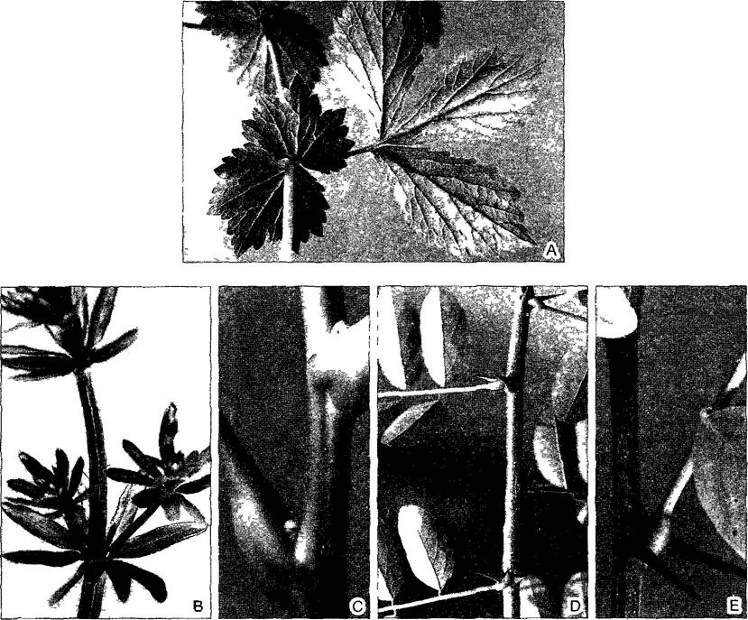
Fig. 4.59. Phyllodes in Acacia heterophylla (after J. Reinke).
Following the twice-pinnate first leaves, pinnate leaves with winged petioles develop, and finally, in subsequent bladeless leaves, flattened petioles called phyllodes take over the functions of the leaves.

Many common plant names are derived from leaf shapes (e.g., golden saxifrage, hepatica, cinquefoil, arrowhead, lady's mantle). Flora identification keys feature diagnostically important differences between plants along with their names. Morphologically, pinnate leaves are of particular interest. In these, the petiole continues into a rachis, which bears several pairs of lateral leaflets and (most frequently) an unpaired terminal leaflet. In fern fronds, multiple pinnation is especially common, where primary pinnae are divided into secondary pinnae, which in turn are divided into tertiary pinnae. If the longitudinal growth of the rachis in once-pinnate leaves is arrested, all pinnae appear to be attached at the apex of the petiole; such leaves are termed palmate. Special forms such as peltate, terete, or ensiform leaves, as well as the tubular carnivorous leaves of pitcher plants, will be examined later in our Discussion of diet development.
A typical leaf is bilaterally symmetrical; its median plane coincides with the direction of the petiole and, correspondingly, the rachis, with the midrib lying within this plane. Deviations from bilateral symmetry are rare and therefore particularly noticeable (such as the oblique leaf in begonia). Typical leaves also most frequently exhibit distinct dorsoventrality: their adaxial (or morphologically upper) side facing the stem (at least initially) differs in many features from the abaxial, lower side. These differences involve, for example, the number of Stomata (most leaves are hypostomatous: over 90% of their stomata are located in the lower epidermis), pubescence, the accumulation of pigments in the vacuoles of epidermal Cells, and, finally, anatomy: palisade parenchyma is predominantly adaxial, whereas spongy parenchyma is abaxial. In leaf vascular bundles—Veins, or leaf nerves (both of these vernacular terms are essentially incorrect)—the xylem is situated on the upper side and the phloem on the lower. Leaf dorsoventrality is a consequence of the polarity of stems, on which leaves are positioned laterally, so that the leaf surface is oriented transversely to the polarity gradient.
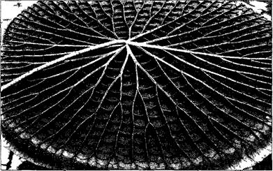
Even upon casual inspection with the unaided eye, the pattern of their venation, i.e., the pattern of vascular bundles within the leaf blade (Fig. 4.60), is readily apparent. Stronger bundles (major veins) supply leaves with Water and facilitate the export of photosynthetic products from the leaves. They are surrounded by a bundle sheath that contacts the mesophyll tissue system and regulates the Metabolic exchange between the vascular bundles and the mesophyll. Such bundle sheaths sometimes extend to the epidermis and also perform a mechanical support function. Major veins often project on the underside of The Leaf as Ribs that strengthen the blade. An extreme case is observed in the giant floating leaves of Victoria amazonica (Fig. 4.61). However, The primary function of leaf veins is to supply water and mineral salts to the particularly active photosynthesizing and transpiring mesophyll cells, as well as to ensure the rapid outflow of photosynthetic products. Within the conducting elements, There is a convective1 flow of contents; beyond the bounds of the bundles, the Transport of substances is restricted to diffusion. The efficiency of diffusive transport decreases as the square of the distance traversed and is effectively insufficient already at distances spanning just a few cells. Water itself flows through vessels a million times more easily than through living tissue. Consequently, minor veins, which establish direct contact with the mesophyll tissue, form such a dense network that within the areoles situated between them, no mesophyll Cell is located further than 7 other cells away from the nearest bundle. (Note that the term "areole" is used here in a completely different sense, cf. Fig. 4.7, C). The total length of the vascular bundles in a beech leaf reaches approximately 30 m.
1 The Mechanism of long-distance transport via conductive Tissues does not correspond to convection in the sense understood by physicists. — Ed. note.
Fig. 4.60. Leaf venation (pattern of vascular bundles within the leaf blade)
A, B—dichotomous (forked) venation in fern leaves: A—Adiantum capillus-veneris (3.5x), B—Asplenium nidus (2.4x); C—combination of parallel and reticulate venation in Maranta, with the leaf blade (only a part is shown) patterned to resemble a leafy stem—presumably a form of mimicry to deter pest insects from laying eggs (1.4x); D—parallel venation in the palm Sabal umbraculifera (0.7x); E—reticulate venation in Boston ivy (Parthenocissus tricuspidata) (3.5x)
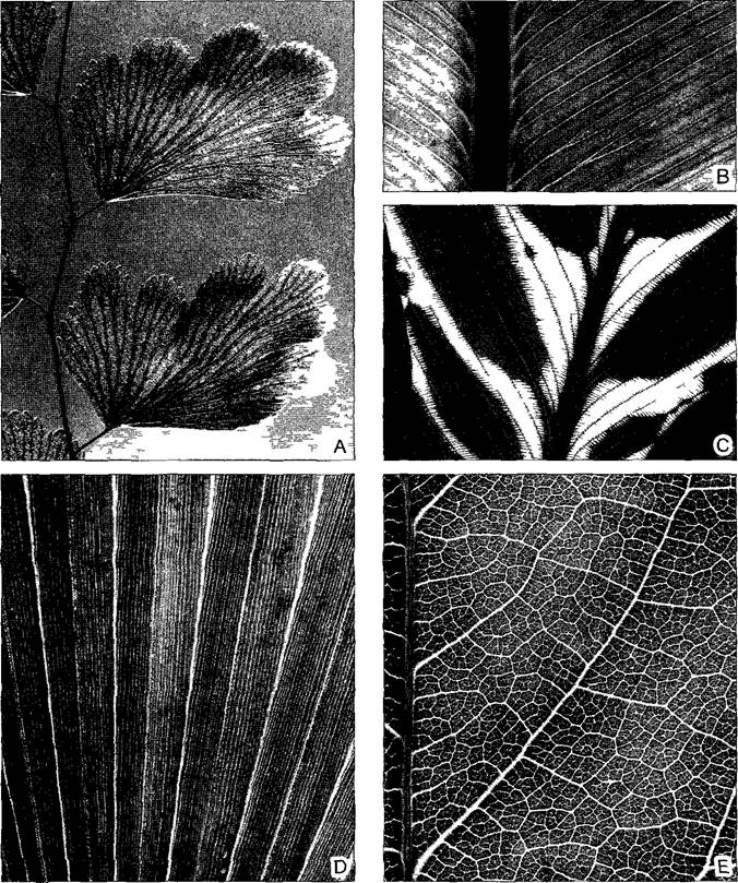
Fig. 4.61. Ribs on the underside of the floating leaf of Victoria amazonica (= regia); the leaf is nearly 2 m in diameter (photo by W. Barthlott)
These functional challenges can be met in various ways. In monocots, parallel venation predominates, where all major veins run longitudinally. This arrangement of vascular bundles is particularly pronounced in the linear leaves of grasses. In the lanceolate leaves of most other monocots, the veins run in smooth arcs corresponding to the equally smooth arcuate leaf margin typical of monocots (see Fig. 4.14, A). In parallel venation, the major bundles are interconnected by smaller transverse cross-veins, so that a regular network of veins is actually present. (This can be readily observed with the naked eye, for example, in the leaves of Clivia.)
True dicots form a more complex network of veins: reticulate venation. This allows for almost any shape of the leaf blade and especially its margin. Differences in venation are also reflected in the arrangement of stomata: in monocot plants, they are most frequently oriented in parallel, whereas in magnoliids and true dicots, they are arranged irregularly (see Fig. 3.13).
A third type of venation, dichotomous, or flabellate, venation, occurs in ferns and in Ginkgo, which is classified among gymnosperms. Here, the stronger vascular bundles branch dichotomously and end blindly at the leaf margin. Therefore, this type of venation, being "open," was formerly contrasted with the supposedly "closed" venation of monocots and true dicots (comparable to closed and open circulatory systems in animals). However, even in reticulate venation, the finest veinlets end blindly in the mesophyll.
4.3.1.2. Development and Special Forms of Leaves
Leaf primordia arise exogenously on the shoot apex as lateral outgrowths through the division of the apical meristem (see Figs. 3.3;
3.5).
In ferns, a two-sided apical cell of the leaf primordium initially arises within the small-celled zone of the flank meristem. From this, a linear marginal meristem develops, in which the original apical cell is no longer distinguishable. Acroplastic growth is typical for most fern leaves, where the apical part of the leaf continues to grow while the cells at the Base of the leaf are already differentiated. The dissection of fern leaves is caused by the fractionation of the marginal meristem due to the local cessation of its cell divisions.
In angiosperms, newly formed leaf primordia show a clear tendency for their bases to expand perpendicular to the shoot axis. This gives rise to a broad leaf base that can completely encircle the stem and lead to The formation of a leaf sheath. A crest-like marginal meristem subsequently forms the leaf blade. In contrast to ferns, basiplastic growth predominates here, meaning that The activity of the marginal meristem ceases first at the apex and last at the base of the leaf blade. Pinnate leaves most frequently arise, as in ferns, As a result of the fractionation of the marginal meristem. The major veins differentiate from the base upward, whereas minor veins develop fully in the distal part of the blade first.
Leaf petioles are formed through intercalary growth, i.e., owing to a meristem located between already formed PARTS OF THE leaf. Similarly, the entire-margined, parallel-veined leaf blades of monocots, especially grasses, owe their origin to a basal intercalary meristem. (Much like the leaves of the peculiar gymnosperm representative Welwitschia, which slowly die off from the tips while continuing to grow at the base for over 500 years; see Fig. 11.211, A.)
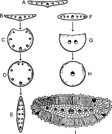
The dorsoventrality of the leaf blade is expressed in the fact that most leaves are bifacial, meaning their upper and lower sides look different (from Lat. facies—appearance, aspect; Fig. 4.62, A–D). There are also equifacial leaves, especially in inhabitants of sunny habitats. Both sides of such leaves are structured identically: for example, they have the same stomatal density and palisade mesophyll beneath the abaxial epidermis (Fig. 4.62, F, I). Such leaves are often thickened or needle-like and oriented edge-on toward the stem. If succulence is added to this, equifacial terete leaves develop, as, for example, in Stonecrop (Sedum; see Fig. 4.70, A). Another way terete leaves are formed is by the lower side of the leaf growing more intensively than the upper side, so that the latter ultimately disappears altogether: unifaciality. Leaf petioles frequently approach such unifaciality, acquiring a stem-like, round cross-sectional shape. However, the leaf blades of certain monocots (rushes, some species of onion, such as chives) are also unifacial and radially symmetrical. A peculiar and special case is found in the leaves of irises (Iris): these are secondarily flattened unifacial leaves, with the flattening occurring not perpendicular to the rhizome axis, but in the same direction ("ensiform leaves") (Fig. 4.62, E).
Fig. 4.62. Cross-sections of various leaf types (schemes A–H after W. Troll and W. Rauh, I after Volkens)
Palisade parenchyma is indicated by stippling, the lower side of the leaf by a thick line, and the xylem parts of the vascular bundles in black. A—normal bifacial flat leaf (cf. Fig. 4.64); B—reverse-bifacial flat leaf (e.g., ramsons, or bear's garlic, Allium ursinum); C, D—origin of a unifacial terete leaf (e.g., Allium sativum, Juncus effusus); E—unifacial ensiform leaf (Iris); F—equifacial flat leaf; G—equifacial needle-like leaf (see Fig. 4.65, A); H—equifacial terete leaf (e.g., Sedum, see Fig. 4.70, A); I—cross-section of an equifacial leaf of the desert plant Reaumuria hirtella, Tamaricaceae (30x)
In peltate — shield-shaped — leaves (from Greek pelte—shield), the petiole attaches not to the lower margin, but almost to the center of the leaf blade (Fig. 4.63). This is due to the fact that, owing to pronounced basiplasty, the marginal meristem of the leaf blade grows vigorously directly from the point of petiole insertion; furthermore, due to its unifaciality, the right and left margins of the blade come into close contact with one another and fuse. The tubular leaves of certain specialized carnivorous plants from the families Sarraceniaceae and Nepenthaceae develop in a similar manner (Box 4.4, Fig. A).
Fig. 4.63. Shield-shaped leaf of nasturtium (Tropaeolum majus) (0.7×), viewed from above (A) and from below (B)
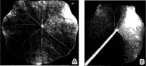
In some plants, the margins fuse not of the same leaf, but of different leaves belonging to the same node (gamophylly). This phenomenon is relatively rare in the vegetative parts of the plant, yet it is widespread in flowers, where we find fused sepals and petals, as well as syncarpous Ovaries.
4.3.1.3. Anatomy
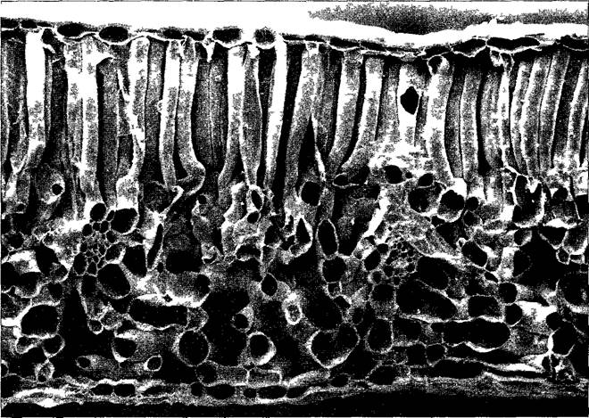
A typical cross-section of a bifacial leaf is shown in Fig. 4.64. Single-layered epidermises enclose the chlorenchyma of the mesophyll, which is differentiated into palisade and spongy parenchyma. The denser, one-, two-, or three-layered palisade parenchyma contains about 4/5 of all leaf METABOLISM/14.html">Chloroplasts, serving as the primary assimilation tissue. In contrast, the very loose spongy parenchyma (see Fig. 3.7, B) functions primarily as a transpiration tissue. Due to numerous, occasionally very large intercellular spaces (accounting for up to 90% of the mesophyll volume), the total surface area of all mesophyll cells is nearly 100 times greater than the leaf surface itself. In hypostomatous leaves, the intercellular System of the spongy parenchyma also facilitates the diffusion of CO2 toward the palisade parenchyma. Epidermal cells contain leucoplasts, sometimes with sparse thylakoids and a low chlorophyll content. Larger vascular bundles are surrounded by an endodermis, referred to here as the bundle sheath. This is usually subtended by a ring of transfer cells corresponding to the pericycle (see 4.4.2.1). Like the endodermis itself, this cell layer exhibits a glandular nature and mediates regulated substance exchange between the bundle and the mesophyll. Sclerenchyma fibers frequently accompany the vascular bundles as well.
Fig. 4.64. Anatomy of a bifacial leaf: transverse section of the leaf of stinking hellebore (Helleborus foetidus, 100×) (SEM micrograph by H.O. Ihlenfeldt)
Beneath the upper epidermis lies the palisade parenchyma, followed below by the loose spongy parenchyma with two transversely cut vascular bundles; the leaf is covered on the underside by the lower epidermis. In such leaves, the epidermis accounts for about 12% of the volume, the Vascular Tissues for 5%, the mesophyll (palisade and spongy parenchyma) for 68%, and intercellular spaces make up about 16%.
A specialized leaf structure is characteristic of so-called C4 plants, whose photosynthesis is adapted to intense sunlight in arid habitats (see 6.5.8). In these plants, the final fixation of CO2 takes place within the bundle sheath cells, which are consequently exceptionally large and rich in Plastids (the "Kranz" type of leaf vascular bundles, see Fig. 6.81). The plastids of bundle sheath cells lack grana1 but contain abundant assimilation starch. In contrast, the chloroplasts of the assimilatory parenchyma possess grana but no starch (chloroplast dimorphism, see Fig. 6.82). Bundle sheath cells and mesophyll cells are interconnected by numerous plasmodesmata. The entire tissue complex acts as a CO2 pump2: the plastids of the bundle sheath cells receive an adequate supply of CO2 even when the stomatal pores are constricted to reduce transpiration, thereby preventing a drop in intercellular carbon dioxide concentration.
1 Only in some C4 plants do the chloroplasts in bundle sheath cells lack grana; in most species, grana are numerous and large. — Ed. note.
2 This is an extremely simplified and distorted description of C4 chlorenchyma function. — Ed. note.
In many plants, the tissue arrangement within the lamina deviates more or less significantly from that shown in Fig. 4.64 (see Fig. 4.62, B–I). Frequently, palisade parenchyma is also present internally beneath the lower epidermis (see Fig. 4.71, A). In vertically oriented grass leaves, the mesophyll is homogeneous, lacking differentiation into palisade and spongy parenchyma, and stomata are distributed with equal frequency on both leaf surfaces. The leaves of aquatic plants (such as waterweed, Elodea) are often only two cell layers thick, whereas plants of extremely humid habitats may possess leaves that are merely a single cell layer thick (Hymenophyllum).
The Internal Structure of an equifacial leaf is illustrated in Fig. 4.65 using a needle leaf as an example. In leaves of this type, the distinction between spongy and palisade parenchyma is generally indistinct and often entirely absent, as in the case presented here. The transverse section of a pine needle reveals polygonal mesophyll cells. The Cell surface area is increased by ridge-like wall thickenings1 (palmate parenchyma). The apparent absence of intercellular spaces is deceptive: disk-shaped layers of assimilation tissue, one cell thick and oriented perpendicularly to the longitudinal axis of the needle, are separated from one another by intercellular spaces. Situated between the assimilation tissue and the epidermis—whose cells die off following pronounced wall thickening—is a dead, lignified mechanical tissue known as the hypodermis. Stomata, whose guard cells require connection with living tissue, are sunken to the level of the assimilation tissue. Several longitudinal resin ducts run through the needle mesophyll. One or two unbranched vascular bundles of the needle leaf are surrounded by transfusion tissue and a common endodermis. The transport of substances between the conducting elements and the mesophyll is mediated by the transfusion tissue, which consists of parenchymal cells—typical Strasburger cells with a distinct glandular character adjoining the phloem—along with dead, short tracheids.
1 In reality, these are not thickenings, but wall invaginations. — Ed. note.
Fig. 4.65. Transverse (A) and longitudinal (B) sections of the equifacial needle leaf of European black pine (Pinus nigra, 40×) (A—after R. von Wettstein, B—SEM micrograph, 285×)
The longitudinal section reveals intercellular spaces. Sk — sclerenchymatous hypodermis, En — endodermis, Pa — assimilation parenchyma, Sp — stoma, H — resin canal, L — vascular bundle with xylem on top, Tr — transfusion tissue, E — epidermis

4.3.2. Leaf Series
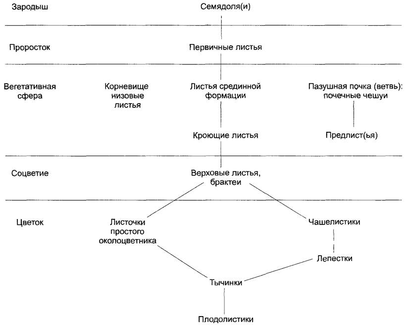
As demonstrated above (Figs. 4.5; 4.6), The Development of phyllomes in angiosperms varies widely during the ontogeny of a single plant—that is, under an identical genotype—spanning a broad spectrum corresponding to the (maximal) scheme outlined below.
A comparison of different leaves (see the leaf series in Fig. 4.66) indicates that their simplified forms, such as cataphylls, bud scales, hypsophylls, and floral phyllomes, arise through the inhibition of upper leaf zone development accompanied by the enhanced Development of the lower leaf zone. The leaf series is a striking manifestation of the organ's capacity for modification through proportional shifts.
Fig. 4.66. Development of the upper leaf zone (Oberblatt) during the transition from bud scales (A–C) to a typical leaf (G) in Siberian crabapple (Malus baccata) (after W. Troell)
D–E — transitional leaves, F — typical leaf shortly before unfolding (A–F — nearly natural size; G — 0.2×); st — stipules; Bg — leaf base; Na — scars left by fallen stipules; St — petiole; La — leaf blade (lamina); o — upper leaf zone

If the cotyledons within a seed serve as "fleshy" storage reservoirs for nutrient reserves, they typically remain enclosed within the rupturing seed coat during germination, thereby either emerging onto the soil surface or remaining underground—hypogeal germination (oak, horse chestnut, pea, scarlet runner bean, etc.). More frequently, epigeal germination occurs, in which the cotyledons are elevated into the light and turn green due to the elongation of the hypocotyl (spruce, beech, maple, mustard, sunflower, common bean, etc.).
1 Prophylls are the very first leaves on a lateral shoot and never succeed bud scales. — Ed. note.
How dramatically a foliar series can be modified is demonstrated by the popular ornamental plant Streptocarpus hybridus. It initially forms only two identical cotyledons, but later one of them enlarges massively into a single, long-lived "leaf" in the axil of which the inflorescence eventually develops.
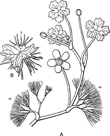
Conversely, a foliar series can become more complex, for instance when juvenile and adult foliage leaves differ, as seen in ivy. Anisophylly refers to a condition where adjacent leaves, sometimes even at the same node, vary in size and robustness due to the dorsiventrality of a plagiotropic shoot (Fig. 4.67). Heterophylly describes a phenomenon where distinctly structured leaves with different functions develop in response to external or internal conditions (Fig. 4.68; for the specific arrangement in the floating fern Salvinia, see Fig. 11.158). Well-known examples include the diverse leaf shapes of juvenile creeping shoots versus upright flowering shoots in ivy.
Fig. 4.67. Anisophylly (after W. Troll, B — after K. Goebel)
A—induced anisophylly in the Norway maple (Acer platanoides): leaves of two adjacent whorls on an obliquely growing branch, arrow indicates the gravity vector (0.25x); B—obligate anisophylly in Selaginella douglasii: each node bears one large ventral and one small dorsal leaf (5x). (The leaves of all spike-mosses are not opposite, but alternate. — Ed. note)
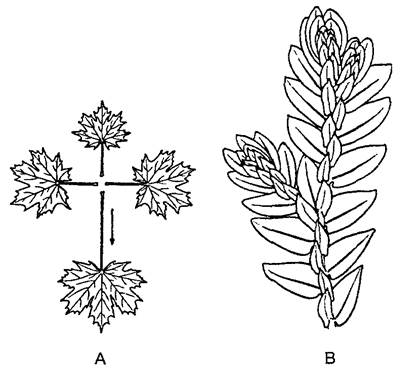
Fig. 4.68. Modification heterophylly in water crowfoot (Ranunculus aquatilis) (after W. Troll)
A—flowering, sympodially growing branch with floating and finely dissected submerged leaves (w); B—transitional leaf form
Individual phyllomes of a foliar series differ not only in shape and function, but also in their lifespan. Cotyledons and perianth segments are typically exceptionally short-lived. Strongly reduced and usually quickly deciduous bracts are referred to as bracts (from Lat. bractea — a thin metal leaf or scale). Foliage leaves generally last longer, but in deciduous plants (broad-leaved trees and, among conifers, larch) they drop at the end of each growing season. Prior to leaf fall, nitrogen-containing compounds in particular are mobilized and translocated out of the leaves. During these dramatic shifts, chloroplasts transform into gerontoplasts1, turning yellow due to retained carotenoids, which typically form fatty acid esters. The leaves or needles of evergreen plants and shrubs function for several years (pine — 2 years; fir — 5–6 years; monkey puzzle tree — up to 15 years). Leaf abscission occurs through the formation of a specialized abscission layer (see Fig. 7.61).
1 In botany, such plastids are conventionally referred to as chromoplasts. — Ed. note
4.3.3. Leaf Modifications
4.3.3.1. Metamorphosis
It has already been mentioned that leaves, much like shoots, can transform into spines (see 4.1.1) and tendrils (see 4.2.6). Examples of such metamorphoses are shown in Fig. 4.69 and 4.7. Leaves frequently function as storage organs, and alongside stem succulents, there are also leaf succulents. Large water-storing cells are located either subepidermally or deep within the leaf (for example, in Lithops — plants commonly known as the "living stones" of South African deserts; see 15.2.6).
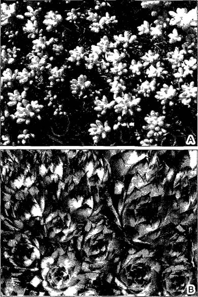
In some plants, the Cells of the mesophyll itself enlarge due to unusually large vacuoles. This represents a morphological correlation with a special adaptation of photosynthesis to sunny, hot, and dry habitats. Such an adaptation is known as CAM metabolism (see 6.5.9). Crassulaceae are "thick-leaved" plants (from Latin crassus, meaning thick), which include, among others, houseleeks (Sempervivum) and stonecrops (Sedum, Fig. 4.70). CAM plants are found not only in the family Crassulaceae, but also in 27 other families, including even succulent ferns. They fix CO2 at night with open stomata in an intermediate form. The malic acid synthesized under these conditions accumulates in the large vacuoles of the mesophyll cells. During the day, the stomata close due to the risk of excessive water loss, but the CO2 released from the malic acid can now be finally assimilated using light energy.
Fig. 4.69. Tendrils (A, B — after H. Schenck; C — after F. Noll): A—tendrils of a pinnate leaf in the garden pea (Pisum sativum); B—leaf tendril of the yellow vetchling (Lathyrus aphaca); C—stem tendril with adhesive disks in Boston ivy (Parthenocissus tricuspidata) (all 0.6×); s—stem; n—stipules; r—leaflets of a compound leaf modified into tendrils; a—axillary peduncle; b—rachis of a compound leaf
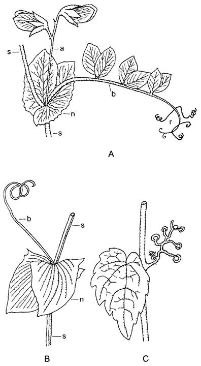
Fig. 4.70. Leaf succulents (0.75×): A—white stonecrop (Sedum album); B—Sempervivum schnittspahmi, rosette with alternate leaves
Leaf metamorphosis is observed primarily in plants adapted to unusual environmental conditions or a specialized lifestyle. In such cases, it is usually not just the leaves, but the entire plant that undergoes corresponding changes, resulting in an adaptive syndrome. Three syndromes affecting leaf structure as well are briefly outlined in the next two sections and in Box 4.4 (Ecological Morphology).
4.3.3.2. Xeromorphic Leaves
For plants of arid regions (steppes, deserts) or dry habitats (rocks, sandy soils; from Greek xeros — dry), water availability is of critical importance. Water supply cannot be stabilized through enhanced water uptake, leaving no alternative but to restrict water loss—that is, transpiration—or even suspend active life entirely during drought periods. As we have already seen, many xerophytes transform their leaves into spines or reduce them to tiny scales, carrying out photosynthesis instead with flattened stems that lack transpiring tissue1 and possess a relatively small surface-area-to-volume ratio. Cuticular transpiration is extremely limited; in many cases, a water-storage system is developed (stem succulents; see Fig. 4.35).
1 This is an incorrect statement, as any photosynthesizing tissue, whether in a leaf or a stem, transpires. — Note by the editor.
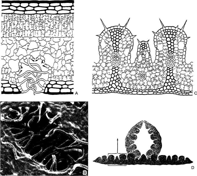
Many xerophytes, however, retain assimilating leaves. These leaves are "xeromorphic," meaning they are structured such that their transpiration is low and/or can be reduced when necessary. Indeed, the leaves of xerophytes differ markedly from those of mesophytes and hygrophytes inhabiting areas with adequate or abundant moisture. While hygrophyte leaves (as well as "shade" leaves, for example, in the beech, see Fig. 7.73) are thin and typically glabrous, with non-sunken and often even elevated stomata relative to the epidermal surface, xeromorphic leaves are usually leathery and rigid (sclerophyllous plants — laurel, myrtle, olive tree) with sunken stomata (Fig. 4.71). Leaves that roll up during a drought effectively isolate their stomata from contact with the surrounding environment. Furthermore, water loss can be restricted by a very thick cuticle and a heavy wax layer, and often additionally by dense pubescence, which creates a boundary layer of stagnant, more humid air right at the leaf surface. The rigidity of xeromorphic leaves, which prevents wilting, is due to the development of sclerenchymatous fibers or solitary stellate sclereids. Xeromorphic leaves are frequently equifacial.
Fig. 4.71. Anatomy of xeromorphic leaves (A, C, D — after O. Stocker; B — SEM micrograph by W. Barthlott)
A—oleander leaf with a multi-layered epidermis (black), three-layered palisade parenchyma, and deeply sunken stomata; hairs prevent air movement within the depressions (crypts) (80x); B—surface view of the crypt (170x); CD—leaves of feather grass Stipa capillata are epistomatic, meaning stomata are located exclusively on the upper side. During drought, the leaves roll inward toward the upper surface, thereby protecting the stomata from ambient air, whereas under favorable water supply, the leaf blade unfolds and becomes flat. The mesophyll is not differentiated into palisade and spongy parenchyma, typical of other grasses (C—80x, D—10x)
Equifacial needle-like leaves (see Fig. 4.65) also exhibit clear xeromorphic features. This is an adaptation to intense absorption of the infrared spectrum of solar radiation and the consequent heating of dark needles under direct sunlight. Because of the relatively low transport capacity of conifer wood tracheids, such warming in needle-like leaves can readily lead to water deficit even under normal conditions. During prolonged frosts, when water uptake from frozen soil ceases, the water balance in sun-warmed needles easily reaches a critical state—a condition known as "frost desiccation."
Although reducing transpiration helps stabilize the water balance, the acute problem of leaf/stem parenchyma overheating remains (Fig. 4.72, 4.73, 4.74). (Transpiration exerts a powerful cooling effect due to the relatively high energy cost of water evaporation—about 41 kJ/mol.) In many plants, excessive heating of leaf blades is prevented by orienting them edge-on to the direction of sunlight. A notable example is the shade-free Australian eucalyptus, whose sickle-shaped leaves hang vertically from branches. Protruding ribs and deeply fissured bark also exert a cooling effect on trunks (see Fig. 4.54, A; 4.55, G-I).
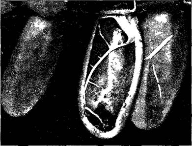
Fig. 4.72. Pitcher leaves of Dischidia major (middle one cut longitudinally) featuring an adventitious ROOT that grows into the leaf cavity through the pitcher opening (0.8x) (photograph by W. Barthlott)
4.3.3.3. Epiphytic Leaves
In contrast to climbing plants, which are invariably rooted in the soil, epiphytes establish themselves directly in the tree canopy from the outset to secure a place in the sun. Trees serve merely as a supporting substrate; these can be replaced by rocks, roofs, or even telephone wires. Thus, the majority of epiphytes are not parasites. However, when growing vigorously, they may "suffocate" their living support. Only a few epiphytes, such as mistletoe, are true parasites.
Plants inhabiting nutrient-poor substrates, particularly those deficient in nitrogen (such as raised bogs), display a remarkable trophic specialization: while capable of living as photoautotrophs, they are additionally equipped with mechanisms to capture and retain small animals, primarily insects. These carnivorous, or insectivorous, plants utilize their prey as an supplementary source of nitrogen, digesting it extracellularly (see 9.1.2). Their leaves are modified in various, often highly ingenious ways to trap animals.
The sticky traps of sundews (Drosera) operate on a relatively simple principle. Their leaves bear emergences known as "tentacles," supplied with a strand of tracheids (cf. Fig. 3.31). Their glandular heads secrete glittering droplets of a sticky substance that attracts small animals. An insect adheres to the gland and, in its attempts to escape, brushes against neighboring glands, becoming increasingly entrapped. The stimulation of the tentacles causes them to bend, pressing the insect firmly against the leaf surface. Here, the prey's body (excluding Chitin) is chemically broken down by secretions from specialized glands and absorbed in liquid form.
The Venus flytrap Dionaea (see Fig. 8.28, E, F) is capable of instantly snapping shut its snap trap by folding the leaf in half. This movement is driven by a hinge mechanism along the midrib and regulated by osmotic processes. The action is triggered the moment an insect, landing on the open leaf, brushes against one of the sensitive trigger hairs (see 8.3.2.4). The folded margins of the leaf blade are fringed with Teeth like a steel trap, allowing the plant to retain even powerful insects such as wasps or bumblebees, which are subsequently digested by secreted Enzymes.
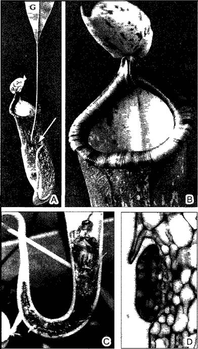
In Nepenthes, Cephalotus, Sarracenia, and Darlingtonia, the pitfall traps take the form of urn-, pitcher-, or sac-like leaves. The pitchers of Nepenthes (Fig. A) contain a watery, acidic digestive fluid secreted by glands lining their interior walls. Lured animals slip on the smooth pitcher rim—paved with waxy platelets—drown in the fluid, and are digested by enzymes.
Fig. A. Trapping pitcher in Nepenthes (C — photo by W. Barthlott)
The pitcher is formed by a sac-like leaf blade. A few milliliters of digestive secretion, produced by specialized peltate glands (D, 260x), accumulate within a pitcher standing several centimeters tall (A, 0.3x). Prey, most commonly insects, land on the brightly colored rim of the pitcher, which is rendered slippery by epicuticular wax platelets (B, 1.2x); nectar glands are located just beneath the inward-rolled margin. Seeking the nectar, an insect inadvertently slips into the trap. During its development, the pitcher lid remains closed to prevent rainwater from entering, but later stays permanently open. The leaf petiole can function as a tendril (arrow in A), suspending the heavy pitcher from branches. The elongated and expanded basal portion of the leaf (G on A) takes over the functions of the leaf blade.
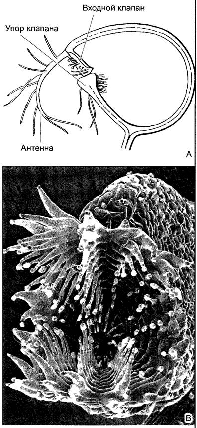
Submerged plants of the genus Utricularia inhabiting stagnant waters bear small, water-filled green bladders on their dissected leaves (Fig. B), which function as suction traps. The "Mouth" of such a trap is initially sealed watertight by a valve. When a small aquatic animal brushes against one of the lever-like trigger hairs on the outside of the valve, the valve opens, and an influx of water sweeps the prey (chiefly small crustaceans, insect larvae, rotifers, and Protozoa) into the approximately 2 mm-sized bladder. This suction action occurs through the release of tension in the bladder walls, which are initially held under elastic compression. The valve then springs back into its original position, resealing the trap.
Fig. B. Trapping bladder of bladderwort (Utricularia) (B—SEM micrograph by W. Barthlott)
A—Cytology/practical/54.html">Longitudinal section of the trapping bladder of U. vulgaris (10x); B—complex antenna-like trigger apparatus of U. sandersonii (100x)
For larger, leafy epiphytes, acquiring adequate water and mineral salts poses a significant challenge. These plants find favorable living conditions only in regions with abundant rainfall and persistently high atmospheric humidity, predominantly in tropical rainforests. Consequently, epiphytes exhibit more pronounced xeromorphic structures the drier the ambient air.
Freely hanging, often green aerial roots typically develop a specialized water-absorbing tissue known as the velamen (Fig. 4.73, A, B). In other epiphytes, upward-growing aerial roots form a dense, branching meshwork that traps moisture and humus. The bird's-nest fern (Asplenium nidus) forms dense rosettes of large fronds, within whose funnel-like spaces humus gradually accumulates. In the staghorn fern (Platycerium), specialized shield-like leaves develop at regular intervals, closely appressed to the substrate to trap water and humus—an example of heterophily (see Fig. 11.162). The modification of leaves is even more pronounced in Dischidia of the Asclepiadaceae family: through exceptionally vigorous planar growth of the blade combined with arrested marginal growth, individual leaves become sac-like with a narrow opening (see Fig. 4.72). These serve as homes for ant colonies, which haul soil into them, while moisture accumulates via water vapor Condensation. An adventitious root arising from the same node grows directly into the cavity of each such leaf. Thus, the plant essentially "pots" itself in self-made flowerpots.
Fig. 4.73. Adaptations of tropical rainforest epiphytic orchids (A, B—SEM micrographs by S. Porembski and W. Barthlott; C—after W. Troll): A—Cross section of an aerial root of Dendrobium nobile; a single-layered exodermis with passage cells is situated between the velamen (Latin for covering), composed of dead cells that fill with water during rain, and the cortex; the cortical parenchyma is separated from the stele (bottom right) by a single-layered endodermis (60x); B—velamen cells with spiral wall thickenings (460x) (analogous to the hyaline cells in the leaves of peat moss Sphagnum, cf. Fig. 11.120, G). C—Coelogyne sp., sympodial system of four shoot generations terminating in pseudobulbs (1–4, 0.2x). (It is more accurate to refer to these structures as tuberoids. — Ed. note.) V—velamen; R—cortex
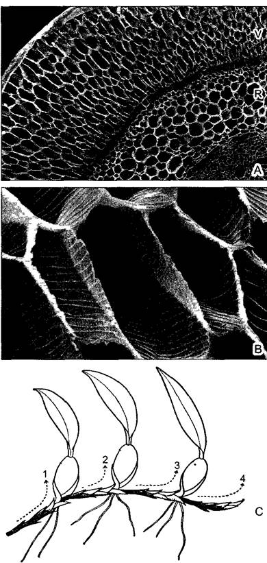
In other cases, stem tubers develop to store water during rainy periods (see Fig. 4.73, C). Various other specific adaptations for efficiently capturing precipitation are also widespread. In bromeliads, the roots function as short, rigid anchoring organs; in some species, such as members of the genus Tillandsia that frequently hang from telephone wires, roots may be entirely absent. These epiphytes acquire water exclusively through absorbing trichomes (scales) on their leaves (Fig. 4.74). Frequently in these plants, the tightly overlapping basal parts of the rosette leaves form cisterns that collect rainwater.
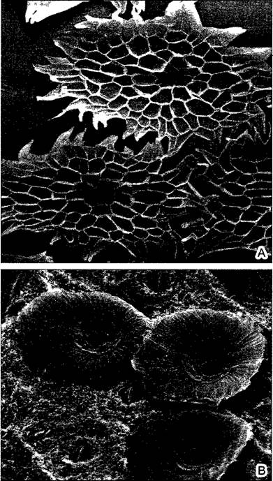
Fig. 4.74. Peltate absorptive trichomes (scales) of epiphytic bromeliads (Bromeliaceae, 170x) (SEM micrograph by W. Barthlott): A—Tillandsia rauhii, B—Acanthostachys. During rain, the dead cells of the trichome shields fill with water, which is then transferred into the leaf via the living cells of the trichome stalk
Generally speaking, the microhabitats created by epiphytes provide shelter for microorganisms, aquatic snails, insects, and even certain frogs. On Jamaica, the freshwater crab Metopaulias depressus even lives in the water-filled tanks of bromeliads.
4.4. Roots
The Root System generally performs two main functions: anchoring the plant in the soil and absorbing water and mineral nutrients from it.
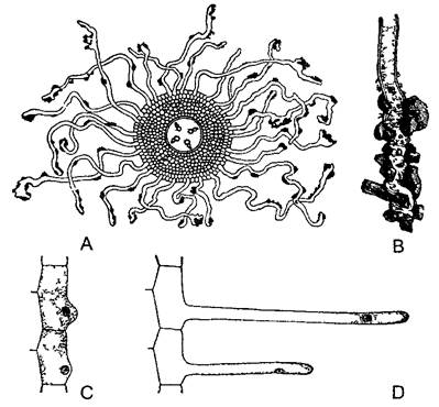
The latter task is often reflected in a tremendous expansion of the root's absorptive surface area. Many cells in the outermost layer, which lacks a cuticle (the rhizodermis = root epidermis1), grow out into long root hairs, ranging from a few millimeters to several centimeters in length (Fig. 4.75). Like the root as a whole, root hairs grow at their tips and are thus capable of forcing their way between soil particles. They are short-lived (3–9 days), and the active root-Hair zone of a growing root is only 1–2 cm long. Calculations show that despite this, a mature rye plant possesses over 10 billion root hairs, with a combined length reaching 10,000 km and a total surface area of 400 m2. This is approximately 50 times greater than the surface area of the above-ground shoot system, including the leaves, which weighs twice as much.
1 The rhizodermis is not homologous to the epidermis. — Ed. note.
Fig. 4.75. Root hairs (A—after Frank, B—after F. Noll, C, D—after Rothert)
A—transverse section through the absorption zone of a root with a tetrarch central cylinder; root hairs with soil particles (10x). B—tip of a root hair at a higher magnification (50x). C, D—rhizodermis in longitudinal section, showing initial and subsequent stages of root hair formation (note the position of the Cell Nucleus) (50x).
Alongside anchorage and the uptake of water and minerals, roots frequently fulfill other roles. For instance, they synthesize vital substances such as Plant HORMONES (Cytokinins, Gibberellins; see 7.6.2, 7.6.3). They also commonly function as storage organs (see Box 4.5).
4.4.1. Root Systems
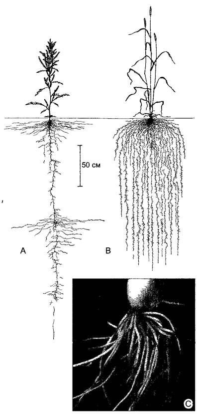
Plant root systems, like all axis systems, vary greatly in structure depending on the preferred habitat. In young plants, or those that spread rapidly via creeping shoots, the root system is often larger than the shoot system (Fig. 4.76). Conversely, root systems are especially poorly developed in many cacti inhabiting arid, hot biotopes where the soil is almost always completely dry (at least during the day). Depending on their vertical penetration into the soil profile, plants can be divided into two groups: those with deep root systems and those with shallow root systems. The deepest root systems are found in plants from environments where the topsoil is dry, but deep underground aquifers are present (e.g., Welwitschia; see Fig. 11.211, A; the taproots of tamarisks reportedly reach depths of up to 30 m, and those of the desert tree Prosopis juliflora in North America even exceed 50 m). In trees, the overall size of the root system is generally determined by the crown spread: the outermost perimeter of the root system extends slightly beyond the PROJECTION OF THE canopy.
Fig. 4.76. Allorhizy and homorhizy (A, B—after L. Kutschera)
A—Rumex crispus, a eudicot, develops a heterogeneous (allorhizic) root system in which the primary root penetrates the soil to a depth of over 3 m. B—secondary homorhizic root system of wheat, featuring the fibrous root system typical of most grasses. C—pronounced homorhizy is found in bulbous plants (e.g., young bunching onion, Allium fistulosum); numerous rather fleshy roots of uniform thickness show virtually no branching (0.7x).
Based on their developmental pattern and definitive structure, root systems are categorized into two types: heterogeneous (allorhizy) and homogeneous (homorhizy).
Heterogeneous root systems. In many plants, the embryonic radicle develops into a main or primary root, forming a taproot that grows vertically downward into the soil. It gives rise to secondary lateral roots of the first order, which grow obliquely or horizontally through the soil and subsequently branch further (lateral roots of the second, third, and higher orders). Higher-order lateral roots grow independently of the gravity vector and can thus permeate the soil in all directions. Such a hierarchically organized root system is termed allorhizic, or heterogeneous (from Greek allos — other, rhiza — root; see Fig. 4.76, A).
Most trees are allorhizic; some retain their original taproot system well into maturity, such as fir, pine, and oak. In other tree species (e.g., larch, birch, linden), In addition to the initial main root, numerous powerful roots develop and spread obliquely through the soil, forming a roughly hemispherical root system beneath the trunk base composed of roughly equal-sized roots, known as a brush-like or spreading system. Shallow-rooted trees (such as spruce and rowan) possess a system of robust secondary roots growing horizontally just beneath the soil surface, from which significantly weaker and shorter roots descend vertically.
In certain tree species, such as Norway spruce and field elm, roots in dense stands can graft together with the roots of neighboring plants. Beneath thick, rope-like root cords that sometimes stretch for many meters, several trees can form a truly interconnected, cohesive community.
Homogeneous root systems. Homorhizic systems consist entirely or predominantly of roots of a single branching order, possessing similar structure, and being unbranched or only weakly branched (from Greek homos — same, similar; see Fig. 4.76, B, C). All pteridophytes possess homogeneous root systems. As spore-bearing plants, they do not produce seeds and therefore lack an embryonic radicle; their axial system has no root pole at all and is initiated unipolarly, meaning that all roots in Pteridophyta are adventitious (primary homorhizy). Furthermore, the arrangement of their roots strongly correlates with leaf position: one or more roots arise beneath the base of each leaf, numbering over 100 in large tree ferns.
Primary homorhizy is typical of pteridophytes. Seed plants, with their bipolar embryos, exhibit secondary homorhizy rather than primary. In monocots, this arises because numerous roots of the same branching order emerge from the lower nodes of the shoot. They functionally Supplement the poorly developed primary root system, which, like the basal part of the shoot, does not undergo primary thickening (see Fig. 11.220). These roots frequently take on an additional supporting function (e.g., in maize; see also Box 4.5, Fig. B). They thus originate through a regenerative process and represent adventitious roots (see Figs. 7.21, 7.24). Eudicots also frequently form adventitious roots, for instance, on runners and in all rhizomatous plants. A particularly striking example is provided by mangroves (Rhizophora; Box 4.5, Fig. B) and the tropical banyan tree (Ficus benghalensis), whose sprawling canopy covers an area of up to 2 hectares (170 m in diameter), supported by hundreds of pillar-like adventitious aerial roots.
The strict association of roots with leaves characteristic of pteridophytes is rarely found in seed plants. Nevertheless, adventitious roots frequently develop at shoot nodes in seed plants as well. However, there are numerous exceptions to this rule1 (cf. Box 4.5, Fig. A).
1 The number of such examples is negligible compared to the number of species that form nodal adventitious roots. — Ed. note.
4.4.2. Root Anatomy
4.4.2.1. Primary Structure
The diagram of a root cross-section in Fig. 4.77 demonstrates radial symmetry at the primary developmental stage. The rhizodermis is bounded on the inside by a tougher, more durable, and frequently weakly suberized cell layer known as the hypodermis; its cell walls often develop Casparian strips, transforming it into an exodermis. This layer encloses the robustly developed parenchyma of the primary cortex, which is delimited internally by the endodermis (see 3.2.2.3). The latter serves as both a morphological and physiological boundary for the stele (central cylinder), which integrates mechanical and conductive elements. The central position of this rigid tissue within a more pliable outer sheath ensures flexibility combined with high tensile strength (cable construction, see 3.2.3), perfectly aligning with the root's function of anchoring the plant in the substrate.
Fig. 4.77. Tissue arrangement in a cross-section of a root.
The stele is surrounded by the pericycle; the xylem is shown in black, the phloem is stippled, and parenchymatous strips lie between them. Here, the xylem is triarch, making the stele triarch. (It is more accurate to consider the hypodermis as an exodermis. — Note by the editor.)
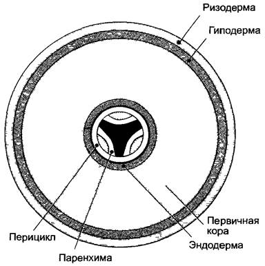
The outermost layer of the stele, the pericycle, consists of thin-walled, Cytoplasm-dense cells that retain their meristematic (dividing) capacity for a prolonged period. Consequently, this cell layer—which, much like the rhizodermis, exodermis, and endodermis, features very few intercellular spaces—is also referred to as the perikambium1 (see 4.4.2.2). The core of the stele, structured as an actinostele2, is typically occupied by the xylem, which extends to the pericycle in the form of two or more radial ridges. Depending on the number of these xylem poles, roots are classified as having 2-, 3-, ..., or polyarch steles3. Pteridophytes, magnoliids, and eudicots predominantly possess 2- to 4-arch steles (Fig. 4.78, A; 4.79, A), whereas monocots frequently feature a polyarch stele (Fig. 4.78, B). Strands of phloem are located in the intervals between the xylem ridges. The xylem and phloem are separated by layers of parenchyma that extend outward to the pericycle alongside the xylem ridges.
1 This term was widely used until the mid-20th century, after which it fell out of use. — Note by the editor.
2 In reality, this should be compared to a radial vascular bundle rather than an actinostele. — Note by the editor.
3 THE CONCEPT OF archness is correctly applied to the xylem of a radial bundle rather than to the stele itself. — Note by the editor.
Fig. 4.78. Stele (A—after D. von Denffer, B—SEM micrograph by H. Falk)
A—cross-section of a tetrarch vascular bundle in the root of creeping buttercup (Ranunculus acer) (160x); B—cross-section of a dodecarch stele in the root of Iris germanica, clearly showing the same tissues as in fragment A, except for the thin-walled phloem areas located directly beneath the pericycle between the 12 xylem poles. Arrow—passage cell; endodermis (as in A) in the tertiary state (120x); Ep—endodermis with passage cells; D, X—xylem with vessels; Ph—phloem with sieve tubes S and darkly rendered companion cells; Pe—pericycle
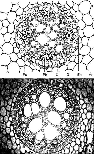
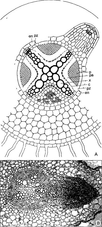
Fig. 4.79. Endogenous origin of lateral roots (A—after O. Stocker): A—cross-section of a eudicot root; above a xylem pole of the stele, dividing cells of the pericycle (top left) give rise to the apical meristem of the future lateral root, which subsequently (top right) grows outward through the cortical tissue (120x); B—cross-section of a Vicia faba root with a pentarch stele and a developing lateral root; meristematic cells appear darker than the highly vacuolated cells of permanent tissues (75x); en—endodermis; pz—pericycle; c—cambium in the parenchyma (pa); x—xylem; ph—phloem
During the development of the stele, its tissues differentiate from the outside inward1—in contrast to stem tissues. Consequently, the protophloem and protoxylem are situated directly beneath the pericycle, while the largest metaxylem vessels occupy the center. Occasionally, metaxylem fails to develop in the exact center of the root. In such instances, as well as in the roots of eudicots, a parenchymatous pith is present at the center of the stele, whereas monocots typically have a regular pith. In particularly robust roots of tall monocotyledonous plants, the stele also contains sclerenchyma.
1 Phloem in the stem also differentiates centrifugally (from inside out) — Note by the editor.
In the hypocotyl, the transition zone between root and stem, the actinostele of the root transitions acropetally into the eustele or atactostele of the shoot (Box 4.3). The architecture of this transition zone varies among different angiosperms.
Typically, as the stele extends upward from the root, it separates into individual vascular tissue sectors, each comprising a single xylem pole and two halves of adjacent phloem strands flanking the xylem laterally. At higher levels, these sectors are displaced toward the periphery of the axial organ; parenchyma (medullary rays) develops between them, with a pith forming in the center. The xylem of each thus isolated vascular bundle rotates such that the protoxylem elements, which previously lay at the periphery of the root stele, now face inward toward the pith, while the metaxylem correspondingly faces outward.
Because the root bears no leaves, it lacks the metamerism characteristic of shoots—namely, the longitudinal segmentation into nodes and internodes. Covered by the root cap (calyptra) (see 3.1.1.2) and thus subapical, the root apical meristem (following the quiescent center) is succeeded by the zone of Cell Division1 and subsequently by an elongation zone measuring 3–10 mm in length. The maximum frequency of cell divisions in the developing primary cortex (periblem) occurs adjacent to the meristem, whereas in the emerging stele (plerome) it happens slightly further back, and in the young rhizodermis (dermatogen or epiblema) it peaks in the region furthest from the initials. Numerous cell divisions also take place within the elongation zone. This zone is adjoined by the root hair zone and, ultimately, the region where lateral roots originate—the branching zone. The Formation of the root hair zone marks the completion of Primary root growth. Longitudinal growth of the root ceases here (Fig. 4.80); otherwise, as the root forces its way through soil particles, it would shear off the lateral root hairs. Consequently, roots elongate exclusively at their extreme tips (see Fig. 7.3).
Fig. 4.80. Rhizophores in radish: A—cross-section (1.2x); B, C—external appearance (0.8x).
Each of the two root orthostichies (which indicate a diarch stele) is actually double, because in Brassicaceae, or crucifers (to which the radish belongs), two closely appressed orthostichies form external to each xylem pole of the stele. The diameter of the taproot, which forms the storage root, is 100 times that of the lateral roots.

1 To clarify: the quiescent center and the zone of cell division constitute the root apical meristem rather than being positioned posterior to it. — Note by the editor.
The elongation zone of a root is considerably shorter than that of an aerial shoot, where it often reaches several centimeters. This is due to the significantly greater resistance encountered by root tips pushing through the soil; correspondingly, aerial roots possess an elongation zone nearly ten times longer than that of subterranean roots.
Box 4.5. Root Metamorphoses
Roots exhibit numerous adaptations tailored to specialized functions. Even the primary function of anchoring a plant under unusual conditions may necessitate root modification. Well-known examples include adventitious climbing roots in lianas (Fig. A) and epiphytes. Stilt roots (prop roots) in mangroves aid in anchoring them within the shifting substrate of tropical Marine tidal zones (Fig. B). Adventitious roots of tall grasses play a fundamentally similar role, albeit under entirely different ecological conditions. Plank roots (buttress roots) develop through enhanced secondary thickening on the upper side of roots that grow horizontally just beneath the soil surface. In certain tall tropical trees, these form buttresses up to a meter high around the trunk (col. pl. 15.1, E, vol. 4).
A distinctive feature is the function of contractile roots, which pull underground shoots—such as rhizomes, tubers, or bulbs—deeper into the soil (Fig. C). The shortening of these roots is driven by longitudinally oriented Cellulose fibrils in the cell walls of the primary cortex, allowing them to shorten (while simultaneously thickening) in response to increased turgor pressure.
Quite a few plants develop specialized storage roots (Fig. D); in many root vegetables, the storage tissues are also at least partially derived from the root (Fig. E). Anomalous secondary thickening1 gives rise to thick, yet sparsely branched root regions; in some cases, globular root tubers form entirely lacking lateral roots. The stored substances predominantly comprise di-, oligo-, and Polysaccharides (sucrose, starch, inulin) 2.
1 The vast majority of storage roots exhibit either normal secondary thickening (dicots) or primary thickening (monocots). — Ed. note.
2 The roots of certain plant species store water. — Ed. note.
Root spines — short, fully lignified, pointed lateral roots situated on adventitious aerial roots. In some palm species, they serve to protect the base of the trunk.
Aerial roots, as previously noted, frequently function to support the shoot system above the substrate. Epiphytes absorb water through their aerial roots when they cannot reach soil moisture (although in some instances they accomplish this via their leaves). Such roots possess a specialized outer tissue layer (velamen, cf. Fig. 4.73, A, B), which develops from the protoderm through periclinal cell divisions. The velamen consists of numerous large cells that die off early, featuring spiral wall thickenings and wall perforations. Much like the hyaline cells in the leaves of peat moss (Sphagnum), these empty cells fill with rainwater, which the velamen absorbs and retains like a sponge.
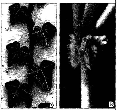
Fig. A—Clinging adventitious roots: A—in English ivy (Hedera helix), the clinging roots do not serve for water and mineral uptake, but function exclusively for attachment to the substrate (here—concrete; 0.7x). A two-ranked leaf arrangement is typical of juvenile forms; B—in trumpet creeper (Campsis radicans), clinging roots develop exclusively at the nodes (2.6x)
In permanently waterlogged soils, extensive root systems suffer from oxygen deficiency due to its low solubility in water. Consequently, many trees and shrubs of tropical swamp forests and mangroves produce upward-growing (negatively geotropic) respiratory roots, or pneumatophores, which project above The surface of the soil (or water) so that the intercellular space system in the cortical tissues remains in contact with the atmosphere. A specialized variant is represented by knee roots, which initially grow upward, but upon reaching the soil surface, bend and re-enter the substrate. As a result of unilateral thickening (similar to buttress roots), air-protruding swellings known as pneumatodes frequently form at these bends.
Fig. B. Stilt roots (photos A—D by Lüttge, B—by Barthlott)
A—Rhizophora mucronata, a true dicotyledonous mangrove plant on a tidal marine shore (Tonga Islands, Southwestern Polynesia); B—the monocotyledonous West African screw pine (Pandanus candelabrum)
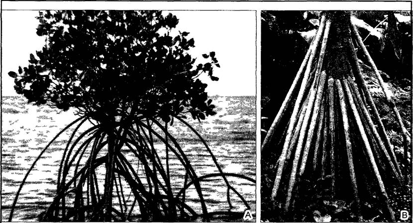
Fig. C. Contractile roots in lords-and-ladies (Arum maculatum) (A—after Rimbach)
A—pulling the corm deeper via root contraction: I—germination, II—onset, III—end of the second year, IV—mature plant, with the corm located 10 cm below the soil surface (0.4x); B—corm and contractile roots, whose outer tissues do not participate in contraction and passively fold into transverse wrinkles (1.8x)1
1 Root shortening is driven precisely by the active contraction of annular zones within the peripheral root tissues. — Ed. note.
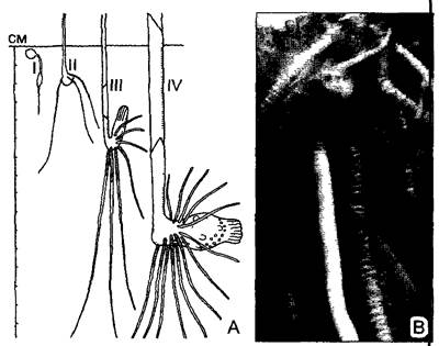
Fig. D. Roots as storage organs (A, B—after R. von Wettstein, C—after Weber)
A, B—root tubers of military orchid (Orchis militans) (0.7x), K1—previous year's tuber, which produced the current year's flowering shoot, with a new root tuber K2 developing in the axil of the lowermost scale-like foliage leaf N on the axillary shoot, W—normal adventitious roots, Kn—bud of the axillary shoot for the next growing season; C—adventitious storage roots of dahlia (0.15x); D—adventitious root tubers of the homorhizous root system in lesser celandine (Ranunculus ficaria), where tubers easily detach at the base and subsequently grow into a new plant (2x); E—less pronounced root tubers of daylily (Hemerocallis) compared to dahlia, though here too lateral roots form exclusively in the distal, non-storage region of the root, a—underground stolon (0.5x)

It has already been mentioned that the roots of epiphytic orchids even take over the functions of leaves (cf. Fig. 4.4). Parasitic and symbiotic lifestyles lead to profound morphological modifications of the roots (cf. 9.2, 9.3).
Hemiparasites — are green plants that remain capable of independent photosynthesis, yet obtain water and mineral nutrients from a host plant, penetrating its xylem with the aid of root haustoria. This category includes, for example, eyebright (Euphrasia), yellow rattle (Rhinanthus), cow-wheat (Melampyrum), and lousewort (Pedicularis) from the Scrophulariaceae family, as well as the evergreen mistletoe. Mistletoe germinates as a parasitic epiphyte on the branches of specific trees; its root system subsequently spreads beneath the bark (phloem) of the host branch, while haustoria penetrate from the roots into the host sapwood, connecting to the branch's water-conducting system via characteristic short vessels (cf. Fig. 4.39, B).
Holoparasites — are plants that lack chloroplasts entirely and derive their Nutrition exclusively at the expense of the host. Toothwort (Lathraea) utilizes root haustoria to draw sap from the xylem of woody hosts. Species of the genus Orobanche, conversely, parasitize the phloem of their victim's roots. The haustoria of these yellowish, reddish, or purplish parasites laterally penetrate the host root and absorb phloem sap so intensively that it causes the distal root segment to die off. Consequently, the parasite appears to be attached directly to the tip of the host root.
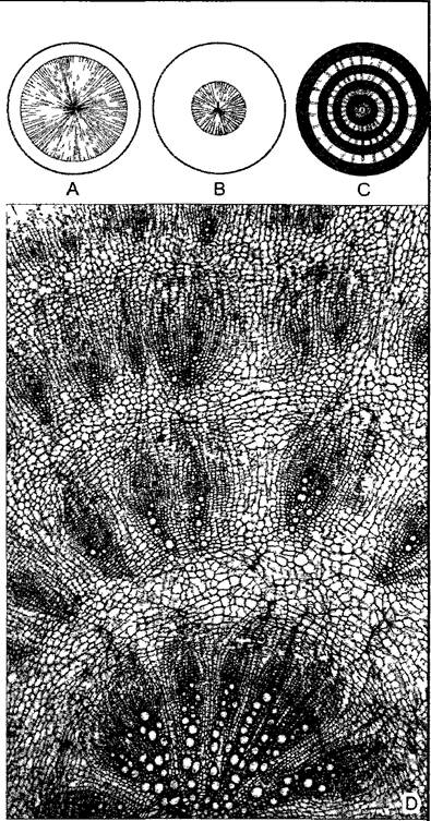
Fig. E. Anatomy of root vegetables (transverse sections) (A—C — after W. Franke)
A—in "woody" root vegetables, xylem is predominantly and powerfully developed, yet consists mainly of wood parenchyma; example: radish. B—in "bast-rich" root vegetables, by contrast, secondary phloem serves as the primary storage tissue; example: carrot. C—in beetroot (cultivated forms of Beta vulgaris: sugar beet, fodder beet, garden beet), alternating concentric rings of xylem (light-colored) containing phloem1 and parenchyma (dark-colored) are formed; this results from anomalous secondary thickening with repeated formation of additional cambia in the cortex1. The appearance under a Light Microscope is illustrated in D (bottom: original central cylinder; 48x).
1 Beetroot forms discrete collateral vascular bundles rather than continuous concentric rings of xylem and phloem. — Ed. note.
2 Accessory cambia develop not in the cortex, but within the proliferating pericycle. — Ed. note
Symbiosis with nitrogen-fixing Bacteria leads to the formation of root nodules, which are localized proliferations of cortical tissues (see 9 2.1). Within specialized vacuoles of enlarged polyploid parenchymatous cells, the prokaryotic symbionts reside as bacteroids.
Much more widespread is the symbiosis with soil Fungi known as mycorrhiza (see 9.2.3). The plant utilizes the exceptionally high absorptive capacity of fungal hyphae primarily for mineral salt uptake. Characteristically, mycorrhizal roots completely lack root hairs.
4.4.2.2. Lateral Roots
Unlike lateral shoots, lateral roots develop endogenously, i.e., from within the root body precisely at the boundary between the vascular cylinder (stele) and the primary cortex (Fig. 4.79). Here, pericycle cells undergo re-embryonation and, dividing periclinally and anticlinally, form the apical meristem of the new root. The first lateral roots always initiate behind the root hair zone. Thus, this represents a true de novo formation of apical Meristems; unlike shoots, there is no fractionation of the parent apical meristem here. Adventitious roots also originate internally within the stem cortex1. Consequently, the vascular tissues of lateral roots establish early connections with The Vascular System of the parent organ, growing outward through the cortical tissues. The emergence point of a lateral root is frequently surrounded by a collar-like protruding edge of the ruptured root or stem cortex.
1 Typically initiated in the cambial region — Ed. note
The endogenous origin of lateral roots and the subapical position of the root apex initials have significant morphological and anatomical consequences. In both instances, the root surface is formed by an internal tissue initially enclosed by another layer. (Indeed, the rhizodermis of primary roots is initially also covered by the short-lived cells of the root cap.) This is clearly the reason why the rhizodermis lacks a cuticle and stomata.
Lateral roots are often arranged in distinct longitudinal rows on the parent root, termed rhizostiches (Fig. 4.80), because the de novo formation of meristems in the pericycle predominantly occurs opposite the xylem poles of the vascular cylinder. Consequently, the number of rhizostiches allows one to infer the archy of the central cylinder directly from the external Morphology of the root.
4.4.2.3. Secondary Growth
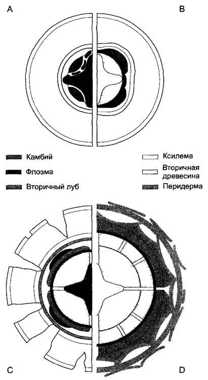
In perennial woody plants, the primary root undergoes extensive secondary thickening, much like a tree trunk (Fig. 4.81). Initially, within the concavities of the parenchyma strips between the primary phloem and xylem, specific cambial patches are formed through re-embryonation, producing secondary xylem inward. The initially uniseriate pericycle outside the xylem poles becomes multiseriate, and the cells of its innermost layer form cambial segments. Joining with the cambial segments located between the xylem and phloem, these complete the cambial ring. Initially, the resulting cambial ring appears star-shaped in transverse section. However, it soon rounds out due to enhanced xylem production beneath the phloem strands. Primary rays are laid down opposite the xylem poles. (True pith rays do not occur in roots.) The delicate rhizodermis typically dies off even before secondary thickening begins and is replaced by the hypodermis. Neither the hypodermis nor the primary root cortex participates in secondary growth; they rupture, die, and are eventually shed along with the endodermis. Therefore, the bark covering heavily thickened older roots does not form through periderm development in the primary cortex (as observed in stems), but rather originates from the pericycle, which persists as a continuous ring even after radial root growth has commenced.
Fig. 4.81. Secondary thickening of the root (transverse sections)
A—formation of a continuous cambial layer via cell re-embryonation in the parenchyma between the xylem and phloem and above the xylem pole of a tetrarch vascular cylinder; B—rounding of the cambial layer due to xylem production beneath the phloem strands; C—onset of secondary phloem formation, with vascular rays developing above the xylem poles; the primary cortex and endodermis die and disintegrate, while a phellogen forms within the now multiseriate pericycle, producing outward layers of cork; D—subsequent annual rings give rise to secondary vascular rays, and successive phellogens within the phloem generate bark scales
The xylem and phloem of the root are histologically structured in the same manner as those of the stem. This applies to the rays as well. The transverse section of a root subject to annual radial growth is nearly indistinguishable from the corresponding cross-section of a stem. Obvious anatomical differences persist only at the center, where the primary structure is retained. However, unlike the hypocotyl at the primary growth stage, it is no longer possible to delineate a distinct transitional zone within a perennial root where it merges into the stem.
Last update: 07/08/2026
Editorial and Educational Adaptation: This material has been compiled based on the primary/original source text. The project team performed an editorial review, corrected technical inaccuracies, structured sections, and adapted the content for an educational format.
What was processed:
- elimination of formatting defects (OCR errors, structural breaks, corrupted characters);
- editorial organization of content;
- standardization of terminology in accordance with academic sources;
- verification of factual statements against the original source text.
All mentions of the author, publication year, and origin of the primary text have been preserved in accordance with the source.