MODERN BOTANY - P. RAVEN - 1990
SECTION IV. DIVERSITY
CHAPTER 15. MULTICELLULAR PROTISTS: RED, BROWN, AND GREEN ALGAE
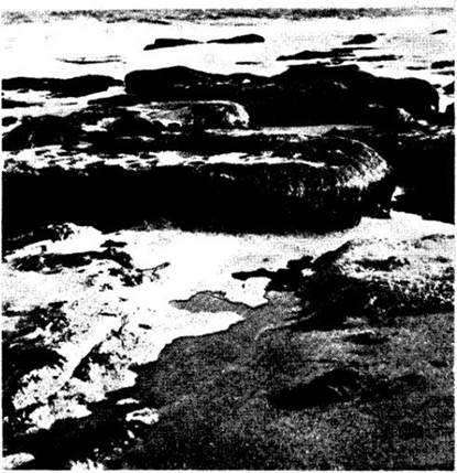
The open ocean, the coastal zone, and the land are the three primary ecological zones that make up our biosphere. In the first two (and most ancient) realms, Algae play a role comparable to that of plants in the much younger terrestrial world (Fig. 15-1). Algae also frequently predominate in freshwater habitats—pools, rivers, and lakes—often serving as the primary contributors to the productivity of these ecosystems. Wherever they grow, their ecological importance is no less than that of plants in terrestrial habitats.
Class="center">Fig. 15-1. Algae on rocks at low tide (coast of North Carolina)
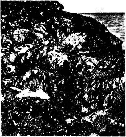
Along rocky marine shores, one can find larger and more complexly organized algae that typically form distinct vertical zones depending on their position in the tidal or intertidal zone, or littoral (Fig. 15-2). Their structural complexity reflects an adaptation to life in this dynamic environment, where twice daily they experience drastic fluctuations in moisture, Temperature, salinity, and light intensity, as well as the pounding of surf and the abrasive action of suspended sand particles churned up by waves.
Fig. 15-2. Rocks along the coast of Cornwall, England. Several vertical zones formed by different species of algae are clearly visible
Algae attached to the substrate outside the surf zone provide shelter for A wide variety of microorganisms, as well as fish and invertebrates that feed on these microorganisms or on one another. Such algal beds can easily rival forests in their extent and density. For instance, massive kelp forests stretch along the coast of California, featuring broad blade-like thalli suspended in the Water up to 15 m long, attached by winding holdfasts ("stems") anchored to the sea floor that reach lengths of 30 m or more. Many large predators, including sea otters and tunas, find prey and refuge in these underwater forests. Such algae are also harvested by humans for food and fertilizer.
This chapter discusses three phyla of algae in which all or some representatives are multicellular. It is these protists that form dense coastal communities or drift freely in the open ocean, such as in the Sargasso Sea. The green algae phylum (Chlorophyta) also includes unicellular forms, many of which make a significant contribution to the productivity of freshwater and marine plankton. This exceptionally diverse phylum is of special interest because it was almost certainly species from this group that gave rise to land plants approximately 430 million years ago. Although green algae resembling the direct ancestors of plants may no longer exist today, many forms retain certain putative ancestral traits.
In addition to green algae—which are more abundant in freshwater than in the sea and are also common in terrestrial habitats—There are two other phyla of predominantly multicellular protists: the red and brown algae (phyla Rhodophyta and Phaeophyta). Both of these groups are almost exclusively marine. Only a few genera of red and brown algae inhabit fresh water. All brown algae are multicellular, whereas some red algae are unicellular.
Characteristics of Red, Brown, and Green Algae
As discussed in the previous chapter, the METABOLISM/14.html">Chloroplasts of red, brown, and green algae probably originated from different Bacteria. In red algae, they contain chlorophyll a and phycobilins, bearing a resemblance to the ancient group of cyanobacteria. Many scientists consider the evidence supporting the cyanobacterial endosymbiotic origin of red algal chloroplasts to be quite compelling. Meanwhile, green algae, with chloroplasts containing chlorophylls a and b, are closely related in this respect to euglenoids and their plant descendants. In their biochemical characteristics, their chloroplasts are similar to the bacterium Prochloron (see Figs. 11-2, 11-17) and may have originated from an Organism of precisely this type. Finally, it is possible that the golden-brown chloroplasts of closely related brown algae,
chrysophytes, and dinoflagellates, which contain chlorophylls a and c, trace their ancestry back to a bacterium similar to the recently discovered species *Heliobacterium chlorum*. The chloroplasts themselves may have originated from Eukaryotic Cells engulfed by algae, but ultimately, their photosynthetic Organelles are of bacterial origin.
Representatives of the phyla examined in this chapter differ significantly in many other characteristics, some of which are summarized in Table 14-1. Although Cellulose has been found in The Cell walls of certain species within each phylum, The chemical composition of these walls varies widely both among different groups and within them. Red algae lack flagella and centrioles at all stages of their life cycle. Instead, they possess structures called *polar rings* associated with the nuclei, which move during prophase of mitosis in a manner similar to centrioles; whether they are homologous to centrioles remains unknown. Similarities between red algae and ascomycetes have long been noted and warrant further investigation using additional traits. The flagellated cells of brown algae, like those of their close relatives the chrysophytes and diatoms, bear two distinct flagella—an anterior tinsel (feathery) flagellum and a smooth posterior one (see Fig. 2-26). By contrast, the flagella of green algae (when present) are almost invariably uniform and smooth.
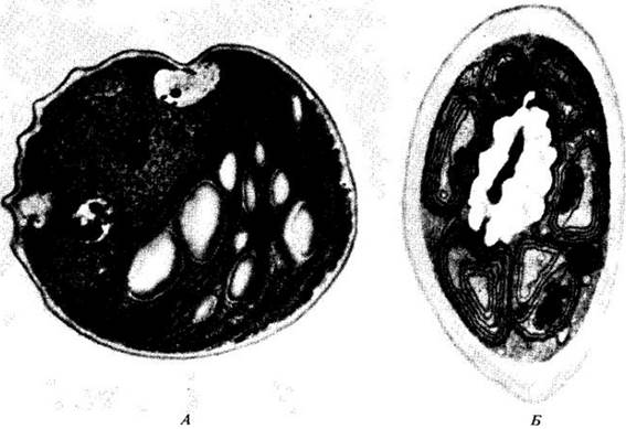
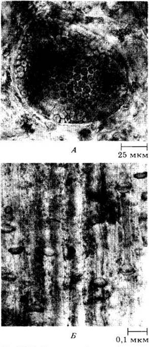
Green algae, much like plants, store nutrient reserves in the form of starch within their Plastids (Fig. 15-3, A). All other groups of photosynthetic eukaryotes—including those discussed in Chapter 14, as well as red and brown algae—store their reserves outside the chloroplasts in the Cytoplasm: red algae store them as starch, whereas brown algae store laminarin, a polyglucoside with different glucose linkages than those found in starch. In addition, brown algae store the sugar alcohol mannitol.
When algal cells divide, their Plasma Membranes typically pinch inward by furrowing, much like those of animals, Fungi, and protists. However, cell plates—resembling those of plants—have been discovered in one species of brown algae and a few genera of filamentous green algae. In most cases (with the exception of red algae), centrioles are present, which give rise to the basal bodies of flagella in motile cells (see p. 36).
Fig. 15-3. In green algae and higher plants, nutrient reserves accumulate as starch within plastids, whereas in other photosynthetic eukaryotes, analogous reserves are formed in the cytoplasm. A. *Chlorella*, a green algal cell with large, pale starch grains inside its plastids. B. A plastid-surrounded mass of starch grains located in the cytoplasm of the red alga *Batrachospermum moniliforme*
On the whole, the Multicellular Organisms belonging to the phyla considered here lack the complexly organized Tissues characteristic of vascular plants. However, certain brown algae possess a central conducting strand composed of cells reminiscent of the sieve elements found in plant stems and leaf blades. The reproductive structures of algae are typically unicellular rather than multicellular with sterile protective jackets, as seen in bryophytes and vascular plants (see Chapter 16).
The three algal phyla examined here are ancient groups that differ sharply in their characteristics. Fossil forms resembling unicellular green algae have been discovered in the deposits of the Bitter Springs Formation in central Australia, dating back approximately 900 million years. During the Cambrian period, at least 550 million years ago, large siphonous (coenocytic) green algae and red algae appeared. Multicellular lineages of green algae also arose during the Paleozoic era. For instance, the earliest charophytes—representatives of their most distinctive group—date back to the Late Silurian (about 420 million years ago). These green and red algae, which are best represented in the fossil record as impressions, possess calcified cell walls. Such walls are known in only a single genus of brown algae, making fossils of this group relatively rare; they have not been preserved as exceptionally well as those of certain green and red algae.
Phylum Rhodophyta
Red algae, or floridean algae, possess complexly organized cells lacking flagella and feature intricate life cycles. They lack centrioles, thereby differing from green and brown algae, in which centrioles are typically present even in vegetative cells. In red algae, their function is performed by polar rings.
This phylum includes about 4,000 species, which are particularly abundant in tropical and warm waters, although many species also inhabit cold Regions of the globe. Fewer than 100 species are found in fresh water, but in the marine environment, their numbers surpass those of all other groups of multicellular seaweeds combined. Members of this phylum are typically attached to rocks or other algae; There are also a few free-floating and unicellular colonial species.
The characteristic coloration of red algae is imparted by water-soluble phycobilins—accessory pigments that mask the color of chlorophyll a. These organisms are exceptionally well adapted to absorbing green, violet, and blue light rays that penetrate to great depths. Their chloroplasts apparently originated from symbiotic cyanobacteria, to which they are biochemically and structurally similar.
The cell walls of most red algae contain rigid microfibrils of cellulose or another polysaccharide, embedded in a mucilaginous matrix, typically made of a sulfated galactose polymer such as Agar or carrageenan (see “Economic Uses of Marine Algae,” p. 257). It is these latter components that make red algae flexible and slippery to the Touch.
In addition, many red algae deposit calcium carbonate within their cell walls. Among them, the particularly rigid and “stony” species form the large family Corallinaceae. They are widespread in the oceans on stable, well-illuminated substrates (Fig. 15-5B, C). Recently, a crustose coralline alga was recorded at a depth of 268 m; this is a record depth for photosynthetic organisms (Fig. 15-6). It is nearly 100 m below the depth to which sunlight typically penetrates.
Coralline algae play a vital role in coral reef formation. Indeed, the productivity of such reefs and their ability to thrive in relatively nutrient-poor tropical waters depend directly on these algae and the symbiotic dinoflagellates (zooxanthellae) living inside the polyps (see Fig. 14-20). Reef-building corallines are usually unjointed (Fig. 15-5C), whereas many other members of this family exhibit a prominent jointed Structure (Fig. 15-5B). Some corallines, much like crustose Lichens, form crusts on rocks (Fig. 15-6). This ancient group is known as far back as the Late Cambrian, meaning its age exceeds 500 million years.
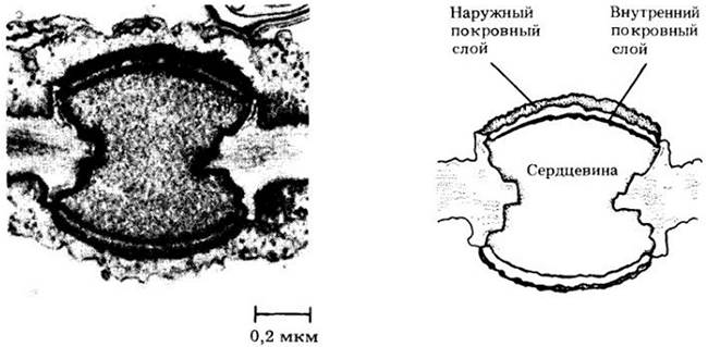
Most red algae consist of filaments that are often densely intertwined and held together by a mucilaginous intercellular matrix. Simpler forms, such as the freshwater alga Batrachospermum, are represented by single filaments not gathered into such bundles (Fig. 15-4). Growth in filamentous red algae originates from a dome-shaped apical cell that divides successively to form the axis of the alga. This axis then produces whorls of lateral branches (Fig. 15-4B). Most red algae are multiaxial with an outwardly parenchymatous three-dimensional body. In such forms, filaments reconnect secondarily to form a network bound by a mucilaginous matrix. Many red algae are characterized by specialized cell-wall pit connections, which are lens-shaped plugs held within the walls by their equatorial constrictions (Fig. 15-8). In a few genera (e.g., Porphyra, see p. 258), a true parenchyma of densely packed cells, similar to that of higher plants, is found.
Fig. 15-4. A. A simple filamentous red alga, Batrachospermum. The soft, mucilaginous, branched filaments of this freshwater protist are found mainly in cold streams, ponds, and lakes worldwide. B. Apex of a Batrachospermum filament with whorls of lateral branches. C. Two cystocarps of Batrachospermum

Fig. 15-5. A. Bonnemaisonia hamifera clearly showing the typical filamentous structure of red algae. Multicellularity apparently evolved independently in red algae from green and brown algae, in which such an Organization is absent. The branched filaments of this red alga bear hooks that allow it to cling to other algae. B. Jointed coralline algae in a tide pool on the California coast. C. The reef-building unjointed coralline alga Porolithon craspedium. D. Irish moss (Chondrus crispus), an important source of carrageenan and other colloids
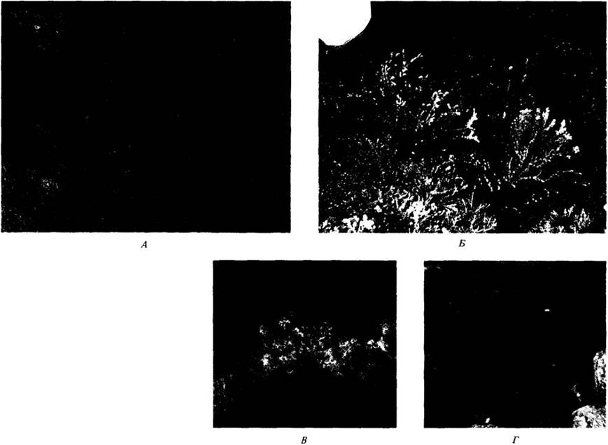
Fig. 15-6. A. Scanning electron micrograph of an unidentified species of red alga collected from a depth of 268 m by Mark Littler and his colleagues from the Smithsonian Institution in October 1984. This alga forms patches about 1 m in diameter on a seamount in the Bahamas at an illumination of approximately 0.0005% of that at the ocean surface, covering about 10% of the rocky substrate surface. Laboratory studies have shown that it absorbs and utilizes light about 100 times more efficiently than its shallow-water relatives. This alga, growing continuously up to the summit of the seamount (depth about 70 m), lives approximately 100 m below the previously established lower depth limit of life for any attached photosynthetic organisms. B. Purple crustose corallines and other deep-water algae from the same seamount. C. The Johnson-Sea-Link I submersible, which was used to obtain the deepest photographs of algae
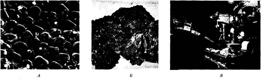
Fig. 15-7. Life Cycle of Polysiphonia, a widespread marine red alga. Reproductive Organs arise at the tips of the branches of haploid gametophytes, which develop from haploid tetraspores. Cells functioning as spermatia emerge from tightly clustered spermatangia. The expanded basal part of the carpogonium contains a nucleus and Functions as an egg cell. Following Fertilization, mitosis produces diploid carpospores within the carposporangium, which are released through an opening in the pericarp—the outer structure developing around them. The pericarp is formed from the carposporophyte. Carpospores germinate into tetrasporophytes, which are similar in Size and Structure to the gametophytes. Tetrasporangia form on the tetrasporophytes, and Meiosis occurs within each to produce four haploid tetraspores. The cycle then begins anew

The life cycle of most red algae is unusual in that it comprises three phases: (1) a haploid gametophyte, (2) a diploid phase—the carposporophyte, and (3) another diploid phase called the tetrasporophyte (Fig. 15-7). The male individual of the gametophyte generation produces spermatangia (antheridia) from which nonflagellated spermatia—male Gametes that float passively in the water—are released. A long, Hair-like outgrowth, the trichogyne—similar in Structure and function to the trichogynes of ascomycetes (Chapter 13)—develops on the female reproductive organ, the carpogonium. A spermatium lands on the trichogyne by chance via water currents and attaches to it. Thereafter, its nucleus enters the trichogyne through a pore, migrates to the egg nucleus, and fuses with it.
In simply organized red algae such as Batrachospermum, the diploid generation—the carposporophyte (Fig. 15-4C)—develops directly from the fertilized carpogonium, upon which terminal spore-producing structures, the carposporangia, arise. In more advanced groups, the diploid nucleus formed inside the carpogonium through syngamy is transferred to a second cell, termed the auxiliary cell, from which the carposporophyte develops. Such a life cycle is observed in the genus Polysiphonia (Fig. 15-7).
Diploid carpospores, formed inside the carposporangium, are released and develop into a free-living tetrasporophyte. Another type of spore-producing structure—the tetrasporangium—arises on it, in which meiosis takes place to form four haploid tetraspores. Upon germination, each of these gives rise to a gametophyte.
In most red algae, the gametophyte, carposporophyte, and tetrasporophyte are very similar—that is, they are isomorphic, as seen in Polysiphonia and corallines. Increasingly, heteromorphic cycles are being described, in which tetrasporophytes have a microscopic filamentous structure or form a thin crust tightly attached to a rocky substrate.
Fig. 15-8. Pit connections between Cells of the red alga Palmaria. They appear as prominent lens-shaped plugs with a protein core and at least partially polysaccharide covering layers. Such connections form between sister cells and neighboring filaments, uniting them into a single red algal body. They also arise between many parasitic red algae and their hosts, possibly facilitating The transfer of assimilates between them
Phylum Phaeophyta
Brown algae are an almost exclusively marine group comprising most of the large protists of temperate regions. Comprising only about 1,500 species, the group is of considerable interest because it dominates rocky coastal substrates in the cold regions of the globe (Fig. 15-9). Large species of the order Laminariales form extensive kelp forests along coasts. In clear water, brown algae thrive from the low-tide line to a depth of 20–30 m and, where the bottom slope is gentle, may extend 5–10 km offshore. Even in the tropics, where brown algae are less common, the genus forms dense floating masses (Fig. 15-10), such as in the Sargasso Sea in the Atlantic Ocean northeast of the Antilles.
Fig. 15-9. Brown algae. A. Durvillaea antarctica during low tide on the rocky intertidal zone of New Zealand. B. A fragment of kelp, showing holdfasts, stipes, and blade bases. C. Fucus vesiculosus densely covering many rocky areas exposed at low tide. When the algae are submerged by water, air-filled vesicles on the blades lift them toward the light. Photosynthetic rates in seaweeds frequently exposed to air can be up to seven times higher in air than in water; conversely, in species that remain submerged almost constantly, this rate is higher when submerged. This partly explains the vertical zonation of marine Algae in the intertidal zone
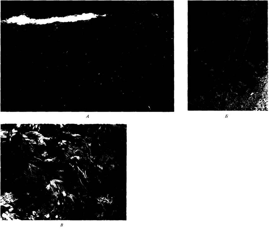
Fig. 15-10. The brown alga Sargassum has a complex organization. This genus, like Fucus, belongs to the order Fucales and exhibits the life cycle shown in Fig. 15-14. Two non-sexual species of this genus form massive, free-floating populations in the Sargasso Sea, while others attach to rocks along shores

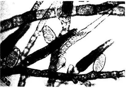
The size of brown algae ranges from microscopic to the largest of all marine seaweeds. The structurally simplest members of the division form branched filaments (e.g., Ectocarpus, Fig. 15-11) that grow via internal intercalary Meristems. In more highly evolved filamentous brown algae, these filaments are aggregated into a solid thallus, whereas the most complex forms possess a three-dimensional cellular structure similar to that observed in plants.
Fig. 15-11. Ectocarpus, a brown alga with simple branched filaments. This micrograph of E. siliculosus shows unilocular sporangia (short, rounded, pale structures) and plurilocular sporangia (elongated dark structures) borne on the sporophytes. Meiosis occurs within the unilocular sporangia (zoospores have already been released from the ones shown here). These algae inhabit shallow waters and estuaries worldwide, ranging from cold Arctic and Antarctic regions to the tropics.
Large brown algae, notably Laminaria, consist of leaf-like blades, a stipe, and a holdfast structure for attachment to the substrate, with the meristematic zone located between the blade and the stipe (Fig. 15-9, B). This growth pattern is particularly important for the commercial harvesting of Macrocystis along the coast of California; when harvesters cut the mature fronds of these algae, the deeper remaining portions readily regenerate. Giant members of the genera Macrocystis and Nereocystis can exceed 60 m in length and grow very rapidly, yielding a large biomass. One of the most important products derived from them is alginate, a slimy intercellular substance used as a stabilizer and emulsifier in the food and dye industries, as well as for paper coating.
Fucus (Fig. 15-9, C) is a dichotomously branching brown alga with air bladders at the tips of its blades. Its thallus differentiation is similar to that seen in kelps. Closely related to Fucus are species of Sargassum (Fig. 15-10), individuals of which often break away from the substrate to form floating masses lacking any holdfast structures. Unlike kelps, Fucus and Sargassum grow through the division of a single apical cell rather than intercalary meristems.
The Internal Structure of kelps is quite complex. In some species, the center of the stipe contains elongated cells modified for nutrient conduction, resembling the phloem elements of vascular plants (Fig. 15-12). These possess sieve plates and can transport assimilates from the blade near the water's surface down to the dimly lit stipe and holdfast located much deeper, at a rate of about 60 cm/h. In many relatively massive kelps, lateral transport also occurs from the outer photosynthetic layers to the inner cells. The transported substances are mainly the sugar alcohol mannitol and Amino Acids.
Fig. 15-12. Some brown algae, such as the giant kelp Macrocystis pyrifera, have evolutionarily developed sieve tubes similar to those of vascular plants. A. Cross section showing The structure of a sieve plate. B. Longitudinal section through the stipe with sieve tubes. In this preparation, the sieve plates appear thickened because they are coated with Cell wall material.
As discussed in Chapter 14, brown algae are closely related to chrysophytes (division Chrysophyta), which is why many botanists now tend to combine these groups into a single division. Their shared features are summarized in Table 14-1. Some chrysophytes strikingly resemble the hypothetical unicellular ancestors of brown algae.
The life cycles of most brown algae involve Morphology/12.html">ALTERNATION OF GENERATIONS with meiosis occurring at the spore-formation stage (see Fig. 10-11, B). Gametophytes form multicellular reproductive structures, plurilocular gametangia, which produce male or female gametes, or flagellated haploid spores that will give rise to new gametophytes. Sporophytes are diploid and form both plurilocular and unilocular sporangia (Fig. 15-11). Diploid zoospores arise in the plurilocular sporangia and develop into new sporophytes. Meiosis takes place inside the unilocular sporangia, producing haploid zoospores that germinate into gametophytes.
In the genus Ectocarpus, the gametophyte and sporophyte are identical in size and shape (isomorphic). In many large brown algae, including kelps, heteromorphic generations alternate—a large sporophyte and a microscopic gametophyte (Fig. 15-13). In Laminaria, unilocular sporangia develop On the surface of mature blades. Half of the resulting zoospores germinate into male gametophytes, and the other half into female gametophytes. The plurilocular gametangia formed on these gametophytes have evolutionarily evolved into unicellular antheridia, each producing a single sperm, and unicellular oogonia, each containing a single egg. The fertilized egg remains attached to the female gametophyte, where it develops into a new sporophyte. In some brown algal genera, the signaling molecules released by female gametes to attract male gametes are open-chain olefinic Hydrocarbons with one or more double bonds.
Fucus has a unique life cycle (Fig. 15-14) with meiosis occurring during gametogenesis (see Fig. 10-11, B). Zygotic meiosis, which is typical of the two Major Classes of green algae, is unknown among brown algae.
Fig. 15-13. Life cycle of the brown alga Laminaria. Like most brown algae, Laminaria exhibits an alternation of heteromorphic generations with a prominent sporophyte. Motile haploid zoospores are produced in sporangia As a result of meiosis. These grow into microscopic filamentous gametophytes, which in turn produce motile sperm and non-motile eggs. In simpler brown algae, the sporophyte and gametophyte are often similar, representing an alternation of isomorphic generations.

Fig. 15-14. In Fucus, gametangia develop within specialized chambers (conceptacles) located in fertile areas (receptacles) at the branch tips of diploid individuals. There are Two Types of gametangia: oogonia and antheridia. Meiosis is immediately followed by mitosis, resulting in 8 eggs within the oogonium and 64 spermatozoa within the antheridium. The eggs and sperm are then released into the water, where fertilization takes place. Because meiosis is gametic, the zygote directly gives rise to a new diploid individual.

Division Chlorophyta
Green algae are the most diverse group of all algae, both in structure and in life cycle. Comprising about 7,000 species, most are aquatic, but some inhabit other environments, including snow surfaces, tree bark, soil, and symbiotic associations with lichens, Protozoans, and hydras. Examples include green algae such as species of the unicellular genus Chlamydomonas growing on snow, or the filamentous Trentepohlia growing on tree branches, which synthesize large amounts of carotenoids for protection against bright light. Due to these accessory pigments, they often appear red or orange. While the majority of green algae are freshwater organisms, a few groups are strictly marine. Many members of the division are microscopic, but some marine species, such as the Mexican Codium magnum, can reach up to 25 cm in width and over 8 m in length.
Green algae share several key features with plants. They contain chlorophylls a and b, store reserve starch inside plastids (a trait unique to them and plants), and possess rigid cell walls composed in some genera of cellulose embedded in a matrix of hemicelluloses and pectic substances. Furthermore, the ultrastructural details of flagellar attachment to the basal body in plant sperm and cells of certain green algae are identical, supporting the hypothesis of a direct evolutionary relationship between the two groups. Based on these and other arguments, it is believed that green algae gave rise to land plants.
Electron Microscopy studies have greatly expanded our understanding of The Diversity of green algae. Members of this division branch into A number of evolutionary lineages that arose independently from unicellular flagellated ancestors. Although their chloroplasts share common features, they exhibit extremely pronounced differences in other traits. Their common ancestor (if such existed) likely possessed a flagellum and surface scales that evolutionarily fused into a continuous cell wall; hence, unicellular flagellate forms are considered the most primitive within the division. They are grouped in the class Micromonadophyceae.
Cell Division in green algae
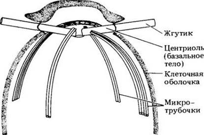
Members of the largest class of green algae, Chlorophyceae, exhibit a unique type of cell division involving a phycoplast (Fig. 15-15). Daughter nuclei move toward each other while the transient spindle breaks down, and a new system of microtubules—the phycoplast—forms parallel to the plane of cell division. This structure presumably ensures The formation of the Cleavage furrow between the daughter nuclei. The nuclear envelope remains intact throughout mitosis. In motile cells, the flagella are inserted at the anterior end. Inside such cells, the flagellar ROOT system consists of four narrow microtubular bundles arranged in a cruciate pattern (Fig. 15-16). These bundles originate from or near the anterior basal bodies.
Fig. 15-15. Cell division in two classes of green algae. In the class Chlorophyceae (A, B), the mitotic spindle is absent (does not persist) and daughter nuclei, positioned relatively close to one another, are separated by a phycoplast. A. Cell division by furrowing. B. Cell division by cell plate formation. In the class Charophyceae (C, D), the spindle is persistent and daughter nuclei are relatively far apart. C. Cell division by furrowing. D. Presence of a phragmoplast and division via cell plate formation. In the Ulvophyceae, the spindle is persistent, as in Charophyceae, but does not form a phragmoplast.
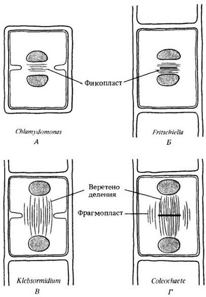
Fig. 15-16. Diagram of the cruciate arrangement of microtubules associated with the centrioles (basal bodies) of flagella; a characteristic feature of green algae of the class Chlorophyceae.
In other green algae, the spindle persists during cell division, and in some cases, a phragmoplast is formed. The microtubules of the phragmoplast are oriented perpendicularly to the plane of division (Fig. 15-15). The spindle remains until it is "ruptured" either by a cell plate, which arises in the center of the cell and grows toward its margins, or by a cleavage furrow. This mechanism is less specialized than that involving a phycoplast. The type of cell division is of great significance in the Taxonomy of green algae.
Classification of Green Algae
The small class of unicellular Micromonadaphyceae, comprising scaled or naked flagellate forms, exhibits many features presumed to be ancestral for this division. In addition to this class, modern phycologists distinguish four other classes of green algae, each of which is considered to represent an independent evolutionary Lineage. One of these, Pleurastophyceae (see p. 258), is a small group and is not discussed here in detail. The remaining three classes are extensive groups encompassing A large number of well-studied organisms.
The three main classes of green algae—Charophyceae, Ulvophyceae, and Chlorophyceae—differ from one another in several essential features. In Chlorophyceae, which form a phycoplast, the spindle fibers always shorten in anaphase, whereas in Charophyceae and Ulvophyceae (which retain the nuclear envelope during mitosis), this typically does not occur. In the latter two classes, the nuclear envelope breaks down at the onset of mitosis, much like in plants, whereas in Chlorophyceae it persists throughout the entire process. Each class includes unicellular flagellate forms that either exist as independent organisms (Chlorophyceae) or serve as reproductive cells in the other two classes. The ancestral forms of each class were undoubtedly unicellular flagellates, and multicellular organisms arose independently within each lineage.
Charophyceae and Chlorophyceae inhabit primarily freshwater habitats, whereas Ulvophyceae are predominantly marine. Motile cells in these three classes vary considerably: in Charophyceae they are asymmetrical, whereas in the other two they are entirely or nearly radially symmetrical. The flagella in Charophyceae are lateral or subapical—meaning they originate near the apex of the cell—and are directed sideways at right angles to its axis. In Chlorophyceae and Ulvophyceae, they are apical and directed forward (Fig. 15-17).
Fig. 15-17. Electron micrograph of the anterior region of a sperm cell of the green alga Coleochaete; a multilayered structure is visible near the Mitochondria. A layer of microtubules extends from this structure to the wall-less posterior end of the cell, functioning as its Cytoskeleton. The flagellar and cell membranes are covered with a layer of small rhomboid scales
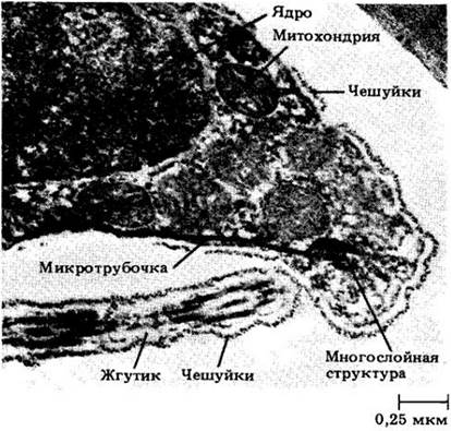
Inside the cells of Charophyceae lies a system of microtubules shaped like a broad, flat band that originates from a multilayered structure near the laterally positioned basal bodies and diverges toward the posterior end of the cell. A similar structure has been discovered in the motile sperm cells of plants. Furthermore, the Peroxisomes of Charophyceae produce the photorespiratory enzyme glycolate oxidase, which, as far as is known, is localized there exclusively in plants, although outside of peroxisomes it has also been found in certain green algae. These and other features closely link Charophyceae to plants rather than to other classes of green algae; therefore, the form most closely related to the plant ancestor should presumably be sought among the representatives of this class.
Sexual reproduction in Charophyceae and Chlorophyceae invariably involves the formation of a resting zygote (zygospore) in which meiosis takes place (see Fig. 10-11, A). In contrast, sexual reproduction in Ulvophyceae is frequently associated with an alternation of generations and meiosis at the spore stage, while resting zygotes are uncommon. The life cycle of Ulvophyceae appears to be the most advanced among these classes, whereas that of Charophyceae is the most primitive.
Class Charophyceae
This class comprises unicellular, few-celled, filamentous, or parenchymatous representatives whose phylogenetic affinity is supported by numerous important Structural and Biochemical characteristics. These include the presence of asymmetrical flagellar cells with lateral or subapical flagella oriented at right angles to the cell axis; meiosis at the zygote stage and the formation of resting zygospores; the presence of a multilayered structure in motile cells; and distinctive features of cell division. All Charophyceae possess a persistent mitotic spindle, and in some, a phragmoplast develops to assist in the Formation of the new cell plate.
Spirogyra (Fig. 15-18) is a well-studied genus of unbranched filamentous algae that frequently form frothy or slimy floating mats in freshwater bodies. Each filament is encased in a watery, mucilaginous sheath. The generic name refers to the presence of one or more spirally twisted ribbon-like chloroplasts within each uninucleate cell. The chloroplasts contain numerous pyrenoids—specialized regions that serve as centers for starch formation in green algae, yet are almost invariably absent in plant chloroplasts. Recent studies have demonstrated that in two other green algae, the enzyme ribulose-1,5-bisphosphate carboxylase is concentrated here (see p. 102). Pyrenoids may also be involved in The conversion of sugars into starch, which typically surrounds these structures.
Fig. 15-18. A. Sexual reproduction in Spirogyra requires the formation of conjugation tubes between cells of adjacent filaments. B. The contents of cells from the minus strain migrate through these tubes into the cells of the plus strain. C. Syngamy occurs within the latter, and the zygote develops a thick, durable wall, transforming into a zygospore. The vegetative filaments of Spirogyra are haploid, and meiosis occurs during the germination of the zygospores, as is characteristic of all Charophyceae
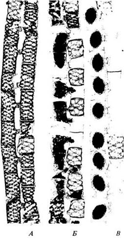
Asexual reproduction in Spirogyra occurs via cell division and filament fragmentation, with flagellate cells being entirely absent at all Stages of the life cycle. Morphologically, sexual reproduction corresponds to isogamy; however, one isogamete acts as a male reproductive cell, migrating through the conjugation tube to fuse with the other isogamete. Meiosis is zygotic, resulting in the formation of resting zygospores, as in all Charophyceae.
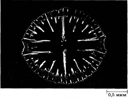
Desmids constitute a large group of freshwater green algae related to Spirogyra; they likewise lack flagellate cells. Some desmids are filamentous, but the majority are unicellular. Their cellular architecture is unusual in that The cell wall consists of two symmetrical halves separated by a narrow median constriction
(Fig. 15-19). Cell division proceeds, much like in Spirogyra, with the formation of a persistent spindle and a cleavage furrow. Some taxonomists estimate that the number of desmid species exceeds 10,000.
Fig. 15-19. In this desmid belonging to the genus Micrasterias, each cell exhibits a deep constriction, as is typical of most members of this large group of unicellular freshwater Charophyceae
Charophyceae Resembling Higher Plants
The two groups of green algae discussed below bear the closest resemblance to plants in the details of their cell division—specifically, the presence of a phragmoplast and The breakdown of the nuclear envelope at THE START OF mitosis. Furthermore, like plants, they are oogamous. However, neither of these groups represents the direct ancestor of plants, as each is overly specialized in certain respects. Plants likely evolved from an extinct representative of the Charophyceae, as indicated by their striking similarities.
The first group to be examined in relation to plant origins is the order Coleochaetales, which includes branched filamentous, disc-shaped, and unicellular forms. The genus Coleochaete, species of which grow epiphytically on submerged freshwater plants, possesses a particularly complex morphology (Figs. 15-17, 15-20). During asexual reproduction, these algae produce zoospores, and their zygotes are enveloped by a layer of sterile cells formed following fertilization. Individual Coleochaete thalli may consist of a mass of dichotomously branching filaments or adopt a disc-like form. Vegetative cells are uninucleate, containing a single large chloroplast and one or more pyrenoids. Cell division may occur at the apices of the filaments or along the margins of the disc, depending on the growth form.
Fig. 15-20. A. Coleochaete from the stem of an aquatic flowering plant inhabiting shallow lake waters. B. Individuals of this Coleochaete species form a parenchymatous disc typically one cell layer thick. The large cells are zygotes protected by surrounding cells. Setae (bristly cells) with basal sheaths project from the disc. The name Coleochaete translates to "sheathed bristle." It is believed that these setae protect the alga from being grazed by aquatic animals
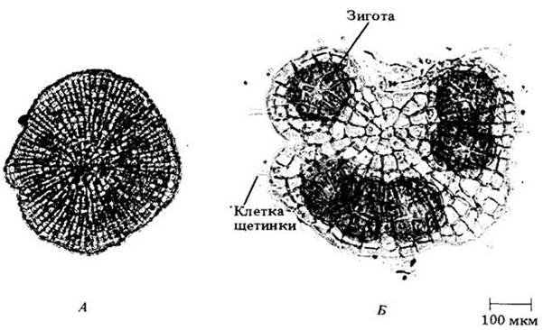
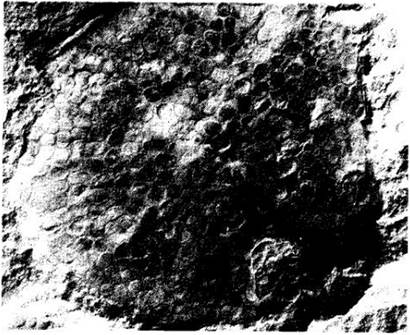
This genus is considered closely related to the putative ancestor of higher plants. Some of its species are parenchymatous and grow through The activity of a marginal disc meristem; reproduction is oogamous, with zygotes protected by a layer of cells. Like certain other Charophyceae, the genus Coleochaete shares similarities with plants due to the presence of a phragmoplast, the respiratory enzyme glycolate oxidase in peroxisomes, and a multilayered structure associated with flagella in reproductive cells (see Figs. 15-17). In addition, fossil forms have been discovered that existed approximately around the time of The Emergence of the first plants and apparently resemble Coleochaete (Figs. 15-21). Although this genus itself is probably not the direct ancestor of plants, its characteristics—including a range of structural and biochemical adaptations beneficial for terrestrial life—provide insight into the potential evolutionary pathway of the earth's flora.
Fig. 15-21. Parka decipiens, a Devonian fossil alga dating back approximately 380 million years, strongly resembles Coleochaete in its form, structure, and chemistry. This organism exhibits numerous features suggesting a close phylogenetic affinity with the group of aquatic protists from which bryophytes and vascular plants eventually arose.
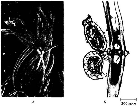
The order Charales represents the second plant-like group of Charophyceae (Fig. 15-22). These distinctive green algae inhabit freshwater or brackish environments. Some species possess heavily calcified cell walls, which has enabled them to be exceptionally well-preserved in the fossil record. Today, there are approximately 250 extant species of charophytes. Their morphology is relatively complex: growth is apical, similar to that of land plants, and the thallus is differentiated into nodes and internodes. Whorls of short branches arise at the nodes, whereas the internodes are coenocytic. Charophyte sperm cells are produced in multicellular antheridia that are structurally more complex than those found in other protist groups, while the egg cells develop in oogonia, which are simpler in structure than the antheridia. Spermatozoids represent the only flagellated cells in the life cycle of charophytes.
Fig. 15-22. A. Chara, a charophyte alga growing in shallow temperate lake waters, clearly displaying its distinctive growth habit. B. A segment of the thallus bearing gametangia: an oogonium at the top and an antheridium below.
Class Ulvophyceae
This class is unique among green algae in comprising predominantly marine forms. Their flagellated cells may be scaly or "naked," much like those of certain Charophyceae, yet they are nearly radially symmetrical and possess apical, forward-directed flagella, characteristic of Chlorophyceae. As in the latter group, Ulvophyceae may have two, four, or numerous flagella, whereas Charophyceae possess only two. Regarding the details of cell division, Ulvophyceae exhibit closed mitosis with a persistent nuclear envelope; the mitotic spindle also persists through cytokinesis. This is the only group of green algae characterized by an alternation of generations and sporic meiosis; unlike the other two classes discussed in detail here, Ulvophyceae rarely form resting zygospores.
Members of the Ulvophyceae may be multicellular, filamentous, or form flat cellular sheets, and can range from parenchymatous to coenocytic. All filamentous or more complex forms of marine green algae exhibit the characteristics of this class, which presumably originated in the marine environment, unlike the other classes of this division.
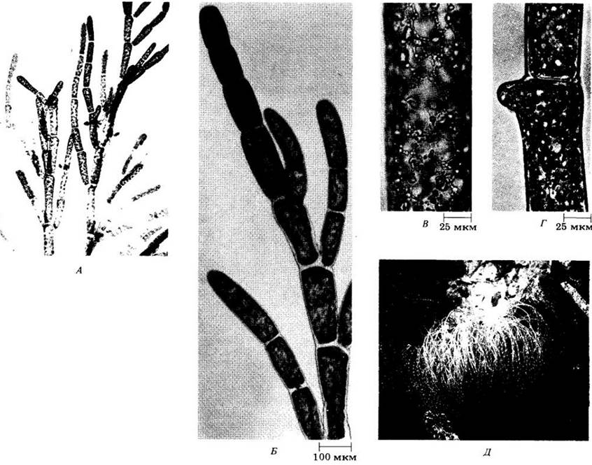
The first evolutionary lineage of the Ulvophyceae encompasses filamentous forms characterized by large multinucleate cells separated by septa. This group includes the genus Cladophora (Fig. 15-23), which is widespread in both freshwater and marine habitats. Its representatives form dense, free-floating or attached mats anchored to rocks or submerged vegetation; the filaments are elongated and branch primarily near their tips. Each cell contains a single peripheral reticulate chloroplast with numerous pyrenoids. Marine species within this genus exhibit an alternation of isomorphic generations, whereas freshwater species lack an alternation of generations—a trait presumably lost in the course of evolution.
Fig. 15-23. Cladophora, a widely distributed alga belonging to the class Ulvophyceae, found in both marine and freshwater environments. Marine species exhibit an alternation of generations typical of most Ulvophyceae, whereas freshwater species do not. A. Branching filaments of Cladophora. B. A section of a branching filament. C. Portion of a cell showing the reticulate chloroplast. D. Initiation of branching in the apical region of a cell. E. An individual Cladophora thallus growing in a slow-flowing stream in California.
Another representative of this lineage is Ulothrix, a common alga found in cold streams, ponds, and lakes. Its filaments are attached to rocks or other substrates by means of a specialized holdfast structure (Fig. 15-24). The vegetative cells along the filament are generally uniform in structure, each containing a single girdle-shaped chloroplast, a pyrenoid, and a nucleus. Asexual reproduction occurs via the formation of quadriflagellate zoospores, whereas sexual reproduction involves isogametes bearing two flagella. Meiosis occurs immediately prior to zygospore germination, rendering the Ulothrix filaments haploid. Although several groups of green algae formerly classified within the genus Ulothrix are actually members of the Chlorophyceae (see Fig. 15-37), the genus itself belongs to the class Ulvophyceae.
Fig. 15-24. Ulothrix, a representative of the class Ulvophyceae featuring an unbranched filamentous structure. The filament on the left, with densely packed cellular contents, consists of sporangia where zoospores are developing. The other filaments are vegetative. The holdfast apparatus is clearly visible on the right filament.

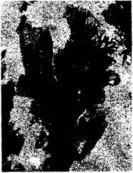
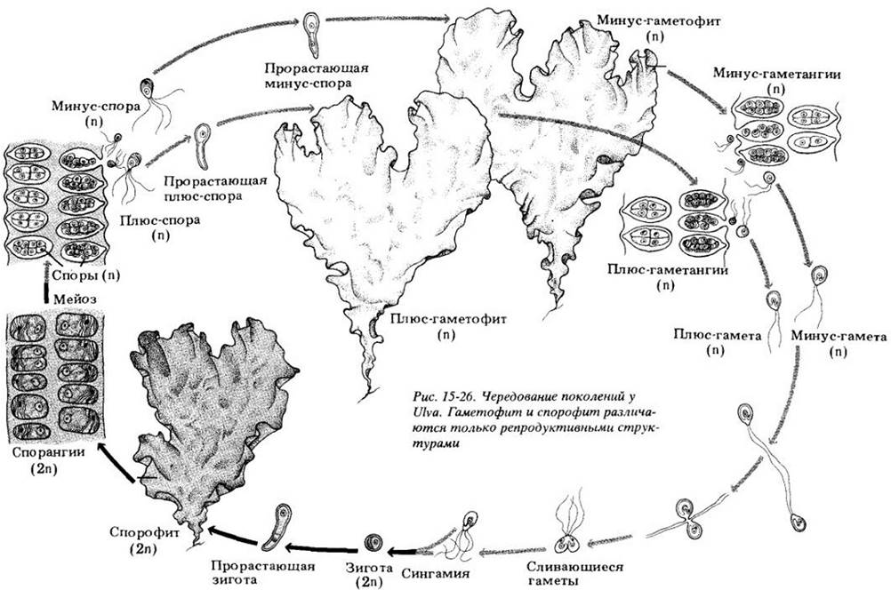
The genus Ulva, commonly known as sea lettuce, exhibits a distinct type of growth. These common marine algae are found along coastlines across all temperate regions of the world (Fig. 15-25). Individual sea lettuce plants consist of a glistening, flat thallus (a simply organized, relatively undifferentiated vegetative body) that is two cell layers thick and can occasionally reach lengths of a meter or more. The thallus is anchored to the substrate by extensions of basal cells. Each cell contains a single nucleus and chloroplast. Ulva is anisogamous and, like most Ulvophyceae, displays an alternation of isomorphic generations (Fig. 15-26).
Fig. 15-25. The sea lettuce Ulva, a widespread representative of the class Ulvophyceae, growing on rocks, pilings, and similar substrates in shallow marine waters worldwide.
Algae belonging to the marine order Siphonales are characterized by very large, branched, coenocytic cells that are rarely septate and arise through repeated nuclear divisions unaccompanied by cytokinesis (Fig. 15-27). This order includes the previously mentioned species Codium magnum. Cell walls in siphonous algae are formed only during the reproductive phase. One such alga, Valonia, common in tropical waters, has been extensively utilized in cell wall research and physiological experiments requiring large volumes of cell sap. Valonia appears unicellular, but is actually a large multinucleate vesicle equipped with rhizoids and young branchlets (Fig. 15-27, B) that can grow to the size of a chicken egg. Another well-known siphonous representative is Acetabularia, widely used in experiments investigating the genetic Regulation of cellular differentiation. The nuclei of siphonous algae are generally diploid, with gametes representing the only haploid cells in the life cycle.
Fig. 15-27. Three genera of siphonous green algae (class Ulvophyceae). A. A species of Codium, abundant along the Atlantic coast. B. Valonia is commonly found in tropical waters, with individuals often approaching the size of a chicken egg. C. Acetabularia, known as "mermaid's wine Glass," is a mushroom-shaped alga. The siphonous algae in the Background belong to Dasycladus; photographed in the Bahamas.
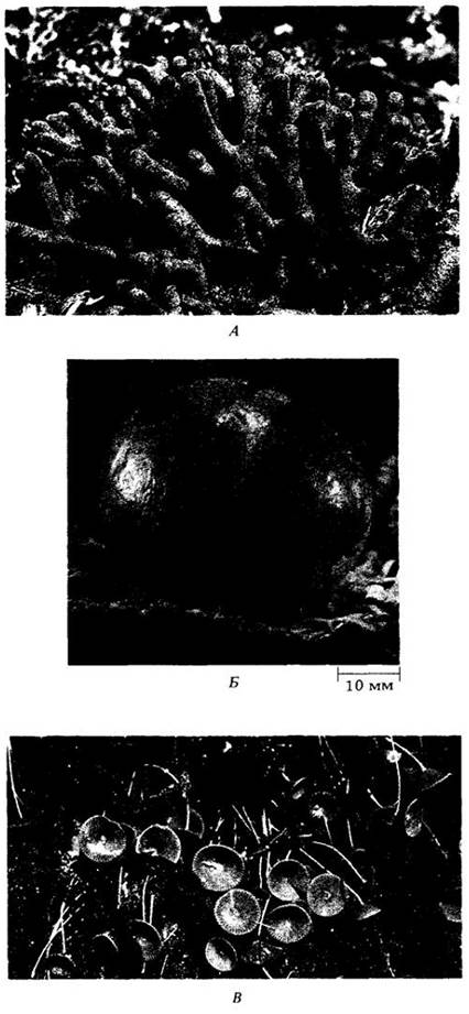
The chloroplasts of certain species within these algae (including Codium) can function as symbionts within the bodies of shell-less marine sea slugs (nudibranchs). These animals consume the algae, and the ingested chloroplasts persist and continue to divide within the cells of the slug's digestive diverticula. When exposed to light, these chloroplasts carry out Photosynthesis so efficiently that, according to some studies, oxygen may be produced in the slug Placobranchus ocellatus at a rate exceeding its consumption via animal Respiration.
It has recently been discovered that a siphonous genus, Halimeda (Fig. 15-28), produces a secondary metabolite that significantly deters herbivorous fish from grazing on the alga, apparently acting as a chemical defense mechanism for these widespread tropical protists. These organisms often constitute the most abundant phototrophic Components of the reef ecosystems they inhabit. Such toxic compounds are likely widespread among algae and will undoubtedly be discovered with increasing frequency in the future.
Fig. 15-28. Halimeda, a siphonous green alga that often dominates reefs in warm waters worldwide. This protist produces unpalatable compounds that deter fish and other marine herbivores.
Class Chlorophyceae
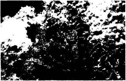
Most green algae belong to this diverse group, which is characterized by cell division involving a phycoplast. This unique feature indicates that no known group of organisms arose from representatives of this class. The Chlorophyceae include flagellated and non-flagellated unicellular algae, multicellular forms, motile or non-motile colonies, and filamentous and parenchymatous species. They inhabit primarily fresh water, although a few unicellular planktonic species are found in coastal marine zones. Some representatives of the class lead a predominantly terrestrial lifestyle, growing in soil or on wood.
Motile Unicellular Chlorophyceae
The simplest forms of Chlorophyceae include unicellular biflagellated organisms of the order Chlamydomonadales. The best-studied among them is the genus Chlamydomonas, which includes some of the most common green algae in fresh waters. Individual cells are small (typically less than 25 µm in length), green, and spherical or pear-shaped (Fig. 15-29). They move rapidly in characteristic jerky motions driven by the beating of two smooth flagella originating from the narrower anterior end of the cell. Moving in opposite directions, these flagella propel the alga through the water Column.
Each Chlamydomonas cell possesses a large, massive chloroplast containing a red pigmented body, the stigma (eyespot), which presumably acts as a shielding device connected to the lightsensing region. Recent studies have shown that the photoreceptor of Chlamydomonas is homologous to rhodopsin, the visual pigment of Multicellular animals. It has been suggested, though not yet proven, that a similar visual pigment is present in the eyespot of dinoflagellates, indicating a very ancient origin. Chlamydomonas individuals can move toward light of a specific intensity. It has recently been suggested that they are also capable of orienting themselves in a magnetic field, but this still requires confirmation, and The Mechanism of such orientation remains unclear.
Fig. 15-29. Chlamydomonas, a unicellular green alga. In this electron micrograph, only the bases of the flagella are visible.
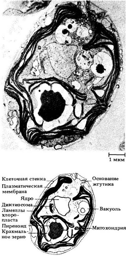
The chloroplast of Chlamydomonas contains a nearly spherical pyrenoid. The uninucleate protoplast is surrounded by a Plasma Membrane, external to which lies a thin glycoprotein cell wall rich in hydroxyproline. Cellulose is absent from the cell wall. At the anterior end of the cell are two contractile vacuoles that collect excess water and expel it to the exterior.
Under certain environmental conditions, Chlamydomonas cells become immotile. In doing so, they typically lose their flagella, and their cell wall gelatinizes. When conditions change, the flagella may reappear, and the cell once again becomes free-swimming.
Chlamydomonas reproduces both asexually and sexually. In the former case, the haploid nucleus typically divides mitotically to form four daughter cells within the parental cell wall. Each daughter cell then secretes a wall around itself and develops flagella. These cells secrete an enzyme that degrades the maternal wall, allowing them to escape, although they often remain inside it for some time even after full development. In ancestral flagellates, such clusters of daughter cells may have been precursors to colonial organisms.
Sexual reproduction, known in several species of Chlamydomonas, occurs through the fusion of individuals belonging to different mating types (Fig. 15-30). In vegetative cells, nitrogen starvation stimulates the formation of gametes, which resemble these cells and aggregate into clusters. Within these aggregates, pairs are formed in which the gametes are initially joined by their flagellar membranes and subsequently by a thin protoplasmic strand at the Base of the flagella. Following the formation of this protoplasmic bridge, the flagella detach, and one or both pairs begin to beat, moving the partially fused gametes through the water. Complete fusion then takes place. Shortly thereafter, the flagella shorten and eventually disappear altogether, and a thick wall develops around the diploid zygote. The thick-walled, resistant zygote (zygospore) undergoes a resting stage, at the end of which meiosis occurs, producing four haploid cells, each with two flagella and its own cell wall. These cells can either reproduce asexually by simple division or fuse with cells of a different mating strain to form a new zygote. Thus, meiosis in Chlamydomonas is zygotic (see Fig. 10-11, A), and the haploid phase predominates in the life cycle.
Fig. 15-30. Life cycle of Chlamydomonas. Sexual reproduction occurs when gametes of different mating types approach each other, adhering first by their flagellar membranes and then forming a thin protoplasmic strand—the conjugation bridge. The protoplasts of both cells fuse completely (plasmogamy), followed by the fusion of their nuclei (karyogamy). A thick wall then forms around the diploid zygote. After a resting period, meiosis occurs, and four haploid individuals are formed. Their asexual reproduction most commonly proceeds via simple division.
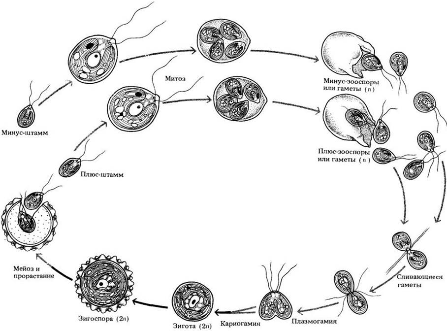
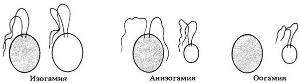
In many species of Chlamydomonas, cells of two distinct mating types (conventionally designated as "+" and "-") are isogamous, meaning they are identical in size and structure. In addition to isogamous species, the genus includes anisogamous species (in which the female gamete is still motile but larger than the male) and oogamous species (with a non-motile female gamete) (Fig. 15-31). Thus, within a single genus, all types of gametic differentiation known among algae are represented.
Fig. 15-31. Types of sexual reproduction based on gamete morphology; each has been discovered in at least one species of Chlamydomonas. In isogamy, gametes are identical in size and shape. In anisogamy, one gamete, conventionally termed male, is smaller than the second. In oogamy, the female gamete is immotile.
In the past, Chlamydomonas was considered a primitive form within the division under consideration; however, this genus possesses many advanced features characteristic of other Chlorophyceae and does not resemble in detail the ancestor of all green algae. Chlamydomonad-like cells resemble certain representatives of specialized evolutionary lineages within this class, which are discussed below.
Non-motile Unicellular Chlorophyceae
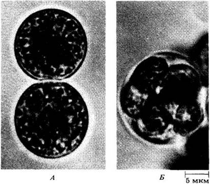
Chlorella is a unicellular green alga lacking flagella, eyespots, and contractile vacuoles; it has a spherical shape and is smaller in size than Chlamydomonas (Fig. 15-32). In nature, Chlorella is widely distributed in fresh and salt water, as well as in soil. Each cell contains a cup-shaped chloroplast, with or without a pyrenoid, and a small nucleus. The only known mode of reproduction is asexual, with each haploid cell dividing mitotically two or three times to produce four or eight offspring, respectively.
Fig. 15-32. Chlorococcum echinozygotum, a close relative of Chlorella and a very common soil alga. A. Two individuals. B. Formation of asexual spores via mitosis within the cell.
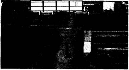
Chlorella, the first alga to be cultivated in the laboratory, has been extensively used in studying key Stages of Photosynthesis. The ease with which it can be cultured makes it an ideal experimental organism. The possibility of using Chlorella as a food source for humanity is currently being investigated (Fig. 15-33). Pilot cultivation facilities have been established in the USA, Germany, Japan, and Israel. Japanese researchers have learned to process this alga into a tasteless white powder rich in Vitamins and protein, which can be mixed with flour in baking products. Recently, the potential of using Chlorella for energy production has been explored; in these experiments, it is grown alongside bacteria that convert the starch synthesized by the alga into Lipids. Such systems could be deployed on barges or platforms in the open ocean or even in space, which explains the current interest in their development. Other algae that produce large amounts of hydrocarbons are also being studied for potential industrial Applications.
Fig. 15-33. These open-air ponds in Sede Boqer (Israel) are used for experiments investigating the potential industrial production of protein from Chlorella. Although the cost of protein produced in this way remains higher than that derived from soybeans—partially due to uneven light penetration in the ponds—experiments are ongoing and, according to experts, may well prove successful.
Non-motile Colonial Сhlorophyceае
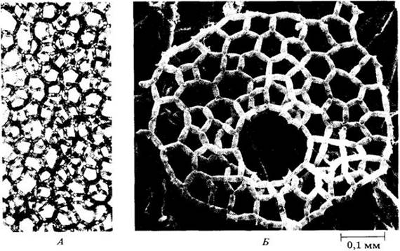
Hydrodictyon, or the "water net", is a non-motile colonial representative of this class (Fig. 15-34). Under favorable conditions, this alga forms large mats in ponds, lakes, and slow-moving streams. Each colony consists of many elongated cells arranged to form a large, hollow cylinder. The cells are initially uninucleate and later become multinucleate. When mature, each cell has a large central vacuole and a peripheral layer of cytoplasm containing the nuclei and a large reticulate chloroplast with numerous pyrenoids. Asexually, Hydrodictyon reproduces by forming uninucleate, biflagellate zoospores. Eventually, these zoospores aggregate in groups of four to nine (most commonly six) inside the cylindrical mother cell, lose their flagella, and develop into daughter colonies. The Sexual process in this genus is isogamous, featuring zygotic meiosis, as is characteristic of all sexually reproducing Сhlorophусеае.
Fig. 15-34. A. The "water net", Hydrodictyon, a colonial member of the Сhlorophyceae. B. Scanning electron micrograph of a flattened young colony of Hydrodictyon reticulatum
Motile Colonial Сhlorophусеае
Among the motile colonial genera of this class, chlamydomonad-like forms are united into colonies that move via the beating of individual cellular flagella (Fig. 15-35). Members of this group are often referred to as volvocine algae, named after the genus Volvox, the largest and most complex colonial organism in the group (Fig. 15-36). In the colonies of some algae of this type, the cells are interconnected by cytoplasmic bridges, which ensure the Integration of the organism as a single functional unit. The simplest representative of this group is Gonium (Fig. 15-35, A, B). Its colony consists of individual cells held together by a gelatinous matrix. Each colony is composed of 4, 8, 16, or 32 cells (depending on the species), forming a slightly curved, shield-like plate. The flagella of the cells beat independently, propelling the entire colony forward. Each cell is capable of dividing to form a new colony.
Fig. 15-35. Several colonial Сhlorophусеае. A, B — Gonium from two Perspectives. C — Pandorina, D — Eudorina. The cells of these algae resemble Chlamydomonas; they are united by a mucilaginous matrix into multicellular colonies that move through the beating of individual cellular flagella. The level of cellular specialization varies among different genera
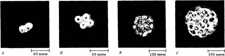
The closely related organism Pandorina forms a densely packed ovoid or ellipsoidal colony of 16 or 32 cells embedded within a matrix (Fig. 15-35, C). The colony is polar, with the eyespots of the cells being larger at one end. Each cell bears two flagella, and since all of them face outward, Pandorina rotates in the water like a ball. When the cells reach their maximum size, the colony settles to the bottom, where each cell divides to form a daughter colony. The latter remain together until all of them have developed flagella. Subsequently, the parental matrix ruptures like Pandora's box (hence the alga's name), releasing the new organisms into the water.
Eudorina is a spherical colonial form comprising 32, 64, or 128 (depending on the species) green flagellated cells (Fig. 15-35, G). It differs from Gonium and Pandorina in that certain smaller cells located at the anterior end (relative to the direction of movement) are incapable of reproducing to form new colonies. Thus, functional cellular specialization is initiated here.
However, the most fascinating representative of these colonial green algae is Volvox (Fig. 15-36). It is a hollow sphere formed by a single layer of 500 to 60,000 (in various species) biflagellate vegetative cells and a small number of reproductive cells. As Volvox spins its way through the water, it resembles a rotating universe of countless stars fixed to an invisible celestial sphere.
Fig. 15-36. Asexual reproduction in Volvox carteri. Approximately 2,000 small, chlamydomonad-like somatic cells are located on the periphery of the transparent sphere; in the mature colony of this species, they are not interconnected, unlike in some other members of the genus. In the specimen shown here, 16 reproductive cells within the sphere have already divided to form young spheres containing about 2,000 tiny somatic cells and 16 reproductive cells. Each daughter colony then "eats" its way out of the parent, emerges into the surrounding water, and the cycle repeats
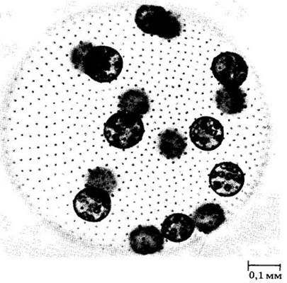
Colonies of this species are polar, meaning they possess distinct anterior and posterior poles. The flagella of each cell beat in such a way that the entire organism advances
(usually toward light during most stages of its life cycle) while rotating clockwise around its axis.
Most cells in this spherical colony are purely vegetative, while a few associated with asexual reproduction are typically positioned in a specific pattern within the posterior hemisphere. In some Volvox species, asexual reproductive cells are not entirely distinct in young spheres but later become prominent through successive cycles of growth and division. Eventually, these dividing cells form daughter spheres that "hatch" from the parent by secreting a specific enzyme that dissolves its transparent matrix. In more advanced members of the genus, such reproductive cells segregate very early in each reproductive cycle and remain structurally and functionally distinct from the vegetative, or somatic, cells at all times (Fig. 15-36). In these species, such as Volvox carteri (Fig. 15-36), a true "division of labor" is observed between two interdependent cell types: the motile somatic cells carry the non-motile reproductive cells to the water's surface, where there is sufficient light and СО2 for photosynthesis. Thus, the most advanced representatives of Volvox are no longer merely colonial, but rather multicellular, differentiated organisms, much like plants and animals.
Sexual reproduction in Volvox is always oogamous, although it varies considerably in detail among different species. In some cases, eggs and sperm may arise within the same sphere, whereas in others, members of a genetically uniform clone (derived asexually from a single individual) develop exclusively into male or female colonies, with bisexual spheres being unknown. However, in more advanced species, sex is genetically determined, and every asexually derived clone is either male or female. In all studied species, sexual reproduction within a population of colonies is synchronized via chemical sex Inducers. These molecules are synthesized by spheres that have reached "sexual maturity" through a mechanism that is not yet fully understood. A single male colony of V. carteri can release enough inducer to trigger the transition to sexual reproduction in over 0.5 billion other colonies of this species.
Volvox is one of the simplest organized multicellular organisms featuring a clear "division of labor" among cells. The sexual inducer of this alga is among the most potent BIOLOGICALLY ACTIVE SUBSTANCES known to date. Consequently, a vast amount of research has focused on this genus (especially V. carteri) in recent years. Like other related algae, Volvox is haploid, meaning its mutant genes are not masked by dominant alleles, and Mutations affecting development can be readily identified. Hundreds of strains with specific hereditary defects have already been isolated and are being used to study The regulation of cellular differentiation by specific genes.
In summary, it should be noted that increased specialization in colonial Chlorophyceae can be traced along several directions. First, there is an increase in cell number and colony size. Second, morphofunctional cellular specialization takes place. Finally, sexual specialization increases, paralleling that observed in the genus Chlamydomonas. Thus, Gonium and Pandorina are isogamous, while Eudorina and Volvox are oogamous. Nevertheless, this evolutionary branch is clearly a dead end, as it did not give rise to a more complexly organized group of organisms.
Filamentous and Parenchymatous Chlorophyceae
Members of the orders Chaetophorales and Oedogoniales exhibit the most complex structure among the algae of this class. Their cells are often specialized in terms of their functions or their position within the protist's body and are interconnected by plasmodesmata, much like plants. Although many genera in these groups form a cell plate during cell division, just as plants do (Fig. 15-37), they also possess a phycoplast, like all Chlorophyceae, and therefore cannot be considered the ancestors of plants. Furthermore, as in all Chlorophyceae, the nuclear envelope persists throughout the mitotic cycle.
Fig. 15-37. Stigeoclonium, a representative of the class Сhlorophусеае, formerly assigned to the genus Ulothrix (Ulvophусеае). Cell division in Stigeoclonium occurs with the formation of a cell plate, which is almost completely formed at the cytokinesis stage shown in this electron micrograph. The nuclei lie close together because the mitotic spindle has disintegrated by the telophase stage of the preceding mitosis. The phycoplast is not visible due to the relatively low magnification
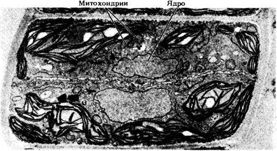
Among the filamentous members of this class, Stigeoclonium (Fig. 15-37) externally resembles Ulothrix (Fig. 15-24), but possesses all the diagnostic features of Chlorophyceae. These two genera, along with other Chlorophyceae, were once lumped together, but detailed electron microscopic studies have now demonstrated their placement in different classes. Unlike Ulothrix, Stigeoclonium features branched filaments.
Fritschiella is a terrestrial alga that shares most microscopic features with Stigeoclonium, but is more complex in structure. Its body consists of subterranean rhizoids, parenchymatous creeping strands near the soil surface, and two types of upright branches protruding from it (Fig. 15-38). Fritschiella also inhabits damp surfaces such as wet walls, tree trunks, and leaves. Like plants, it has adapted to a terrestrial lifestyle and exhibits certain traits likely characteristic of ancestral plant forms.
Fig. 15-38. Fritschiella, a terrestrial alga of the class Chlorophyceae. In ADAPTATION TO A terrestrial lifestyle, it has evolutionarily and independently acquired features characteristic of plants

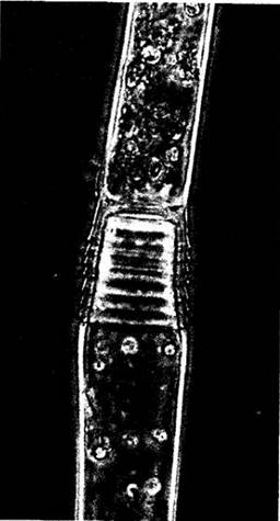
Oedogonium is an unbranched filamentous alga attached to the substrate by a specialized holdfast. It exhibits highly distinctive features, particularly regarding cell division. The cells of this genus are uninucleate with a peripheral reticulate chloroplast. When a cell divides, a ring-shaped structure is formed from cell wall material at its upper (apical) end. Following nuclear division, one of the daughter nuclei migrates to the upper end of the cell, where the wall abruptly ruptures precisely at this ring. The ring then elongates into a cylinder, forming the wall of the upper daughter cell located closer to the apex of the filament. A new transverse septum is formed in the plane of the phycoplast immediately behind the edge of the ruptured parent wall. As the ring elongates, the margins of the original mother cell roll outward, forming characteristic scars (Fig. 15-39) that indicate the number of previous divisions.
Fig. 15-39. Oedogonium, an unbranched filamentous alga of the class Chlorophyceae. Portion of a vegetative filament showing apical scars
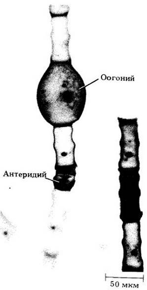
Asexual reproduction in Oedogonium occurs via zoospores, which are produced singly per cell. Each zoospore bears a crown of approximately 120 flagella. Sexual reproduction is oogamous (Fig. 15-40). Each antheridium produces two multiflagellate sperm, while each oogonium produces a single egg cell. Meiosis is zygotic, as in all Chlorophyceae.
Fig. 15-40. Oogamous sexual reproduction in Oedogonium. Each oogonium produces a single egg cell, and each antheridium produces two multiflagellate sperm
Last update: 07/08/2026
Editorial and Educational Adaptation: This material has been compiled based on the primary/original source text. The project team performed an editorial review, corrected technical inaccuracies, structured sections, and adapted the content for an educational format.
What was processed:
- elimination of formatting defects (OCR errors, structural breaks, corrupted characters);
- editorial organization of content;
- standardization of terminology in accordance with academic sources;
- verification of factual statements against the original source text.
All mentions of the author, publication year, and origin of the primary text have been preserved in accordance with the source.