BIOLOGY Volume 3 - A Guide to General Biology - 2004
23. THE CONTINUITY OF LIFE
23.8. Protein Synthesis
«DNA makes RNA, and RNA makes protein»
From everything discussed in the previous sections, it is evident that the only molecules synthesized under the direct control of DNA are Proteins. Proteins can be structural, such as keratin and Collagen, or functional, such as Insulin, fibrinogen, and, most importantly, Enzymes responsible for the Regulation of cellular METABOLISM. It is precisely the set of enzymes contained within a given Cell that determines its cell type. In this way, DNA controls cellular activity.
The «instructions» required for the synthesis of enzymes and all other proteins are encoded in DNA, almost all of which is located in The Nucleus. However, as demonstrated in the early 1950s, synthesis actually takes place in the Cytoplasm and involves Ribosomes. It became clear that a mechanism must exist to transfer Genetic information from the nucleus to the cytoplasm. In 1961, two French biochemists, Jacob and Monod, postulated the existence of a special form of RNA, which they named Messenger RNA (mRNA). Their idea proved to be correct. The sequence of events occurring during Protein Synthesis is encapsulated in the short phrase: «DNA makes RNA, and RNA makes protein».
Class="center">23.8.1. The Role of RNA
RNA is found in all living Cells in the form of single-stranded molecules. It differs from DNA in that it contains ribose as its pentose sugar (instead of deoxyribose) and uracil as one of its pyrimidine bases (instead of thymine). Analysis of cellular RNA has shown that there are Three types of RNA involved in the synthesis of protein molecules: messenger RNA (mRNA), Transfer RNA (tRNA), and ribosomal RNA (rRNA). All three types of RNA are synthesized directly on DNA, and The amount of RNA in each cell is directly proportional to the amount of protein produced by that cell.
23.8.2. Messenger RNA
Analyses have shown that mRNA accounts for 3–5% of total cellular RNA. It is a single-stranded molecule transcribed from one of the DNA strands in a process called Transcription. During mRNA synthesis, only one strand of the DNA molecule is copied. The synthesis of mRNA will be described later. The base sequence of mRNA is a complementary copy of the corresponding DNA strand; its length varies depending on the length of the polypeptide chain it encodes. Most mRNA molecules remain in The Cell for only a short time. In Bacteria, this may be just a few minutes, whereas in developing erythrocytes, mRNA can serve as a template for Hemoglobin synthesis for several days.
23.8.3. Ribosomal RNA
Ribosomal RNA accounts for approximately 80% of total cellular RNA. It is encoded by genes located in the DNA of several Chromosomes situated in the nucleolar region known as the nucleolar organizer. The base sequence of rRNA is similar across all organisms, from bacteria to Higher Plants and animals. rRNA is found in the cytoplasm, where it associates with protein molecules to form cellular Organelles known as ribosomes (Sec. 5.10.4).
Protein synthesis takes place on the ribosomes. Here, the «code» carried by the mRNA is translated into the Amino Acid Sequence of a polypeptide chain.
23.8.4. Transfer RNA
The existence of transfer RNA (tRNA) was postulated by Crick and demonstrated by Hoagland in 1955. Each amino acid has its own family of tRNA molecules. They deliver Amino Acids from the cytoplasm to the ribosome. Thus, tRNA acts as an intermediary molecule between the triplet code in mRNA and The amino acid sequence of the polypeptide chain. tRNA accounts for approximately 15% of total cellular RNA; these RNA molecules have the shortest polynucleotide chains, averaging about 80 NUCLEOTIDES. Each individual cell contains more than 20 different tRNA molecules (60 have already been identified). All tRNA molecules share a similar basic Structure (see Fig. 23.25).
The 5' end of a tRNA molecule always bears guanine, while the 3' end features the CCA base sequence. The nucleotide sequence in the rest of the molecule varies and may contain «unusual» bases such as inosine and pseudouridine. The base sequence of the anticodon triplet (Fig. 23.25) strictly corresponds to the amino acid carried by that specific tRNA molecule. Each amino acid is attached to one of its specific tRNAs through the action of the enzyme aminoacyl-tRNA synthetase. This results in The formation of an amino acid-tRNA complex known as aminoacyl-tRNA, in which the bond energy between the terminal nucleotide A in the CCA triplet and the amino acid is sufficient to subsequently form a peptide bond with the adjacent amino acid. In this manner, the polypeptide chain is synthesized.

Fig. 23.25. One of the proposed structural models of transfer RNA (tRNA). The molecule consists of 80 nucleotides, but only 21 Base Pairs are formed through complementary base pairing.
23.8.5. Summary
The process of protein synthesis consists of two main stages, schematically represented in Fig. 23.26.
1. Transcription — the synthesis of mRNA on a DNA template. In this process, a specific region of DNA (a Gene) is copied to produce mRNA.
2. Translation — The conversion of the base sequence in an mRNA molecule into the amino acid sequence of a protein molecule.

Fig. 23.26. Schematic diagram of the main Stages of Protein Synthesis.
23.8.6. Transcription
Transcription is the process by which the base sequence of a DNA strand is "copied" into a complementary mRNA base sequence. The DNA double helix unwinds as the relatively weak Hydrogen Bonds between the bases of the two single DNA strands break. Only one of these strands serves as a template for the synthesis of a complementary single strand of mRNA. This molecule is formed by the joining of free ribonucleotides under the action of RNA polymerase, following the base-pairing rules of DNA and RNA (Tables 23.5 and Fig. 23.27).
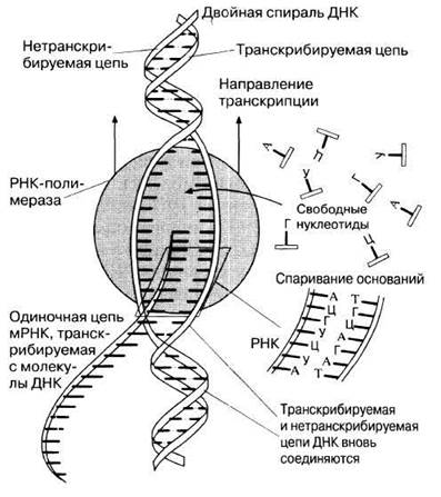
Fig. 23.27. Diagram of the transcription mechanism. In the presence of RNA polymerase, the DNA double helix unwinds due to the breaking of hydrogen bonds between complementary bases. A single-stranded mRNA polynucleotide chain is then built from free ribonucleotides along the exposed single DNA strand. The mRNA bases align opposite their complementary DNA bases. (After E. J. Ambrose, D. M. Easty, Cell Biology, 1977, 2nd ed. Nelson.)
The exact mechanism by which DNA bases are transcribed into RNA bases was demonstrated in experiments using synthetic DNA consisting of only a single type of nucleotide — thymidine (TTT...). When this DNA was introduced into a cell-free system containing RNA polymerase and all four nucleotides (A, U, G, and C), an mRNA molecule containing exclusively complementary adenine nucleotides was synthesized.
The synthesized mRNA molecules exit the nucleus through nuclear pores and deliver genetic information to the ribosomes. Once a sufficient number of mRNA molecules have been produced via transcription, RNA polymerase detaches from the DNA, and the two DNA strands re-associate (like a closing zipper), restoring The Double Helix.
23.8.7. Translation
Translation is the process by which the base sequence of an RNA molecule is translated into the amino acid sequence of a polypeptide chain. This process takes place on ribosomes. Several ribosomes attach to an mRNA molecule like a string of beads, forming a structure known as a polyribosome or polysome. Polysomes can be visualized using an Electron microscope (Fig. 23.28). The advantage of polysomal Organization is that it enables the simultaneous synthesis of multiple Polypeptides (Section 23.8.3). Each ribosome consists of two subunits — a large one and a small one — and resembles a mushroom in shape (Fig. 5.27). The first two mRNA codons (a total of 6 bases) enter the ribosome, as shown in Fig. 23.29, A. The first codon binds an aminoacyl-tRNA molecule, which contains a complementary anticodon and carries the first amino acid (usually Methionine) of the synthesized polypeptide. Next, the second codon binds an aminoacyl-tRNA molecule containing its complementary anticodon (Fig. 23.29, A and B). The function of the ribosome is to hold the mRNA, tRNA, and enzymes involved in translation in the correct orientation until a peptide bond is formed between adjacent amino acids.
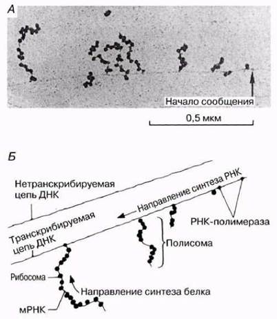
Fig. 23.28. The process of transcription and polysome formation in bacteria. Note that because bacteria lack a nucleus, RNA does not need to be separated from DNA. A. Electron micrograph of a region of bacterial DNA showing stages of mRNA formation and ribosome attachment. B. Schematic representation of the structure shown in micrograph A.
As soon as a new amino acid is added to the growing polypeptide chain, the ribosome moves one codon along the mRNA. The tRNA molecule that was previously bound to the polypeptide chain leaves the ribosome and returns to the cytoplasm to form a new aminoacyl-tRNA complex (Fig. 23.29, C).
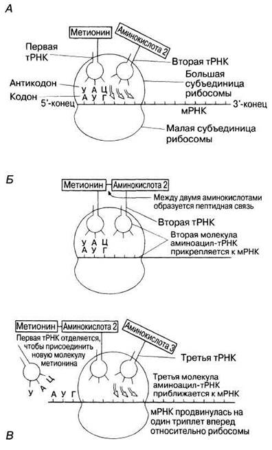
Fig. 23.29. A and B. Stages of the attachment of aminoacyl-tRNA molecules via their anticodons to mRNA codons, and the formation of a peptide bond between adjacent amino acids. C. Relative Movements of the mRNA and ribosome, which expose a new triplet frame for the binding of an aminoacyl-tRNA molecule. The first tRNA molecule detaches from the ribosome and returns to the cytoplasm, where enzymes reactivate it, enabling the formation of a new aminoacyl-tRNA complex.
Such sequential "reading" and "translation" of the code encoded in the mRNA by the ribosome continues until the process reaches one of the stop codons (termination codons). These codons are the triplets UAA, UAG, and UGA. At this stage, the polypeptide chain, whose Primary Structure was determined by the DNA, leaves the ribosome, and translation is completed. The MAIN STAGES OF translation are:
1) attachment of mRNA to the ribosome;
2) activation of the Amino Acid and its attachment to tRNA;
3) initiation (start of synthesis) of the polypeptide chain;
4) elongation (extension) of the chain;
5) termination (end of synthesis) of the chain;
6) subsequent utilization or degradation of the mRNA.
The translation process is schematically illustrated in Fig. 23.30.

Fig. 23.30. Schematic representation of translation. The anticodon of each individual aminoacyl-tRNA molecule pairs with its complementary mRNA codon on the ribosome. In the case shown here, this will be followed by the formation of a peptide bond between leucine and Glycine, thereby adding another amino acid to the growing polypeptide chain.
Once The polypeptide chains have detached from the ribosome, they may immediately acquire their characteristic secondary, tertiary, or quaternary structure (Section 3.5.3). If the ribosome is attached to the ER, the protein enters the ER and is subsequently transported to its destination. The initial part of the growing polypeptide chain consists of a "signal sequence" of amino acids that matches a specific receptor in the ER membrane, ensuring the attachment of the ribosome to the ER. The growing protein molecule enters the ER via the receptor (Fig. 23.31). After the protein is inside the ER, the signal sequence is cleaved off, and the protein molecule folds into its final conformation.
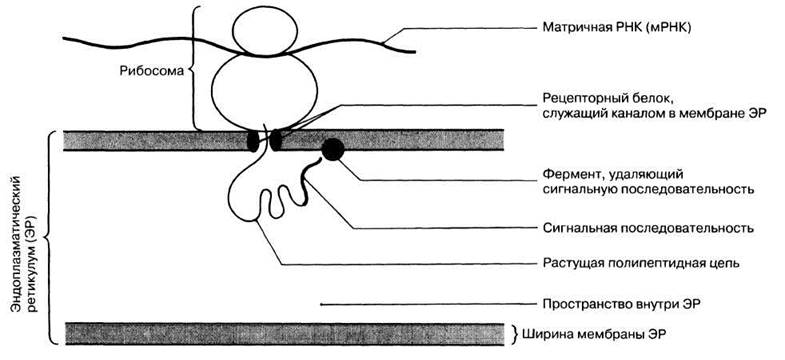
Fig. 23.31. Entry of a newly synthesized protein into The Endoplasmic reticulum.
Evidence that the incorporation of an amino acid into a polypeptide chain is determined by complementary base pairing between the mRNA codon and the tRNA anticodon, rather than by the amino acid itself, was provided by the following experiment. A Cysteine-tRNA molecule normally pairs via its ACA anticodon with the UGC mRNA codon. When this complex was treated with Raney nickel catalyst, the cysteine was converted to the amino acid Alanine. When this novel alanine-tRNA complex (carrying the cysteine-tRNA anticodon) was placed in a cell-free system containing poly(UGC) mRNA, the synthesized polypeptide chain consisted exclusively of alanine. This experiment confirmed the vital role of the mRNA codon–tRNA anticodon pairing mechanism in the Introduction/27.html">Translation of the Genetic Code.
The entire sequence of events comprising protein synthesis is schematically illustrated in Fig. 23.32.
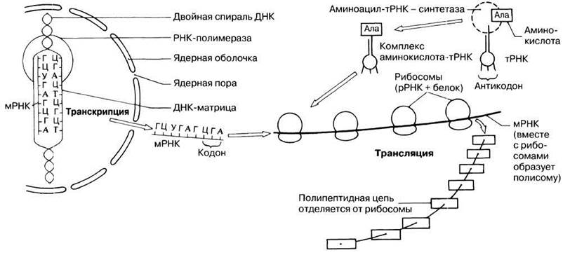
Fig. 23.32. A simplified generalized diagram of the major structures and processes involved in protein synthesis.
23.8.8. Non-coding DNA
Human DNA contains approximately 3000 million base pairs and an estimated 100,000 genes, although these figures remain largely provisional. The challenge is that a large proportion of DNA (roughly 95%) appears to lack any obvious function, as it is non-coding. In other words, it codes for neither proteins nor RNA. It is sometimes referred to as "junk DNA," though assuming a lack of function merely because that function is unknown is unwise. It is possible that some of this DNA represents vestigial genes that no longer serve any useful purpose. Other portions may perform structural roles, such as participating in chromosome packaging. Approximately 30–40% of this DNA consists of highly repetitive short base sequences. This fraction includes satellite DNA, whose role in "genetic fingerprinting" is described in Section 25.7.12. Certain regions of DNA, known as introns, are discussed below.
Introns and exons
In 1977, biologists were surprised to discover that eukaryotic genes are longer than their corresponding mRNA transcripts. One would naturally expect an mRNA molecule to match the exact length of the gene, as it serves as a copy of it. It turned out that immediately after transcription, certain segments of the mRNA molecule are excised before translation takes place. The Regions of the gene encoding these discarded RNA segments were termed introns. The remaining regions of the gene that code for the functional protein were named exons (Fig. 23.33). The size and arrangement of introns vary widely and are characteristic of a given gene. Prokaryotes do not possess introns.

Fig. 23.33. Exons, introns, and intron splicing.
One potential function of introns came to light when it was discovered that different introns can be removed from the same pre-mRNA in different cell types. Thus, a single gene may utilize Alternative Splicing to encode distinct, albeit related, proteins, thereby expanding its functional repertoire. A prime example is the Calcitonin gene. This gene can yield two different mRNA forms depending on which introns are excised. One form is produced in The Thyroid Gland and encodes a 32-amino-acid calcitonin protein. Calcitonin is a hormone that lowers Blood calcium levels. The other mRNA form is synthesized in the Hypothalamus and encodes a 37-amino-acid protein known as calcitonin gene-related peptide (CGRP), which shares similarities with calcitonin and exhibits potent vasodilator activity. It is also released from nerve endings in certain regions of the Peripheral Nervous system.
Last update: 06/08/2026
Editorial and Educational Adaptation: This material has been compiled based on the primary/original source text. The project team performed an editorial review, corrected technical inaccuracies, structured sections, and adapted the content for an educational format.
What was processed:
- elimination of formatting defects (OCR errors, structural breaks, corrupted characters);
- editorial organization of content;
- standardization of terminology in accordance with academic sources;
- verification of factual statements against the original source text.
All mentions of the author, publication year, and origin of the primary text have been preserved in accordance with the source.