Chordate Zoology - textbook - Y. V. Tsaryk - 2013
Chapter 3. SUBPHYLUM VERTEBRATES, or CRANIATES, VERTEBRATA, seu CRANIATA
3.1. DIVISION AGNATHA. CLASS CYCLOSTOMATA. STRUCTURAL FEATURES
The division Agnatha is also known as entobranchiates, as the gills (gill pouches) of these animals are lined with folded endoderm. It comprises fossil and extant vertebrates in which the notochord Functions as the primary supporting axial rod throughout their lifespan.
The Class Cyclostomata includes 38-45 species of modern Jawless vertebrates, which are grouped into two subclasses: lampreys (Petromyzones) and hagfishes (Myxini). This is the most ancient class of living vertebrates. They are characterized by a cartilaginous Skeleton with poorly differentiated individual elements, primitive gills (5-16 pairs of endodermal gill pouches), a simply structured circulatory and Reproductive System (Gonads lack independent gonoducts), a small Brain, etc. Lampreys inhabit seas, brackish waters, rivers, and streams; hagfishes live exclusively in marine environments (dying when salinity drops below 25%). They swim by undulating their bodies in a snake-like fashion. Most of them are parasites and predators, feeding on the soft Tissues, Blood, and Lymph of aquatic animals, primarily fish.
Appearance and integument. Representatives of the class have an eel-like body (Fig. 3.1). The integument lacks an exoskeleton. The Skin consists of a multilayered epidermis and dermis, containing numerous unicellular glands that secrete mucus. Mucus protects the body from damage, facilitates movement through Water, and, in hagfishes, AIDS in penetrating the prey's body. Small pores of the lateral line Organs are located on the HEAD and trunk.

Fig. 3.1. External appearance of a lamprey (A) and a hagfish (B)
Paired fins are absent. In lampreys, approximately in the middle of the dorsal region of the body, there is an anterior dorsal fin, followed by a posterior one, which is connected to a small caudal fin. The caudal fin is protocercal (symmetrical in both external appearance and internal Structure). The dorsal and caudal fins are supported by a system of long, thin cartilaginous rays called pterygiophores. On the ventral side, at the boundary between the trunk and the tail, the anus is located, followed by the urogenital papilla on a small cone. The Mouth opening is located deep within the preoral funnel. The myochordal complex is well-developed in both larvae and adults.
Skeleton. The visceral region is cartilaginous, and jaws are absent. The Axial Skeleton is formed by the notochord. The notochord is surrounded by a thick sheath, which also protects the Spinal Cord. In lampreys, rod-like cartilages (two pairs in each body segment) are embedded within this sheath, representing the rudiments of vertebrae. Hagfishes lack such "cartilages."
The neurocranium of cyclostomes is at an evolutionary stage characteristic of the early embryonic development of other vertebrates. It consists of expanded parachordals that enclose the brain from below and from the sides (in hagfishes, the lateral parts are undeveloped). From above, the brain is covered by a thin Connective Tissue membrane.
The occipital region of the braincase is undeveloped. Anteriorly, an unpaired but bilobed olfactory capsule is adjacent to the neurocranium, while paired auditory capsules are attached to the posterior lateral walls (Fig. 3.2).
The visceral skeleton is unique: it comprises the Skeleton of the preoral funnel, the branchial basket, and the pericardial Cartilage. An annular cartilage and several unpaired cartilages support the walls of the preoral funnel and the muscular Tongue. Some of these cartilages fuse with the neurocranium. The branchial basket is developed only in lampreys and is absent in hagfishes. The pericardial cartilage, which encloses The Heart laterally and posteriorly, is fused to the branchial basket. The suborbital arch, which fuses with the neurocranium, also belongs to the visceral skeleton.
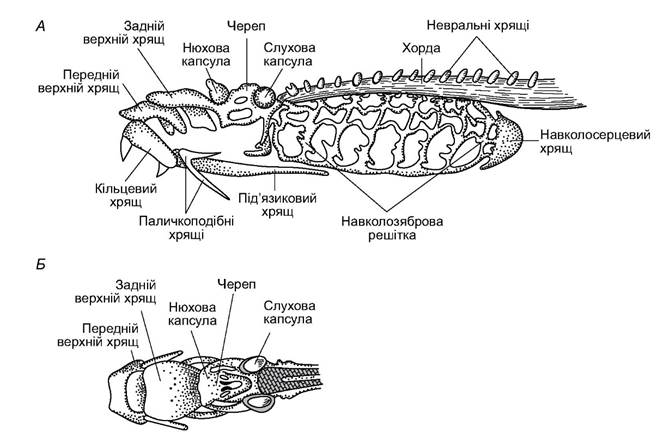
Fig. 3.2. Skeleton of a lamprey: A - dorsal view; B - lateral view
The caudal and dorsal fins are supported by long and thin cartilaginous rays.
The Muscular System of cyclostomes is significantly more powerful than that of acraniates. It consists of Muscle segments called myomeres, separated from each other by connective tissue partitions known as myosepta. Each myosepta forms a zigzag line resembling the Latin letter W. On the head and in the branchial region, beneath the myomeres of the somatic Muscles, there are visceral muscles that form a complex system of Muscles of the preoral funnel, tongue, and gill pouches (Fig. 3.3). These muscles enable attachment to the prey, drilling a hole in its integument, sucking food, and driving the flow of water through the gill pouches.

Fig. 3.3. Sagittal section of the anterior end of a lamprey's body
Digestive System. The digestive tract begins with the preoral funnel, which is more developed in lampreys than in hagfishes. Small skin folds are present along the margin of the funnel, facilitating better attachment. Hagfishes have two pairs of tentacles around the mouth. Keratinized epithelial outgrowths on the inner surface of the funnel form horny Teeth and tooth plates; their size, shape, and arrangement are of taxonomic significance.
Deep within the funnel lies a circular mouth opening, bounded below by the tip of a powerful tongue, which bears 1-2 large horny teeth or a complex horny tooth plate. Lampreys suck in small prey along with water. When attached to large prey, lampreys rasp through the skin using the tip of their tongue; hagfishes burrow into soft tissues using their lingual teeth.
In lamprey larvae, a ciliated groove called the endostyle is located at the beginning of the Pharynx, which secretes mucus. The beating of the endostyle's cilia and the Movements of the velum—a fold at the boundary between the Oral Cavity and the pharynx—propel a mucus cord extending to the intestine. Food particles enter the pharynx with water, are trapped by the mucus stream, and are directed to the intestine, while water passes into the gill pouches and is expelled through their external openings. This feeding mechanism is characteristic of the lamprey larva, the ammocoete. It is highly similar to the feeding of the lancelet. This similarity is an example of recapitulation—the repetition of ancestral traits during The Development of descendants.
During the metamorphosis of the lamprey larva, the lower part of the posterior pharyngeal wall expands (Fig. 3.4, 4), dividing the pharynx into two isolated compartments: the Esophagus and the respiratory tube. The internal openings of the gill pouches open into the respiratory tube. From the side of the oral cavity, the entrance to the respiratory tube is guarded by a respiratory fold, the velum.
In a free-swimming lamprey, the velum is deflected, closing the lumen of the esophagus; water enters the respiratory tube through the mouth and then passes into the gill pouches. When the lamprey is attached to prey, the velum closes the entrance to the respiratory tube, allowing the victim's blood to enter the lamprey's esophagus and intestine.
Due to muscular contraction and the elasticity of the branchial basket, the volume of the gill pouches changes, drawing water into them; in this process, the respiratory tube facilitates the redistribution of water among the gill pouches.
In all cyclostomes, the intestine does not form loops and opens via the anus. In lampreys, the absorptive surface of the intestine is increased by a large fold—the spiral valve—which runs along the entire length of the gut.
Cyclostomes possess a large, cone-shaped Liver located posterior to the heart. The Gallbladder opens into the intestine via a Bile duct. In lampreys migrating to spawn, which cease feeding during this period, the gallbladder is reduced. The Pancreas is represented by islets of Cells embedded in the intestinal walls.

Fig. 3.4. Pharynx of an ammocoete (A) and a lamprey (B)
Cyclostomes are capable of consuming large amounts of food. For example, hagfishes can increase their body mass 7 to 8 times during 7–10 hours of feeding. Satiated animals can survive without food for long periods. Lampreys prey on fish of various sizes. Hagfishes are also predators, attacking fish and cephalopods. They locate their prey using their SENSE OF SMELL. They hunt at night, burrowing into the bottom substrate during the day.
Respiratory system. Even in cyclostome embryos, gill slits develop, connecting the pharyngeal cavity with the external environment. The inner surface of the gill pouches features numerous folds. Hagfishes have 6 to 16 pairs of gill pouches, while lampreys have 7 pairs. In hagfishes, the external ducts of the gill pouches on each side of the body open externally via a common aperture located mid-body; in lampreys, the gill pouches open through separate external pores.
Respiration is driven by the rhythmic action of the branchial wall muscles. Water enters through the mouth into the pharynx (or the respiratory tube in adult lampreys) and then into the gill pouches. During feeding, water enters and exits directly through the external gill openings. Cutaneous respiration also occurs, facilitated by capillaries in the skin.
The Circulatory system OF cyclostomes is similar to that of the lancelet. However, cyclostomes possess a true heart consisting of an atrium and a ventricle (Fig. 3.5). They have a single circulatory system, with only venous blood flowing through the heart. Large Veins empty into the thin-walled sinus venosus, from which blood enters the atrium and then the muscular ventricle. The ventral aorta arises from the ventricle, giving off paired afferent branchial Arteries to the interbranchial septa, each supplying blood to half of the adjacent anterior and posterior gill pouches.
In the capillaries of the gill pouch folds, the blood is oxygenated and releases carbon dioxide. The capillaries merge into efferent branchial arteries, which empty into the unpaired dorsal aorta located beneath the notochord.
Carotid arteries branch from the anterior end of the dorsal aorta to supply blood to the head, while other arteries run to the myomeres, digestive tract, and other organs.
Venous blood from the caudal region collects in the caudal vein, which splits into two posterior cardinal veins.

Fig. 3.5. Diagram of the circulatory System of the river lamprey (ventral view)
Blood from the head is carried by two anterior cardinal veins. Both the anterior and posterior cardinal veins empty into the sinus venosus. Venous blood from the lingual muscles and the lower head region flows through the inferior jugular vein, which also drains into the sinus venosus. Blood from the intestine is collected by the subintestinal vein, which breaks up into a capillary network in the liver, forming the hepatic portal system. Its capillaries then merge into a short hepatic vein, which likewise empties into the sinus venosus.
Cyclostomes lack a Spleen. Hematopoiesis occurs in the walls of the esophagus and intestine, as well as in the Kidneys, liver, and adipose tissue along the notochord. The total blood volume constitutes 4–5% of the animal's body mass. There are 130,000–170,000 erythrocytes per 1 mm3 of blood, containing 3 to 8% Hemoglobin.
Excretory system. Cyclostomes possess kidneys, the excretory organs characteristic of all vertebrates, which use a filtration apparatus to excrete excess water and Metabolic waste products from the body (Fig. 3.6). Thus, the kidneys participate in water-salt balance and, together with the skin, maintain the osmotic Stability of the internal environment.
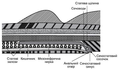
Fig. 3.6. Cytology/practical/54.html">Longitudinal section of the posterior trunk of a lamprey
During ontogeny, the embryonic pronephros is replaced by paired trunk kidneys (mesonephroi) characteristic of adult individuals. The kidneys are located on the DORSAL SIDE OF the Abdominal cavity as ribbon-like structures covering the upper portion of the gonad. Ureters run along the lower margin of the kidneys and empty into the urogenital sinus, which opens externally via a pore at the tip of the urogenital papilla located just behind the anus. Microanatomically, the kidneys of cyclostomes differ significantly from those of Gnathostomes. A glomus—a cord-like cluster of arterial capillaries that produces filtrate—runs along the entire length of the kidneys. The filtrate flows through intercellular spaces into short renal tubules, where substances valuable to the body are partially reabsorbed. Unlike in other vertebrates, the anatomical Integration of the filtration glomerulus and the capsule that collects the filtrate has not yet occurred in cyclostomes.
In marine hagfishes, the Osmotic Pressure of the blood is isosmotic to that of seawater. This osmotic pressure is maintained by inorganic salt ions and a high concentration of urea in the blood. In freshwater environments, the osmotic pressure in the blood of lampreys is stabilized by increasing the volume of water excreted from the body (up to 45% of the animal's body mass per day).
Reproductive system. All cyclostomes are dioecious. In hagfishes, sexual differentiation occurs just before reaching maturity. In mature individuals, the unpaired gonad (the Ovary in females) occupies almost the entire abdominal cavity. Cyclostomes lack specialized genital ducts. Mature Gametes rupture the gonad wall, enter the body cavity, pass through the genital pores into the urogenital sinus, and are discharged through the urogenital pore. Fertilization is external.
Half of all lamprey species are anadromous: they live in the sea and migrate into rivers to spawn. During spawning migration, anadromous lampreys do not feed, relying instead on accumulated fat reserves (up to 20% of their body mass).
Freshwater lampreys (such as the Ukrainian brook lamprey, Eudontomyzon mariae) lay up to 5,000 eggs; anadromous species (such as the European river lamprey, Lampetra fluviatilis, and the Caspian lamprey, Caspiomyzonwagneri) lay up to 40,000; and the Arctic lamprey, Lampetra japonica, lays up to 125,000 eggs. Sea lampreys can produce up to 240,000 eggs.
During spawning, lampreys experience an enlargement of their dorsal fins, degeneration of the intestine, disappearance of the gallbladder and bile duct, and cessation of the oral funnel glands. Most lampreys are semelparous (monocyclic), meaning they die after spawning. Some marine lampreys are iteroparous (polycyclic).
At depths of 100 m or more, female hagfishes lay 15–30 oval eggs, 2.0–2.5 cm in length (Fig. 3.7). The eggs are enclosed in a horny shell and are rich in yolk. Tufts of filaments ending in small hooks are located at the poles of the eggs, allowing them to cling to each other and attach to the substrate. All hagfishes are iteroparous and do not feed during the spawning period.
The development and growth of lampreys and hagfishes differ significantly. Lampreys have eggs with a small amount of yolk. About 3-12 days after fertilization, a larva about 1 cm long, called an ammocoete, hatches from the egg.
Unlike adults, it lacks a suctorial funnel and horny teeth, has a well-developed upper lip, poorly developed dorsal fins, and underdeveloped eyes (Fig. 3.8). The larvae have a large pharynx with gill slits and an endostyle. In their lifestyle, they resemble the lancelet. The larval period lasts 3-4 years, followed by metamorphosis, during which the suctorial funnel is formed, horny teeth develop on its walls and tongue, the pharynx divides into an esophagus and a respiratory tube, powerful tongue muscles develop, the eyes enlarge, and the ammocoete transforms into a lamprey.

Fig. 3.7. Hagfish eggs (after V.K. Soldatov)

Fig. 3.8. Adult brook lamprey Lampetra planeri (A) and its larva - ammocoete (B)
The development of hagfishes occurs without metamorphosis: the young differ from adults only in size.
Nervous system and Sense Organs. In cyclostomes, compared to other vertebrates, The Nervous System is primitive. The brain is relatively small, its divisions lie in a single plane and do not overlap (Fig. 3.9). The Forebrain is small; its floor is formed by the corpora striata. The olfactory parts are somewhat larger than the forebrain. This is associated with The Importance of the chemical sense for cyclostomes when searching for prey.
On the sides of the Diencephalon, the habenular ganglia—primary visual centers—are visible. On the roof of the diencephalon, There are two outgrowths which, in some species, acquire the characteristics of eyes (having pigment, photosensitive, and ganglion cells). In the anterior part of the diencephalon, There is a parietal organ; above it lies the pineal organ, which in other vertebrates is modified into the Pineal Gland (epiphysis).
A pair of optic nerves emerges from the anterior part of the floor of the diencephalon. An optic chiasm is not formed in cyclostomes. Behind it lies the infundibulum, to which the Pituitary Gland (hypophysis) is adjacent. The lateral walls of the Midbrain form small optic lobes. Behind the midbrain is a very small Cerebellum, which has the appearance of a transverse ridge and borders the Rhomboid fossa anteriorly. Its poor development reflects the simplicity of cyclostome movements. The Medulla Oblongata gradually transitions into the ribbon-like spinal cord. Due to the underdevelopment of the occipital region, the ninth and tenth pairs of Cranial Nerves emerge behind the auditory capsules outside the Skull. In lampreys, the dorsal roots of the spinal cord are not joined with the ventral ones, whereas in hagfishes (as in gnathostomes), such a connection exists. The sympathetic nervous system is developed.
The spinal cord and Peripheral Nervous System possess high autonomy: a decapitated lamprey or hagfish performs swimming movements upon stimulation.
The relatively low level of Development of the Central Nervous System is also reflected in the development of individual sense organs.

Fig. 3.9. Lamprey brain: A - dorsal view; B - ventral view
In cyclostomes, the organs of chemical sense are well developed. A nasal passage starts from the single nostril, leading to the olfactory capsule and continuing as a long pituitary outgrowth, the blind end of which terminates beneath The Skull and the anterior end of the notochord. The pituitary gland invaginates into the middle part of the pituitary outgrowth through an opening in the skull floor covered by a thin membrane. The olfactory capsule is divided into two parts by a fold. This suggests that in the ancestors of cyclostomes, the nostrils and olfactory capsules were paired. Inside the capsule, there is a pigmented olfactory sac. The inflow and outflow of water to and from the olfactory sac occur due to Changes in the volume of the pituitary outgrowth. In hagfishes, the pituitary outgrowth opens into the oral cavity, so even an animal buried in mud is able to fill its gill pouches with water through the nostril.
Another sense organ is the lateral line (seismosensory organ). It is clearly visible in the anterior part of the body, running along the dorsal side to the beginning of the dorsal fin as a chain of small tubercles.
The paired eyes have a structure typical of vertebrates but are covered by a thin transparent skin. Lampreys can see the outlines of objects at a close distance. In hagfishes, the eyes are less developed.
The Organ of Hearing and Balance is represented by the Inner ear, located in the auditory capsule. Lampreys develop two vertical semicircular canals, while hagfishes have only one.
On the head of the sea lamprey, there is an electric organ that regularly generates electric impulses every 50-80 ms. As a result, an electric field with a voltage of up to 1 mV is formed around the animal's head. By detecting changes in this field, the animal can sense the approach of other organisms or objects.
Nerve endings are located within the skin, which perceive thermal, tactile, and chemical stimuli.
Last update: 19/08/2026
Editorial and Educational Adaptation: This material has been compiled based on the primary/original source text. The project team performed an editorial review, corrected technical inaccuracies, structured sections, and adapted the content for an educational format.
What was processed:
- elimination of formatting defects (OCR errors, structural breaks, corrupted characters);
- editorial organization of content;
- standardization of terminology in accordance with academic sources;
- verification of factual statements against the original source text.
All mentions of the author, publication year, and origin of the primary text have been preserved in accordance with the source.