Vertebrate Zoology - V. M. Konstantinov 2011
Chordates
Jawless vertebrates
Class Cyclostomata
The Class Cyclostomata is represented by two orders: Myxiniformes (hagfishes) and Petromyzoniformes (lampreys). Compared to lower Chordates, cyclostomes exhibit significantly more advanced features. The anterior section of the neural tube is differentiated into a Brain with five distinct regions, ensuring finer control over various body Functions and a more active lifestyle. In appearance and biology, they resemble fish, yet A number of traits reveal their greater primitiveness, isolation, and placement in a separate vertebrate Lineage. At the same time, they possess highly peculiar characteristics associated with their semi-parasitic mode of life.
Cyclostomes lack jaws and paired limbs, which underscores the primitive nature of their Organization. The olfactory capsule is unpaired and opens outward via a single nostril. The branchial apparatus is represented by peculiar gill pouches bearing endodermal lamellae (hence one of the names for cyclostomes: pouch-gilled vertebrates). The Inner ear in lampreys has two semicircular canals (whereas hagfishes have only one). As adaptations to a semi-parasitic existence, they feature an oral funnel, horny Teeth, and naked Skin exceptionally rich in glands.
Organization Features of Cyclostomes
(using the common or river lamprey as an example)
External appearance. The body of the lamprey is elongated, eel-like, and devoid of paired limbs. At the anterior end of the HEAD lies a broad oral funnel fringed with cutaneous lobes. Inside the funnel and at the tip of the powerful Tongue are horny teeth. The skin is tough and naked; the epidermis is extremely rich in glandular Cells that secrete abundant mucus over the body surface. The single nostril is located dorsally on the head between the eyes in lampreys, whereas in hagfishes it is situated at the very anterior tip of the head. A photosensitive spot lies just posterior to the nostril. Along the sides of the anterior body region runs a series of rounded (rather than slit-like, as in fish) gill openings. The caudal fin is homocercal and diphycercal (protocercal).
Skeleton. The Axial Skeleton in the trunk and tail regions is represented by a notochord enclosed within a thick connective-tissue sheath that surrounds both the notochord and the overlying neural tube. Within this connective-tissue sheath is a paired series of small cartilages representing the rudiments of the upper vertebral arches. These cartilages abut the dorsal margin of the notochord, enclosing the Spinal Cord within a nascent neural canal.

Fig. 22. Skull and Skeleton of the branchial apparatus in a lamprey:
1 — notochord; 2 — rudiments of the upper vertebral arches; 3 — olfactory capsule; 4 — otic capsule; 5 — skeleton of the oral funnel; 6 — lingual Cartilage; 7 — branchial arches; 8 — pericardial cartilage
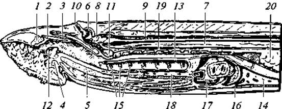
Fig. 23. Sagittal section through the head region of a sea lamprey:
1 — annular cartilage; 2 — anterior dorsal cartilage; 3 — posterior dorsal cartilage; 4 — annular cartilage; 5 — hyoid cartilage; 6 — olfactory capsule; 7 — notochord; 8 — brain; 9 — spinal cord; 10 — nostril; 11 — pituitary pouch; 12 — tooth of the lingual plate; 13 — Esophagus; 14 — Liver; 15 — three anterior gill openings; 16 — atrium; 17 — ventricle; 18 — ventral aorta; 19 — dorsal aorta; 20 — Ovary
The chondrocranium is very primitive, represented by a cartilaginous plate underlying the brain. In the center of this plate lies an aperture through which the pituitary process passes. Cartilaginous otic capsules flank the plate laterally (without fusing with it), while a cartilaginous olfactory capsule is situated anteriorly. Ahead of the neurocranial plate lies a system of cartilages supporting the oral funnel and tongue (Fig. 22).
The visceral skeleton in lampreys is represented by nine pairs of articulated branchial arches interconnected by four longitudinal cartilaginous strands. This cartilaginous lattice Supports the branchial apparatus. Posteriorly, this cartilage system is adjoined by a cartilaginous pericardial pouch. In hagfishes, the branchial basket is rudimentary.
Digestive System. Digestion begins with a wide pre-oral funnel armed with horny teeth. The powerful tongue bears the largest tooth, known as the "rasp". The true Mouth opening lies at the bottom of the funnel and leads into the Pharynx. In lampreys, the pharynx is divided into two parts: a dorsal one for the passage of food, and a ventral one that ends blindly and communicates with the gill pouches (Fig. 23). The Stomach is undeveloped, and the esophagus passes directly into the intestine, from which it is separated by a valve. The intestine lacks divisions and forms no loops. Within it runs a mildly undulating fold called the spiral valve, which increases the intestinal surface area and slows the passage of food, thereby ensuring more thorough digestion. Cyclostomes are characterized by secreting digestive fluids directly into the body of the prey, where chemical breakdown of food begins externally (extraintestinal digestion).
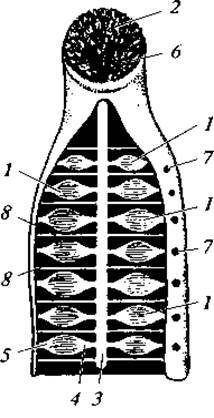
Fig. 24. Mouth and respiratory Organs of a sea lamprey:
1 — gill pouches; 2 — horny teeth; 3 — respiratory tube; 4 — internal and 5 — external gill passages (leading into the gill pouches and outward); 6 — mouth; 7 — external gill openings; 8 — interbranchial septa
A liver is present, developing as an outpocketting of the anterior intestine (compare with the hepatic caecum in amphioxus). The Pancreas is in a rudimentary state.
Respiratory system. Respiration is carried out by gill lamellae; however, the branchial apparatus is unique. Narrow, rounded external gill ducts lead into spacious, lens-shaped gill pouches, the walls of which are covered with numerous folds densely laced with Blood Vessels. On the opposite side of the gill pouches lie internal gill ducts leading into the pharynx (Fig. 24). It is important to emphasize that the gill pouches and their lamellae in cyclostomes are of endodermal origin, whereas in all other gill-breathing modern vertebrates, gill lamellae develop from ectodermal rudiments.
Owing to their parasitic feeding habit of attaching to the host's body, the respiratory Water current enters through the external gill openings into the gill pouches and exits via the same route. In hagfishes, the respiratory mechanism exhibits certain specific features described below. The number of gill pouches in cyclostomes varies from 5 to 15 (numbering 7 in the river lamprey). Circulatory system. The basic structural plan of these organs mirrors that of amphioxi (Fig. 25). However, cyclostomes possess a two-chambered Heart consisting of an atrium and a ventricle. Positioned at the Origin of the ventral aorta, it helps boost blood flow. Adjoining the atrium is a thin-walled sinus venosus into which all major Veins empty. Afferent branchial Arteries depart from the ventral aorta. Efferent branchial arteries, which collect blood from the gills, merge into an unpaired aortic ROOT. From this root, carotid arteries extend anteriorly. Posteriorly, the aortic root continues into the main arterial trunk—the dorsal aorta. The latter runs immediately beneath the axial skeleton (specifically, the notochord). Blood flows from the dorsal aorta to all Organs of the body. Venous blood from the head is collected into paired anterior cardinal (jugular) veins that empty into the sinus venosus, which also receives blood from the posterior cardinal veins draining the trunk. Blood from the intestine is gathered into the subintestinal vein; upon reaching the liver, it forms a hepatic portal system. From the liver, blood flows via the hepatic vein into the sinus venosus. There is no renal portal system. Hagfishes possess three auxiliary hearts within the Venous system: in the head, liver, and tail regions. These enhance Venous Circulation.
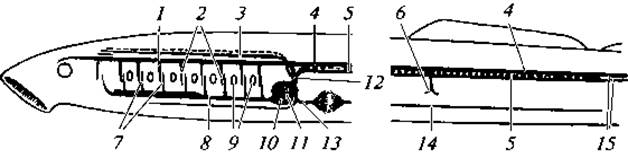
Fig. 25. Diagram of the lamprey circulatory system:
1 — root of the dorsal aorta; 2 — efferent branchial arteries; 3 — anterior cardinal (jugular) vein; 4 — dorsal aorta; 5 — posterior cardinal vein; 6 — intestinal artery; 7 — afferent branchial arteries; 8 — ventral aorta; 9 — gill slits; 10 — ventricle; 11 — atrium; 12 — sinus venosus; 13 — hepatic vein; 14 — subintestinal vein; 15 — caudal vein and artery
Nervous system. The brain comprises the typical vertebrate divisions: Forebrain, Diencephalon, Midbrain, and Hindbrain (Medulla Oblongata), whereas the fifth division, the Cerebellum, is virtually undeveloped. The brain is relatively small in size. All divisions lie in a single plane, i.e., they do not form the flexures typical of more highly organized vertebrates (Fig. 26). The roof of the brain lacks Nervous Tissue and is entirely epithelial.
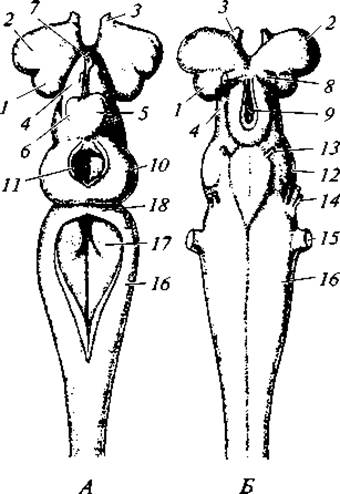
Fig. 26. Brain of a lamprey. Dorsal view (A) and ventral view (B):
1 — cerebral hemispheres; 2 — olfactory lobes; 3 — Olfactory nerve; 4 — diencephalon; 5 and 6 — right and left habenular ganglia; 7 — parietal organ covering the Pineal Gland; 8 — Optic nerve; 9 — infundibulum; 10 — optic lobes; 11 — opening in the roof of the midbrain; 12 — floor of the midbrain; 13 — Oculomotor nerve; 14 — Trigeminal nerve; 15 — Vestibulocochlear nerve; 16 — medulla oblongata; 17 — Rhomboid fossa; 18 — rudimentary cerebellum
There are 10 pairs of Cranial Nerves. Spinal Nerves emerge via two roots—dorsal and ventral—which, unlike those in other vertebrates, do not join to form a common mixed nerve.
Sense Organs. These organs have a very simple Structure. The Organ of Hearing is represented by the inner ear—the membranous labyrinth—along with two (in lampreys) or one (in hagfishes) semicircular canal(s). The eyes are poorly developed. Lampreys have an underdeveloped cornea, whereas in hagfishes the eyes are degenerate due to a parasitic lifestyle. The Olfactory Organ is unpaired. Lateral line organs run along the sides of the body within the skin. They are represented by shallow pits containing the nerve endings of the Vagus nerve (X cranial nerve pair) at their base.
Excretory organs. These are represented by mesonephric Kidneys, bordered at their anterior end by remnants of the pronephros (some researchers believe that a functioning pronephros persists in the genus Bdellostoma among hagfishes). The Ureters are the Wolffian ducts, which empty into the urogenital sinus.
Gonads. The gonads are unpaired and lack specialized ducts. Gametes are released into the body cavity through ruptures in the gonadal wall, from where they enter the urogenital sinus and pass to the exterior through the duct of the urogenital papilla.
Systematics and ecology of cyclostomes
Extant cyclostomes are divided into two well-defined orders—Petromyzontiformes (lampreys) and Myxiniformes (hagfishes). Some taxonomists elevate them to the rank of subclasses.
Last update: 13/08/2026
Editorial and Educational Adaptation: This material has been compiled based on the primary/original source text. The project team performed an editorial review, corrected technical inaccuracies, structured sections, and adapted the content for an educational format.
What was processed:
- elimination of formatting defects (OCR errors, structural breaks, corrupted characters);
- editorial organization of content;
- standardization of terminology in accordance with academic sources;
- verification of factual statements against the original source text.
All mentions of the author, publication year, and origin of the primary text have been preserved in accordance with the source.