General Microbiology - Schlegel H. 1987
Constancy, variation, and transmission of traits
Transmission of traits and genetic recombination
In eukaryotic organisms, haploid sets of genes unite during Fertilization to form a diploid zygote. Following a few or many mitotic divisions in the developing diploid Organism, a reduction division (Meiosis) takes place, recombining the Chromosomes derived from the two parental sets and once again producing Cells with haploid Gene sets (Gametes). This sexual method of genetic reshuffling is contrasted with parasexual processes, which include the recombination of traits in prokaryotes. Bacteria are almost always haploid, possessing only a single gene set. Zygotes do occur in bacteria, but they are never the product of the fusion of entire cells. As a rule, only a portion of the genetic material is transferred from a donor Cell to a recipient cell, resulting in an incomplete zygote (merozygote). The recipient chromosome pairs with the donor chromosome fragment, and they exchange individual segments. Subsequent nuclear and cellular divisions yield a cell containing only the recombined chromosome (Fig. 15.11). Three mechanisms of trait transfer are known in bacteria: conjugation, Transduction, and transformation.
Class="center">
Fig. 15.11. General Homologous Recombination: exchange between a donor DNA fragment (highlighted in red) and the recipient bacterium chromosome. According to one model, homologous double-stranded DNA molecules come into close proximity and exchange segments of one strand, after which Replication or repair processes generate a recombinant chromosome.
As a result of these three processes, DNA is transferred from a donor bacterium to a recipient bacterium. These processes differ in how the DNA is transported. Following DNA transfer, recombination takes place within the recipient cell, during which the donor DNA integrates into the bacterial chromosome of the recipient. A cell in which recombination has occurred is referred to as a recombinant.
15.3.1 Genetic Recombination
Currently, at least three different mechanisms are known for the in vivo recombination of foreign DNA entering a bacterial cell with the bacterial chromosome (or a plasmid): 1) general homologous recombination, 2) Site-Specific Recombination, and 3) illegitimate (non-homologous) recombination.
General homologous recombination. In this case, incoming foreign DNA recombines with cellular DNA via the reciprocal exchange of corresponding segments. Aside from differences caused by Mutations, recombination partners must share the same nucleotide sequence, meaning they must be as homologous as possible. Homologous recombination is controlled by the recA gene; mutants with a defect in this gene (rec-) are incapable of homologous recombination.
Several models exist for this mechanism. It is hypothesized that base pairing occurs between unwound, single-stranded regions of two DNA Double helices. The second strand is likely generated through replication or repair.
Site-specific recombination. This process occurs independently of homologous recombination and is therefore possible even in rec- mutants. It involves the insertion of a short double-stranded DNA molecule into a specific site within a long double helix, causing the smaller partner to lose its autonomy. A typical example of site-specific recombination is the integration of bacteriophage lambda (λ) (Fig. 4.14).
Genetic experiments indicate that upon entering the prophage state, the phage integrates into the host cell chromosome at a specific site—between the gal Operon and the biotin region (Fig. 15.12). Phage insertion is preceded by its attachment to a specific region of the bacterial DNA. It was previously thought that this is determined by a high degree of nucleotide Structure/154.html">Sequence Homology, but this homology turned out to be minimal; instead, a phage-encoded protein known as integrase appears to play a major role here. Within a specific region of the phage DNA (attP) and the corresponding region of the bacterial DNA (attλ), this protein catalyzes the breakage and cross-rejoining of the phage and host cell genomes.

Fig. 15.12. Site-specific recombination, illustrated by the integration of bacteriophage lambda into the host cell chromosome. Utilizing a specific protein, the circular phage DNA attaches via its attP site to the attλ site on the bacterial DNA located between the bio and gal genes; subsequently, through the breakage and cross-rejoining of the DNA double strands, the phage becomes integrated into the chromosome. (See also Fig. 4.14.)
Non-homologous recombination. Recombination processes involving DNA segments that do not exhibit significant genetic homology are referred to as non-homologous recombination. Much like site-specific recombination, it represents an integrative form of recombination—meaning a joining of DNA molecules rather than an exchange. Non-homologous recombination is independent of the recA gene. The following entities are capable of such recombination: 1) insertion sequences (IS elements); 2) Transposons (Tn); 3) bacteriophage μ (mu). The Molecular Mechanism of non-homologous recombination is not yet fully understood.
As established approximately 15 years ago, certain mutations spontaneously arising in Escherichia coli are attributable to the insertion of foreign DNA. Such mutations occur within structural and regulatory genes across the entire chromosome. This foreign DNA consists of so-called insertion sequences (IS elements), which are found in both bacterial chromosomes and Plasmids. IS elements comprise 800–1400 nucleotide pairs; they do not encode recognizable phenotypic traits, and little is known about their Functions. Their mutagenic effect is simply the result of foreign DNA integration disrupting the METABOLISM/31.html">Transcription process (p. 447). It is hypothesized that IS elements play a crucial role in genetic rearrangements.
Transposons are DNA sequences capable of integrating into numerous sites within The Genome and 'jumping' from a plasmid to the bacterial chromosome, to another plasmid, or to a temperate phage. Transposons harbor genes that determine clearly identifiable traits, specifically resistance to Antibiotics such as penicillin, tetracycline, or kanamycin, making them easier to detect than IS elements. Flanking the resistance genes within the transposon are two identical sequences oriented either in the same or in opposite directions. Portions of these repeating DNA base sequences are identical to IS elements. The arrangement of these flanking DNA segments can be determined through electron microscopic analysis of heteroduplexes (Fig. 15.13). Bacteriophage mu resembles IS elements and transposons due to its unusual behavior upon integration into the bacterial chromosome. It exhibits typical phage properties while simultaneously functioning as a giant transposon.
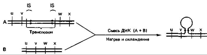
Fig. 15.13. Identification of transposons via Electron Microscopy of heteroduplexes. To visualize the transposon, DNA from wild-type bacteria (B) and transposon-bearing bacteria (A) is heated, causing the double-helix strands to separate ('melting'). Subsequent slow cooling of the mixture allows complementary bases of individual DNA strands from A and B to pair, forming DNA heteroduplexes. If oppositely oriented complementary IS elements are present at the ends of the transposon, these regions also pair to form a stem, with the central portion of the transposon protruding laterally as a single-stranded loop.
15.3.2 Conjugation
The transfer of genetic material via direct contact between two cells is called conjugation. Morphological evidence had long suggested that a form of mating might occur in bacteria; however, it was only through experiments with multiple mutants that the transmission of genetic material via direct intercellular contact was conclusively proven. In 1946, Lederberg and Tatum conducted a landmark experiment using two E. coli K12 mutants, each auxotrophic for two different Amino Acids (Fig. 15.14). One double mutant required amino acids A and B but was capable of synthesizing C and D (A- B- C+ D+); the other mutant was complementary to it (A+ B+ C-D-). These mutants failed to grow on a minimal nutrient medium and did not form colonies. However, when a mixed suspension of both mutants was plated onto the same minimal medium, colonies appeared. Cells from these colonies possessed the hereditary ability to synthesize all amino acids, meaning they belonged to the A+ B+ C+ D+ type (prototrophic). Such cells arose at a frequency of 1:106; they were genetic recombinants that combined the Genetic information of two reciprocally defective (mutually complementary) parental cells. The Use of multiple mutants as initial strains ruled out the possibility of revertants, as the probability of simultaneous reversion at two genes is on the order of 10-14–10-16 per generation. Direct contact between parental cells served as a necessary prerequisite for recombination.
Directed gene transfer from Cell to Cell. Crossing experiments in which one of the parental strains was streptomycin-resistant led to the Conclusion that genetic material is transferred in only one direction. When cells were plated onto streptomycin-containing medium after crossing, recombinants emerged exclusively when one of the strains (the recipient strain) was streptomycin-resistant and survived. The behavior of the other parental strain was inconsequential; it could be streptomycin-sensitive and perish on that medium—it was sufficient that it had time to perform its function as the donor of genetic material. From this, it could be deduced that genetic material transfer is unidirectional—flowing from the donor ('male' strain) to the recipient ('female' strain)—and that the entire recombination and segregation process takes place within the recipient strain cells. Recombinants inherit the majority of their traits from the recipient, receiving only genomic fragments from the donor.
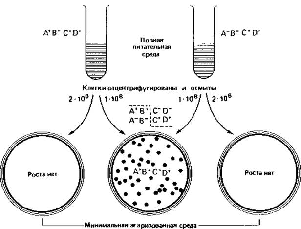
Fig. 15.14. Recombination via conjugation of two Escherichia coli K12 mutants with different pairs of biochemical defects.
The F factor and the Hfr state. Investigations into bacterial mating revealed that a cell's ability to act as a donor is linked to a specific factor transmitted from one cell to another during conjugation—the sex factor F (derived from fertility). Cells lacking the F factor (F- cells) can function solely as recipients. During conjugation, i.e., direct cell-to-cell contact, the frequency of F factor transfer approaches 100%. Consequently, recipient cells are converted into potential Donors as a result of conjugation, although chromosomal traits are not yet transferred.
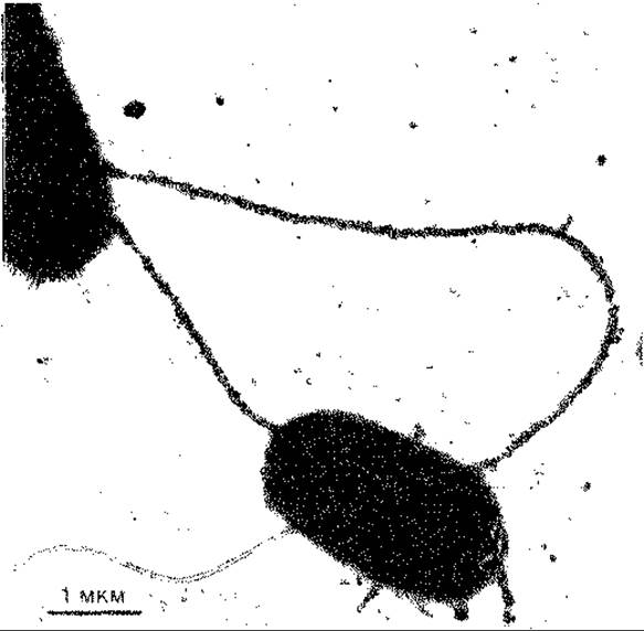
Fig. 15.15. Escherichia coli cells connected by F-pili. The F-pili of the Hfr cell are labeled with donor-specific RNA phages MS-2. The numerous type I pili of the recipient cell (top left) are short and do not adsorb Bacteriophages. Electron micrographs obtained after negative staining with phosphotungstic acid. (Curtiss R. et al., J. Bacteriol., 100 [1969], 1091.)
The F factor is a circular, double-stranded DNA molecule with a Molecular Weight of 45×106 Da. As an extrachromosomal, autonomously replicating DNA element, it is classified as a plasmid. This molecule carries the genes responsible for conjugation, including those that determine specific cell surface structures such as sex pili, or F pili (Fig. 15.15), which are essential for the process. Presumably, they facilitate mutual recognition upon contact between a donor and a recipient cell, enabling The formation of a conjugation bridge through which DNA is transferred into the recipient cell. It remains unclear whether this DNA "injection" occurs directly through the F pili themselves.
In an F+ population, only a small fraction of cells are capable of acting as donors of chromosomal DNA. These have been shown to be the cells in which the F factor has integrated into the bacterial chromosome (Fig. 15.16). When clones of such donor cells are used in mating experiments, recombinants are formed approximately a thousand times more frequently than with conventional F+ cells. These donor cells, which ensure a high frequency of recombination, are designated as Hfr cells (short for high frequency of recombinants). The F factor inserts into the bacterial chromosome only at specific, limited sites—a process comparable to the integration of phage λ (lambda) into a host cell chromosome (see Figs. 4.14, 15.12, and 15.16).
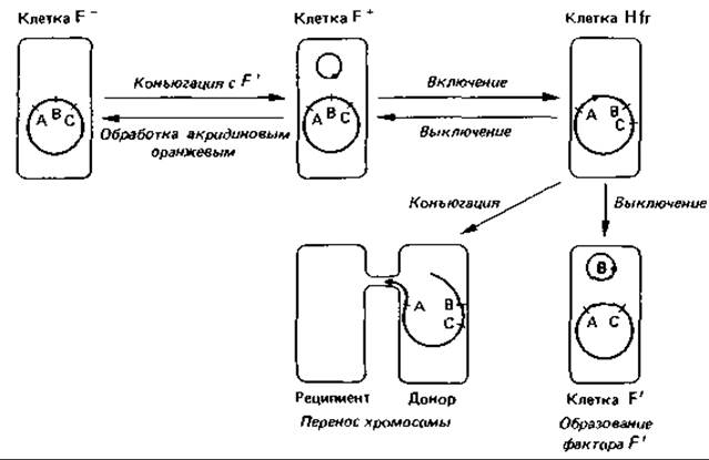
Fig. 15.16. Relationships between mating types in Escherichia coli. An F- cell can function exclusively as a recipient. Upon conjugation with an F+ or Hfr strain, it may acquire the F factor and thereby convert into an F+ cell. In an F+ cell, the F factor exists as an autonomous circular DNA molecule, which can be eliminated by Treatment with acridine orange. The Integration of the F factor into the bacterial chromosome transitions The Cell into the Hfr state. Because the factor can integrate at various sites and in different orientations, the starting point and direction of chromosomal transfer (indicated by red arrows) vary accordingly. In the event of aberrant excision of the F factor from the chromosome, it may give rise to an F' factor carrying a segment of chromosomal DNA.
The transfer process. When an Hfr cell population is mixed with an excess of F- cells, nearly every Hfr cell finds an F- partner and undergoes conjugation. Samples are withdrawn from such mixtures at regular intervals, vigorously agitated in a blender to forcibly disrupt mating pairs, and then plated on Agar to select for recombinants. Subsequent Analysis of the recombinant strains reveals which genes were transferred from the donor to the recipient cells. Studies have demonstrated that each gene is transferred at a precise, characteristic time point following the initiation of conjugation (Fig. 15.16). The temporal sequence of gene transfer corresponds to their linear order on the bacterial chromosome as determined by genetic mapping. This indicates that any given Hfr strain represents a homogeneous population whose cells all transfer their chromosome to the recipient in an identical fashion—starting from a specific locus (the origin) and proceeding in a fixed direction. The farther a gene is located from the chromosomal "origin," the later it is transferred and the lower the probability of its entering the recipient cell, even if conjugation is not artificially interrupted. The transfer of the entire E. coli chromosome takes approximately 100 min at 37°C. Experiments based on the "interrupted mating" technique have made it possible to construct genetic maps.
Different Hfr strains, isolated independently from the same F+ strain, differ in two primary aspects: the specific point on the chromosome that serves as the "origin" and the characteristic sequence of gene transfer. These experimental findings align with the concept that the F factor, during integration (i.e., transition to the Hfr state), can insert into the bacterial chromosome at any of roughly 20 potential gene loci. During transfer, the bacterial DNA replicates starting from the site of F factor insertion, and the newly synthesized strand, 5' end first, is pushed into the recipient cell. This transfer process is followed by homologous recombination within the recipient cell between the donor DNA and its own resident DNA. The relationships among F-, F+, and Hfr cells are illustrated in Fig. 15.16.
Genetic mapping. By applying the interrupted mating technique described above—which determines the temporal sequence of gene transfer from the donor cell—researchers can map the relative positions of genes on the bacterial chromosome (Fig. 15.17). The rate of transfer remains constant throughout the process, and the times of entry into the recipient cell reflect the physical distances between genes on the chromosome. While this method lacks the resolution to distinguish intervals of less than one minute, finer-scale mapping can be achieved through linkage analysis via transduction (gene transfer mediated by bacteriophages).
For Escherichia coli K12, the locations of over a thousand genes—predominantly structural genes encoding Enzymes—are currently known. The order of genes on the bacterial chromosome has also been established for Salmonella typhimurium, Streptomyces coelicolor, Bacillus subtilis, and several other bacteria.
Gene transfer mediated by F' factors. The integration of the F factor into the bacterial chromosome is a reversible process. The F factor can be excised from the chromosome, converting the Hfr cell back into an F+ cell (Fig. 15.16). This excision process occurs at roughly the same frequency as integration. Precise excision involves a break at the exact site of prior insertion; however, in rare instances, the break occurs nearby, leaving an adjacent stretch of chromosomal DNA attached to the F factor. This hybrid element containing a small chromosomal fragment is known as an F' factor. The generation of an F' factor is analogous to the formation of specialized transducing phages (Section 15.3.3).

Fig. 15.17. Genetic Map of the Escherichia coli chromosome. Numbers denote gene positions, indicating the time (in minutes after the initiation of conjugation in nutrient broth at 37°C) at which specific genes are transferred to the recipient cell. Red arrows inside the circle show the direction of gene transfer during conjugation with various Hfr strains (the direction of chromosomal movement is opposite to the arrowhead). Red arrows outside the circle indicate the direction of transcription for individual genes within specific operons (e.g., P, O, Z, Y, A in the lac operon). Gene designations: azi, azide resistance; bio, biotin requirement; gal, galactose utilization; his, Histidine requirement and genes for its biosynthetic enzymes; ilv, isoleucine and valine requirement; lac, lactose operon with genes: P, promoter; O, operator; Z, β-galactosidase; Y, galactoside permease; A, thiogalactoside transacetylase; proA, Proline requirement (block prior to glutamate semialdehyde); recA, capacity for genetic recombination and DNA Repair; thr, Threonine requirement; trp, Tryptophan requirement. (Bachmann B. J., Bacteriol. Rev., 40 [1976], 116.)
A cell carrying an F' factor is termed a primary F' cell. The DNA incorporated into the F' factor can now be transferred by F' donor cells to F- strains with the same high frequency (100%) as the standard transfer of the F factor from F+ to F- recipients. By contrast, the same chromosomal fragment would be transferred by an Hfr strain to an F- strain at a maximum frequency of only 1%. When an F' factor is transferred from its original primary parental cell to a normal F- cell, it creates a secondary F' cell, in which a small segment of the bacterial chromosome is duplicated (i.e., present in a diploid state).
Prevalence of conjugation among other bacterial groups. Gene transfer via conjugation, originally discovered in Escherichia coli, is widespread among enterobacteria.
By transferring F factors from E. coli K12 into Salmonella and Shigella cells, novel genetic systems have been successfully constructed, and analogous systems have been identified within the Pseudomonas group. Conjugation in enterobacteria is a highly evolved process; successful transfer requires merely suspending a mixture of partner cells in liquid medium and letting it stand undisturbed for a short period. In contrast, for many other bacteria, conjugation can only be induced by thoroughly mixing and spreading the colonies of both partners onto a solid medium, where they are allowed to grow for several days. When these cells are subsequently isolated on selective media, many exhibit novel combinations of traits, strongly suggesting that large segments of their bacterial chromosomes have been exchanged. Conjugational processes have been extensively studied in Streptomyces coelicolor, Nocardia species, Rhizobium, and other bacteria. Gene exchange via conjugation and plasmid-mediated mobilization are likely ubiquitous phenomena throughout the prokaryotic world.
Plasmids
Many (if not all) bacteria harbor extrachromosomal DNA elements. These closed, double-stranded circular DNA molecules, which are small compared to the bacterial chromosome, are called plasmids. Under standard growth conditions, bacteria can dispense with them entirely: cells "cured" of plasmids by UV irradiation, mitomycin C, or acridine Dyes grow vigorously on standard nutrient media. Plasmids are typically identified by specific phenotypic traits they confer upon the host cell. Certain plasmids equip the cell to conjugate with other cells, thereby ensuring their own dissemination through direct intercellular contact. We have already encountered the prototype of such a plasmid (p. 457) when examining the F factors of Escherichia coli.
Fertility factors (F factors). As noted previously, these are plasmids capable of integrating into the bacterial chromosome much like the DNA of temperate phage lambda. They mobilize the host's genetic information and mediate its transfer to another cell—a conjugational process thoroughly characterized in E. coli.
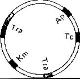
Fig. 15.18. Genetic map of the antibiotic-resistance plasmid RP4. This plasmid features two regions containing large and small tra fragments, which are responsible for conjugation. Genes conferring resistance to the antibiotics ampicillin (Ap), tetracycline (Tc), and kanamycin (Km) are distributed across different segments of the plasmid.
Resistance factors (R factors). Bacteria resistant to specific antibiotics were first discovered in Japan during the 1950s, isolated from dysentery patients (Shigella strains) undergoing antibiotic therapy. A notable feature of these strains was their multi-drug resistance, which could be readily transmitted to other bacteria such as Escherichia coli. It is now well established that resistance (R) factors harbor genes conferring resistance to agents such as sulfonamides, streptomycin, chloramphenicol, and tetracycline. While some R factors confer resistance to as many as eight antibiotics simultaneously, others provide tolerance to toxic heavy metals such as mercury, nickel, cadmium, or cobalt. An R plasmid carries two major Functional groups of genes: (1) genes responsible for plasmid transfer via conjugation (tra genes), which constitute the so-called resistance transfer factor (RTF); and (2) genes responsible for the actual resistance phenotype, which comprise only a minor fraction of the plasmid (Fig. 15.18).
The resistance transfer factor (RTF) comprises all genes required for the intercellular transmission of the R factor, a process typically achieved through conjugation. Thus, R factors, much like F factors, are infectious in a broad biological sense. The molecular architecture of the RTF region is homologous to the corresponding region of the E. coli F factor. Certain R factors exhibit a broad host range, allowing them to transfer across several distinct bacterial genera and thereby accelerating their dissemination. In some instances, R factors have been observed to cotransfer chromosomal genes that they had apparently mobilized.
The Biochemical Mechanisms of Antibiotic Resistance conferred by R factors can differ fundamentally from those of chromosomally mediated resistance. A clear illustration is resistance to streptomycin. When chromosome-mediated, streptomycin resistance stems from an alteration in the 30S ribosomal subunit, eliminating the drug's target site (Section 2.2.2). Conversely, R-factor-mediated resistance relies on the enzymatic inactivation of the antibiotic via adenylation. Enzymatic chemical modification is a frequent basis for plasmid-mediated antibiotic resistance; for example, chloramphenicol is inactivated by Acetylation, kanamycin and neomycin undergo phosphorylation and acetylation, and penicillin is cleaved by penicillinase. Because genetic recombination can also occur among R factors, novel gene combinations may arise that confer expanded resistance profiles. R factors pose a major challenge in Chemotherapy; their existence underscores the hazards of indiscriminate antibiotic use, as they can spread rapidly through bacterial populations like infectious pathogens.
Bacteriocins. Many bacteria synthesize Proteins that kill or inhibit the growth of closely related species or strains. These highly specific agents, known as bacteriocins, are encoded by specialized plasmids called bacteriocinogenic factors. Bacteriocins have been isolated from Escherichia coli (colicins), Pseudomonas aeruginosa (pyocins), Bacillus megaterium (megacins), and numerous other bacteria.
Other plasmid-determined traits. Plasmids can also carry genes encoding specialized biochemical capabilities that confer selective advantages under specific environmental conditions. Genes for enzymes required to degrade camphor, salicylic acid, naphthalene, octane, 2,4-dichlorophenoxyacetic acid, and various other unusual substrates are frequently plasmid-borne. We have already mentioned the tumor-inducing plasmid of Agrobacterium tumefaciens and its metabolic activities in plant cells (Section 4.3). THE SPECTRUM OF plasmid-encoded traits is extensive, encompassing Nitrogen Fixation, Nodulation, synthesis of indoleacetic acid, diacetyl, Hydrogenase, and sugar uptake. Because some of these traits are also occasionally determined by chromosomal genes, genetic exchange between the chromosome and plasmids clearly occurs with some frequency. Plasmids have likely played a pivotal role in the evolution of prokaryotes.
Incompatibility. Many bacteria harbor multiple plasmids of varying sizes. The stable coexistence of different plasmids within a single bacterial cell indicates that they are compatible. Conversely, two closely related plasmids cannot coexist stably in the same cell—they are incompatible. All plasmids are categorized into incompatibility groups, such that plasmids belonging to the same group are mutually incompatible.
15.3.3 Transduction
Transduction is the process by which DNA is transferred from a donor cell to a recipient cell via bacteriophages. Typically, the phage carries only a small fragment of the host DNA. There are two MAIN TYPES OF transduction: non-specific (generalized), in which potentially any fragment of the host DNA can be transferred, and specialized, which affects only strictly defined DNA segments. In generalized transduction, host cell DNA is incorporated into the phage particle either In addition to its own genome or in place of it, whereas in specialized transduction, certain phage genes are replaced by host genes. In both cases, transducing phages are generally defective—for example, they frequently lose The ability to lyse the host cell. Trait transfer via transduction has been observed in numerous bacteria, including species of Salmonella, Escherichia, Shigella, Bacillus, Pseudomonas, Staphylococcus, Vibrio, and Rhizobium. However, not all phages are capable of mediation, and DNA cannot be transferred in this manner into all bacteria.
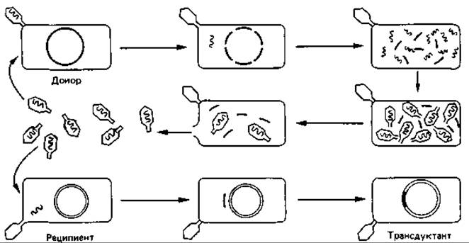
Fig. 15.19. Generalized transduction: one of the mechanisms of DNA transfer from one bacterial cell to another.
Generalized Transduction. The transfer of bacterial chromosome segments by phages was discovered in 1951 by Lederberg and Zinder in Salmonella typhimurium. In the definitive experiment (Fig. 15.19), the donor strain B+ was infected with temperate bacteriophage P22. Following host cell lysis, free phages were isolated and incubated with the recipient strain B-, which was genetically distinct from strain B+ by at least one marker. The authors found that plating the incubated cells on a suitable medium yielded recombinants possessing the traits of the donor strain B+.
The processes underlying this non-specific DNA transfer are quite complex. During the reproduction of phage P22 within Cells of the donor strain B+, bacterial chromosomal fragments may be packaged into capsids instead of phage DNA. Consequently, the phage lysate contains a mixture of normal and defective phages. Infection of the recipient strain B- with a normal phage typically leads to cell lysis. However, some cells are penetrated by defective transducing phages whose DNA can recombine with the recipient chromosome. An exchange of homologous DNA segments occurs, which may result in the replacement of a defective recipient gene with an intact donor gene.
Because only small DNA fragments are transduced, the probability of recombination affecting a specific trait is very low, ranging from 10-6 to 10-8. It follows that a single particle of Salmonella phage P22 or a generalized transducing phage PI of Escherichia coli can transduce only a single gene (or a few very closely linked genes) in any given event. The amount of bacterial DNA comparable to the phage genome accounts for only 1–2% of the total DNA contained within the bacterial cell. An exception is bacteriophage PBS 1 of Bacillus subtilis, which can transduce up to 8% of the host genome.
Specialized Transduction. The best-known example is transduction mediated by phage λ (see Section 4.2.2). Typically, it transduces only specific genes, namely gal and bio. As previously discussed, upon transitioning to the prophage state, this phage integrates into a specific site on the host bacterial chromosome—between the gal and bio genes. The excision of phage DNA from the bacterial chromosome (e.g., as a result of UV irradiation) can occasionally be imprecise, meaning that a fragment of it remains in the chromosome while adjacent host cell genes are packaged into the phage DNA. This is presumably caused by Illegitimate Recombination.
When cells carrying a defect in a specific gene, such as gal-, are infected with a specialized transducing phage, recombination may occur, replacing the bacterium's own defective gene with the intact transduced gene, thereby generating gal+ recombinants (transductants).
Gene transfer by bacteriophage Phi 80 proceeds in a similar manner. Its DNA integrates into the chromosome near the genes encoding enzymes responsible for tryptophan Biosynthesis. For this reason, Phi 80 is particularly well-suited for the transfer of trp genes.
A prerequisite for successful gene transfer in specialized transduction (unlike generalized transduction) is the integration of the phage into the host cell genome.
In some instances, it has been demonstrated that the transduced DNA fragment does not undergo recombination with the recipient chromosome, but instead remains extrachromosomal. Under these conditions, the cell becomes heterozygous for the transferred genes. The transferred DNA is transcribed (as evidenced by the Synthesis of the corresponding gene product) but is not replicated. Consequently, upon Cell Division, the donor fragment is passed on to only one of the daughter cells (abortive transduction). If the recipient is auxotrophic and the transferred fragment corrects the corresponding defect, only those cells that have inherited the fragment can grow, forming microscopic colonies when plated on agar.
15.3.4 Transformation
Genes can also be transferred from cell to cell without any intercellular contact or vectors. This mode of gene transfer, mediated by free soluble DNA isolated from donor cells, is termed transformation. Among bacteria, this mechanism of trait transfer was discovered earliest, and this breakthrough played a pivotal role in The history of science.
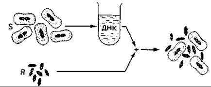
Fig. 15.20. Transformation in Streptococcus pneumoniae (schematic diagram). The capacity for capsule formation is transferred to a non-encapsulated strain (R) using intact DNA extracted from an encapsulated strain (S). (Nultsch W., Allgemeine Botanik, 3rd ed., Stuttgart: Thieme, 1968.)
Discovery of the Role of DNA as Genetic Material. In 1928, Griffith described The conversion of a non-encapsulated R-strain of Streptococcus pneumoniae (pneumococcus) into a capsule-forming strain, i.e., the S-form (Fig. 15.20). Griffith injected mice with a small number of avirulent R-cells combined with heat-killed S-cells. The R-cells were derived from an S-strain (SII) whose capsular substance belonged to a different serological type than that of the heat-killed S-strain (SIII). Virulent encapsulated bacteria of type SIII were subsequently isolated from the Blood of the deceased mice. This demonstrated that the heat-killed type SIII cells transferred the hereditary capacity for type III capsule synthesis to the R-cells, which in turn passed it on to their progeny. The Nature of the “transforming principle” was successfully identified by Avery, MacLeod, and McCarty in 1944. They proved that this factor was indeed DNA. This discovery provided decisive evidence that genetic information resides in DNA rather than protein.
Competence. Subsequently, gene transfer using purified DNA was demonstrated in Haemophilus influenzae, Neisseria, Rhizobium, Bacillus subtilis, Acinetobacter calcoaceticus, Escherichia coli, Pseudomonas, and many other bacteria. Traits such as resistance to various poisons and prototrophy for specific Amino acids can be transmitted via this pathway. Transformation is restricted to bacteria whose cells can take up high-molecular-weight, double-stranded, i.e., intact, DNA. Extremely low DNA concentrations are required for transformation: 0.1 µg per 1 mL of recipient cell suspension is sufficient to transform competent cells (which account for up to 15% of the cell population at maximum). Competence refers to the physiological capability of cells to take up DNA. Competence is dependent on the physiological state of the cell, peaking in the middle of the exponential growth phase and rapidly declining to a minimum thereafter. It is hypothesized that the cell surface undergoes modifications throughout the growth cycle and that DNA uptake can occur only during a specific, relatively brief phase. Bacteria previously considered non-competent (such as E. coli) have been successfully rendered competent by modifying their cell surface, for instance, through calcium chloride treatment. It is anticipated that trait transfer via transformation will continue to grow in significance.
Although competent cells can take up virtually any DNA, recombination occurs exclusively if the DNA originates from a closely related species, as this allows for homologous pairing and exchange between the resident and incoming foreign DNA.
15.3.5 Restriction and modification
Bacteriophages typically exhibit host Specificity: they infect only a single bacterial strain or a limited range of related strains, species, or genera. This specificity is primarily governed by the receptor Properties of the bacterial cell surface (Section 4.2.1).
In addition, bacteria possess other systems that determine the specificity of their interactions with phages. One such system is known as restriction. Its mechanism can be illustrated by the following example: if bacteriophage A is propagated on strain A of Escherichia coli and the resulting lysate is used to infect another strain B, the phage will grow significantly more poorly in cultures of strain B than on strain A. If the few phage particles produced on strain B are subsequently used to infect another culture of strain B, phage reproduction will proceed normally once again; however, if these phages are first passaged back through the original strain A, their growth on strain B will once again be severely impaired. Thus, restriction (the limitation of phage replication) depends on the host in which the phage was previously propagated. It is mediated by the Enzymatic Cleavage of the invading phage DNA by host-strain-specific enzymes. Such enzymes are termed Restriction Endonucleases. Through their nucleolytic action, they prevent foreign DNA from establishing itself within the bacterial cell.
Obviously, a bacterial cell must protect its own DNA from the action of its restriction endonucleases. This protection is achieved by the methylation or glucosylation of specific bases in the DNA, typically adenine or cytosine. This process is known as modification. Phages replicating within cells of a given bacterial strain also exploit this mechanism. During its synthesis within this cellular environment, the phage DNA receives the same “imprint” as the host's own DNA: in the presence of the modifying enzyme, the phage DNA is modified in the exact same manner as the host DNA. It becomes similarly methylated and acquires properties that shield it from the restriction enzymes of that bacterial strain.
Restriction Endonucleases (Restrictases). These enzymes may be encoded not only by the bacterial genome, but also by bacteriophages and plasmids. All of them cleave double-stranded DNA. Restriction Enzymes are classified into several groups. Class I enzymes recognize a specific nucleotide sequence but cleave DNA non-specifically outside of this recognition site. This group includes the restriction endonuclease from bacteriophage P1. Class II enzymes are distinguished by their ability to break DNA strands strictly at defined positions within the recognition sequence. This results in the generation of highly specific, discrete DNA fragments. Class II restriction endonucleases are widely used in molecular cloning, which will be discussed in the following section.
A typical example of a class II restrictase that cleaves DNA specifically is the enzyme EcoRI, the action of which is described below. The sequence recognized by this enzyme consists of six Base Pairs forming a palindrome (the sequence is identical in both strands when read in opposite directions).
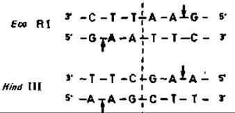
The DNA cleavage sites indicated by the arrows lie outside the axis of Symmetry. As a result of this stagger in the cuts, single-stranded overhangs consisting of four base pairs are formed.
The number of known and characterized restriction enzymes is steadily increasing, as these enzymes play a crucial role in both fundamental and applied research in Molecular Genetics. For example, the DNA fragments generated by their action (restriction fragments) can be ordered to construct a physical map of the genetic material.
15.3.6 Molecular Cloning Method
The exchange of chromosomal DNA segments in bacteria discussed in the previous sections was largely restricted to the limits of a single species. However, as we have already mentioned, the transfer of extrachromosomal DNA molecules capable of autonomous replication can overcome this barrier.
In molecular cloning (Fig. 15.21), plasmids are used as carriers (vectors) to introduce foreign DNA—which may even be of eukaryotic origin—into a bacterial cell and replicate it therein. For this purpose, the plasmid and the corresponding foreign DNA are treated with a specific restriction enzyme, such as EcoRI. As a result, linear DNA fragments with single-stranded ends consisting of AATT or TTAA sequences are produced from both DNA preparations. If the fragments of both DNAs obtained in this way are mixed, complementary single-stranded ends will join through base pairing. The nicks in the sugar-phosphate backbone are covalently sealed by adding polynucleotide ligase. This step completes the Construction of a recombinant DNA molecule—the so-called hybrid plasmid (chimeric DNA).

Fig. 15.21. Production of hybrid DNA by inserting a eukaryotic DNA fragment into a bacterial plasmid (simplified diagram). Foreign DNA and plasmid DNA are cleaved *in vitro* using the same restriction endonuclease. This yields fragments with "sticky" ends (single-stranded terminal regions with complementary bases). Mixing such fragments and treating them with ligase produces plasmids with incorporated eukaryotic DNA. These hybrid DNAs can be introduced into suitable bacteria and propagated to yield mass cultures of transformed cells. The foreign DNA can then be isolated from such a clone.
For the genetic information of the hybrid plasmid to be expressed, the protein-synthesizing machinery of the bacterial cell is required. Therefore, the plasmid is introduced into the bacterial cell via transformation (see above). If the hybrid plasmid is present in the cell in a high copy number, the foreign DNA will be replicated multiple times along with the plasmid. The progeny of a cell containing the hybrid DNA is genetically homogeneous, forming a clone.
The cloning method has found numerous new Applications. Microbes can be used to produce large quantities of foreign DNA for research purposes. If The genes of such DNA are expressed within the bacterial cell, it enables the microbiological production of substances such as Hormones and enzymes. While this novel technique holds great benefit for humanity, the construction of novel DNA molecules involves inherent risks if handled improperly; therefore, the Practical Application of the described method requires strict adherence to A number of safety precautions.
15.3.7 Protoplast Fusion
Somatic cell Hybridization was developed some 15 years ago to conduct genetic research on cell cultures. Building upon this, a technique of genetic recombination via artificially induced protoplast fusion was established and has already been successfully applied to Fungi and plants. The primary product of such fusion is a cell that combines the genomes of both parental cells.
Only recently has this method of genetic recombination been tested on bacteria as well: protoplasts were obtained and their fusion was then induced by treatment with polyethylene glycol. Under specific experimental conditions, morphologically intact cells were regenerated from the fused protoplasts, yielding stable recombinants that exhibited traits from both parental strains. So far, this method has been successfully applied only to Gram-positive bacteria, such as *Bacillus* and *Streptomyces*. It is expected to find broader application in the future.
Unlike the DNA transfer mechanisms described earlier—namely conjugation, transduction, and transformation, in which DNA is transferred unidirectionally from a donor to a recipient—genetic information transfer via protoplast fusion is not unidirectional.
Last update: 13/08/2026
Editorial and Educational Adaptation: This material has been compiled based on the primary/original source text. The project team performed an editorial review, corrected technical inaccuracies, structured sections, and adapted the content for an educational format.
What was processed:
- elimination of formatting defects (OCR errors, structural breaks, corrupted characters);
- editorial organization of content;
- standardization of terminology in accordance with academic sources;
- verification of factual statements against the original source text.
All mentions of the author, publication year, and origin of the primary text have been preserved in accordance with the source.