BOTANY VOLUME 1 - CELL BIOLOGY. ANATOMY. MORPHOLOGY - 2007
1. MOLECULAR BASICS — THE BUILDING BLOCKS OF CELLS
1.2. Nucleic Acids
Nucleic Acids are heteropolymer molecules that serve either for information storage (deoxyribonucleic acid, DNA) or for information transfer and realization (Ribonucleic Acids, RNA). In addition, certain RNAs perform a structural function in ribosome assembly (ribosomal RNA, rRNA). In all cellular organisms—both PROKARYOTES AND EUKARYOTES—double-stranded DNA serves to store hereditary information and transmit it across generations through template-directed Replication. DNA molecules are distinguished by their ability to serve as a template for The formation of new DNA molecules with the exact same base sequence (replication; see 1.2.3). In all organisms except Introduction/7.html">RNA-containing Viruses and Viroids (see 1.2.5), only DNA possesses this replicative duplication function. Since reproduction, perpetuation of the species, and inheritance are the fundamental criteria of life, METABOLISM/36.html">DNA replication lies at the core of all vital processes. Furthermore, DNA molecules can serve as templates for RNA sequences, and through RNA, also convey information regarding the Amino Acid Sequence in Proteins. Through this genetic function of DNA, hereditary information can be expressed: hereditary factors (genes; from Greek genos—birth, race, descent) are manifested as a specific phenotype (the set of outwardly expressed traits of organisms; from Greek phainein—to make visible).
1.2.1. Structure of Nucleic Acids
Nucleic acids are polynucleotides, unbranched polymers composed of monomeric units called NUCLEOTIDES. A single nucleotide consists of a base linked by an N-glycosidic bond to a sugar—forming a nucleoside—along with one to three phosphoric acid (phosphate) residues, thus distinguishing nucleoside mono-, di-, and triphosphates (Fig. 1.3).
Class="center">Fig. 1.3. Nucleotides consist of three structural elements: a pyrimidine or purine nitrogenous base, a sugar (pentose), and phosphoric acid.
The nitrogenous base is linked to the sugar via an N-β-glycosidic bond of pyrimidine or an N9 bond of purine. Phosphoric acid forms an ester with the primary hydroxyl group of the pentose. Up to two additional phosphate residues can be attached to the α-phosphate group via anhydride bonds. A glycoside composed of a nitrogenous base and ribose is called a nucleoside, while one composed of a base and 2-deoxyribose is called a deoxynucleoside (d-nucleoside). Thus, nucleotides are nucleoside mono-, di-, or triphosphates, as illustrated below using adenosine and its nucleotides as an example. Ribose occurs as the sugar in ribonucleic acid, whereas 2'-deoxyribose occurs in deoxyribonucleic acid (DNA). The carbon atoms of the sugar are designated 1', 2'... 5'; C1 forms the glycosidic bond. To simplify complex structural formulas, such as in rings, hydrogen atoms attached to carbon atoms are conventionally omitted (compare the lower formula with those shown above). This convention is frequently used in other illustrations throughout the book for clarity.

The bases found in DNA are Purines—adenine (A) and guanine (G)—as well as Pyrimidines—cytosine (C) and thymine (T). In RNA, thymine is replaced by uracil (U). The term "base" (or nitrogenous base) refers to the alkaline nature of these heterocyclic, nitrogen-containing Aromatic Compounds. The linkage to the sugar is established via N1 of the pyrimidine or N9 of the purine. The sugar moieties are pentoses—ribose in RNA and 2-deoxyribose in DNA, both occurring in the β-D-furanose form (for sugar nomenclature, see 1.4.1). Nucleosides are designated as adenosine, guanosine, uridine, and cytidine when referring to RNA, and deoxyadenosine, deoxyguanosine, thymidine (deoxythymidine), and deoxycytidine when the nucleic acid contains 2-deoxyribose (DNA). Through the formation of an ester bond between the primary hydroxyl group at the C5 atom of the pentose and phosphoric acid, a nucleoside monophosphate is produced. One or two additional phosphoric acid molecules can attach to this α-phosphate group, forming an energy-rich anhydride—a nucleoside diphosphate or nucleoside triphosphate. Nucleoside triphosphates are utilized in the initial stage of DNA or RNA Biosynthesis. In addition, these high-energy group-transfer compounds perform numerous other metabolic Functions. For instance, adenosine triphosphate (ATP) serves as the primary energy donor for many enzymatic reactions (see 6.1).
Through the α-phosphate residue, a nucleotide can be covalently linked to the pentose of a second nucleotide with the elimination of Water, initially yielding a dinucleotide. From this, oligo- and ultimately polynucleotides can be formed. In doing so, nucleic acids form phosphodiester bridges between the 5' and 3' carbon atoms of adjacent pentoses (to distinguish between the atoms of nitrogenous bases and sugars, the carbon atoms of the sugar in nucleosides are denoted by primed numbers). As shown in Fig. 1.4, the type of nucleic acid is determined by the sugar-phosphate backbone of the molecule, which consists of ribose (or deoxyribose) monomers linked by 5',3'-phosphodiester bridges. One end of the molecule features a free 5'-OH group (the 5' end of the nucleic acid), while the other end features a free 3'-OH group (the 3' end). The nitrogenous bases are attached to this sugar-phosphate backbone via glycosidic bonds.
Fig. 1.4. Short sequences of DNA and RNA molecules.
Thymine does not occur in RNA and is replaced therein by uracil. The direction of synthesis and reading of formulas proceeds from left to right, from the 5' to the 3' end of the molecule.

This Primary Structure of nucleic acids is defined by the characteristic linear sequence of nitrogenous bases, which is always read in the 5' → 3' direction, corresponding to the direction of synthesis. The base sequence (the triplet code; see 7.3.1) carries the information for Cell/13.html">Protein Structure. The size of a nucleic acid molecule is conventionally measured by the number of Base Pairs for DNA (bp) or the number of nucleotides for RNA (nt).
1.2.2. Structure of Deoxyribonucleic Acid (DNA)
In only a few phages and viruses does DNA occur as a single-stranded molecule (ssDNA). In many Viruses and Phages, as well as in all cellular Cells, DNA is represented as a double strand consisting of two helically twisted, antiparallel molecules (dsDNA). This structure is conventionally referred to as the DNA double helix. The sugar-phosphate backbones are directed outward, whereas the flat heterocyclic base rings are arranged nearly perpendicular to the longitudinal axis of The Double Helix on its interior (Fig. 1.5). The opposing bases of both strands lie at the same level and form Hydrogen Bonds in the region of the helical axis (see 1.1). This, of course, requires a spatial (steric) complementarity between the adjacent sections of the heterocycles (Fig. 1.6). A purine base (A or G) always pairs opposite a pyrimidine base (T or C), and only the A-T and G-C base pairs are complementary.
Fig. 1.5. The Watson–Crick model of the DNA double helix (B-form): A—diagram; B—space-filling model.

Fig. 1.6. Specific pairing of nitrogenous bases through hydrogen bonding between the two DNA strands.
The molecular STRUCTURE OF THE bases permits the pairing of exclusively A with T and G with C. Two Hydrogen bonds are formed in the A-T pair, whereas three are formed in the G-C pair.

Accordingly, the base sequences in the two strands of the DNA double helix are also complementary, which firmly holds them opposite each other. Thus, the base sequence 5'-GATTACA-3' in one strand must correspond to 3'-CTAATGT-5' in the opposing strand. It follows from the structural principle of the DNA molecule that the molar ratio of purine to pyrimidine bases in the double helix is always equal to 1:1, meaning there is as many C as G, and as many A as T (i.e., A + G = T + C). Conversely, the base ratio (A + T):(G + C) can vary. Although this ratio is constant for the DNA of a given species and even serves as a specific taxonomic marker, it differs already among closely related species and strains. In prokaryotes, the base ratio (A + T):(G + C) spans a wide range (0.3–3.5), whereas in eukaryotes it is close to 1. The melting Temperature ($T_m$) of DNA also depends on the base ratio (A + T):(G + C). DNA melting, or Denaturation, refers, for instance, to the heat-induced Separation of the two DNA strands. During this process, the hydrogen bonds between opposing bases are disrupted. A G-C pair, with its three hydrogen bonds, is more stable than an A-T pair with only two hydrogen bonds; consequently, DNA sequences rich in A-T pairs dissociate at lower temperatures than those rich in G-C pairs.
The base sequence within the chains of a DNA molecule is referred to as its primary structure, and the helical conformation as its Secondary structure. DNA-binding proteins frequently recognize the secondary structure at specific sites along the DNA double helix (see 7.2.2.3). The double helix model shown in Fig. 1.5, proposed in 1953 by J. D. Watson and F. H. C. Crick based on X-Ray Diffraction data obtained by M. Wilkins and R. Franklin, demonstrates the predominant B-form of DNA—
a right-handed helix. As was believed at the time, the DNA double helix has a diameter of approximately 2 nm (20 Å). A single complete turn of the helix (3.4 nm along the axial direction) accommodates 10 base pairs, meaning each base is rotated by 36° relative to its neighbor. More precise measurements conducted later revealed that one turn of B-DNA actually contains 10.5 base pairs spanning a length of 3.6 nm. Alongside the B-form, a right-handed A-form of DNA also exists, which differs in the conformation of its sugar ring. Under special conditions, a left-handed Z-form of DNA can also occur. It consists of a chain of alternating purine and pyrimidine bases. Z-DNA is thought to potentially play a role in Gene regulation. The DNA double helix is flexible, meaning it can bend slightly with a minimum bending radius of about 5 nm (for example, in nucleosomes) (see Fig. 2.21). Within cells, the DNA double helix does not exist in a random state, but rather forms a tertiary structure that, with the participation of numerous proteins, can give rise to the highly compact superstructures of eukaryotic Chromosomes (see 2.2.3.2).
1.2.3. DNA Replication
Due to base complementarity, the two strands of the DNA double helix bear a positive/negative relationship to each other. Thus, the very structure of the hereditary material inherently allows for its identical duplication, or replication. The strands unwind, and a second, complementary strand is synthesized along each template strand (Fig. 1.7). This model of semiconservative replication has been repeatedly confirmed experimentally. It has been demonstrated that all eukaryotic chromosomes replicate in a semiconservative manner. Furthermore, it was established that unreplicated chromosomes contain only a single DNA double helix (single-stranded model; see 2.2.3.2), whereas replicated ones (following the S phase) contain two Double helices.
Fig. 1.7. Experimental proof of semiconservative DNA replication by Meselson and Stahl
In the presence of the heavy nitrogen isotope 15N in the culture medium used to grow Escherichia coli cells, 15N-containing DNA is produced with a specific density of 1.724 g cm3 (determined via CsCl density gradient centrifugation). If cells containing both 15N and 14N are subsequently grown synchronously, then after the completion of the First and Second cycles of DNA replication, The ratio of the resulting densities (see numbers in the figure above) is found to be 1:1. The segregation at the end of the second cycle into Two Types of DNA—of intermediate density (where one of the strands lacks the heavy 15N isotope) or lower density (where both strands lack 15N)—confirms the validity of the semiconservative DNA replication model.

In reality, of course, the replication process is considerably more complex than shown in Fig. 1.7. On the one hand, the unwinding of DNA strands to form a Replication fork induces rapid Rotation of the DNA molecule around its axis—up to 300 rev/s—due to its helical coiling. The random formation of knots and breaks in the double helix is prevented by specialized relaxation Enzymes (topoisomerases I): they introduce single-strand breaks and subsequently seal them. This temporarily creates free rotation points that relieve torsional stress and prevent dangerous DNA breaks without involving adjacent PARTS OF THE molecule in the rotation.
On the other hand, the two strands of the double helix are antiparallel, meaning that at the replication fork, both the 3' and 5' ends must be elongated. However, due to the specific mechanism of the catalyzed reactions, DNA polymerases (and RNA polymerases) are capable of extending chains exclusively from the 3' end. In practice, only the strand oriented toward the 3' end is extended continuously (the leading strand), whereas on the opposite strand (the lagging strand), the second chain is synthesized discontinuously in short segments, which are subsequently covalently joined by ligases (semidiscontinuous replication). Ligases are enzymes capable of covalently linking free 3' ends to free 5' ends. They play a crucial role in the repair mechanisms of damaged DNA strand regions (Fig. 1.8) but are also involved in replication. In organisms with defective ligases, some sequences in the lagging strand remain unjoined, leading to the formation of isolated Okazaki fragments, named after the researcher who discovered this phenomenon.
Fig. 1.8. Repair of a double-stranded DNA break following UV-induced formation of thymine dimers
1—Identification of the damaged region, unwinding of the DNA strands, and excision of the lesion. This process involves numerous proteins, including TFIIH, a general Transcription factor in eukaryotes (see 7.2.2.2) that also functions during mRNA synthesis. This explains why DNA damage in transcribed regions (= regions being copied into RNA) is repaired more rapidly than in non-transcribed genes. 2—Filling in the damaged gap in the 5' to 3' direction starting from the free 3' end with the help of DNA polymerase. 3—Covalent sealing of the free 3' end to the 5'-phosphate of the original strand by DNA ligase, thereby restoring an intact DNA chain.

Unlike RNA polymerases, DNA polymerases can only extend pre-existing 3' ends. Therefore, In addition to a single-stranded DNA template, they require a primer to initiate DNA Synthesis. Short RNA sequences are synthesized at regular intervals—corresponding to the length of an Okazaki fragment—on the lagging strand by a specialized RNA polymerase (primase); DNA polymerases then take over at the resulting 3' ends. Subsequently, the primers are degraded, and the resulting gaps in the sequences are filled in by repair polymerases and ligases.
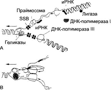
The molecular structure of the replication fork is currently understood to be as schematically depicted in Fig. 1.9. In principle, this model applies to DNA Replication in Prokaryotes, as well as in Mitochondria, Plastids, and Eukaryotic Cell nuclei, where double-stranded DNA is invariably found. However, while relatively short circular DNAs of Organelles and prokaryotes (see 7.2.1) possess only a single start site for replication — THE ORIGIN OF replication — from which the process proceeds in opposite directions along the DNA circle, the linear DNAs of eukaryotic chromosomes (ranging from centimeters to decimeters in length) feature multiple replication origins; otherwise, the complex process of chromosome duplication would take weeks or months despite the high efficiency of polymerases, which must copy the hereditary material with extremely high fidelity. A nucleotide sequence synthesized from a single replication origin is called a replicon. The circular DNAs of Bacteria and eukaryotic cell organelles are monorepliconic1, whereas the linear DNAs of eukaryotic chromosomes are polyrepliconic. Other differences in the duplication of eukaryotic chromosomes compared to bacterial replication (Fig. 1.9) pertain to DNA polymerase: instead of prokaryotic DNA polymerase III, the functional eukaryotic DNA polymerase α possesses its own primase activity, enabling the synthesis of RNA primers on both the leading and lagging strands. However, DNA polymerase α is incapable of synthesizing long DNA stretches and is replaced by the main replicative enzyme (DNA polymerase δ) once the primer has been extended by approximately 30 nucleotides.
1 The question of the number of replicons in the Mitochondrial Genome remains unresolved. It is possible that large Mitochondrial Genomes with complex multicircular structures contain multiple replicons. — Ed. note.
Fig. 1.9. DNA replication in Escherichia coli; replication fork moving in the direction of the arrow (after A. Kornberg, from H. Kletnig and U. Maier): A—Unwinding of the DNA double helix by specific helicase enzymes with temporary stabilization of the separated strands by single-strand binding proteins (SSB). At the leading end (pointing downward in the figure), continuous Synthesis of the new complementary [antiparallel] strand immediately begins (arrowhead: growing 3' end) catalyzed by DNA polymerase III. On the lagging strand (at the top), by contrast, the polymerase operates in the opposite direction (though still 5' → 3'); it extends the 3' ends of RNA primers, which in turn are synthesized at regular intervals by primases (specialized Enzymes of the primosome) (discontinuous replication). Finally, the RNA primers are replaced, gaps are filled via reparative synthesis (DNA polymerase I), and the remaining single-strand nicks are covalently sealed by ligase. B—Hypothetical model of the "replisome," in which all enzymes and protein factors of the replication machinery are organized into a complex. The antiparallel orientation of the parental DNA strands is locally preserved through the formation of loops on the lagging strand.
DNA synthesis proceeds approximately 5 times faster than RNA Synthesis during transcription (see 7.2.2.2). Because Genes are transcribed during replication, it is hypothesized that DNA polymerases temporarily pause their activity until the synthesis of the RNA molecule is completed.
1.2.4. Ribonucleic Acids (RNA)
Unlike DNA, which occurs as a single strand only in certain viruses and phages and is double-stranded in all other cases, RNA molecules are predominantly single-stranded. Through intramolecular base pairing (Fig. 1.10, A, C), they form stabilized secondary structures and, often As a result of association with proteins, tertiary structures. Consequently, there are several structural variants of RNA and ribonucleoprotein complexes (RNPs). The only exception is Transfer RNA (tRNA), which never associates with proteins. Because single-stranded RNA is readily degraded by nuclease enzymes, intramolecular base pairing and protein association enhance molecular stability. Furthermore, secondary and tertiary structures are essential for the biological function of RNA molecules. Thus, as previously mentioned, RNA differs from DNA in the following respects:
• presence of ribose instead of 2-deoxyribose;
• presence of uracil instead of thymine;
• significantly smaller size of RNA molecules compared to DNA.
Fig. 1.10. Ribonucleic acids
A—Cloverleaf structure of a tRNA molecule, exemplified by Yeast Tyrosine-transfer RNA (tRNATyr). The structure is stabilized by a specific molecular conformation folded in such a way that hydrogen bonds form between paired nitrogenous bases within each of the three loops, while unpaired bases are located in the outer Regions of the loops. Many tRNA bases are modified; some key modifications are shown in B. In addition, methylated bases are present. A triplet of bases, known as the anticodon, binds to the complementary mRNA base triplet, the codon. The triplet sequence in the mRNA determines (via codon-anticodon interactions) The amino acid sequence in the synthesized protein (Genetic Code, see 7.3.1.1; Protein Biosynthesis, see 7.3.1.2). The D-loop is named after dihydrouracil (UH2), which occurs here instead of uracil; the T C-loop is named after the invariant base sequence 5'-T-
 -C-3'. Different tRNAs feature a V-loop of variable size (derived from 'variable'). The amino acid attachment loop and the anticodon loop are essential for the recognition of the respective tRNA and its cognate amino acid by Aminoacyl-tRNA synthetases. At the same time, the anticodon loop positions the anticodon so that it can engage in complementary pairing with the mRNA codon within the ribosome. The T
 C and D loops are presumed to be of particular importance for binding tRNA to the ribosome. C—Example of a circularly closed RNA molecule: a viroid causing potato spindle tuber disease (PSTV, Potato Spindle Tuber Viroid). It consists of 359 covalently closed nucleotides. Its structure is stabilized by intramolecular base-pairing interactions, which are depicted as single lines for clarity.

Structural diversity dictates the functional diversity of various RNA types (Table 1.2). Most short-lived Messenger RNA (mRNA) molecules represent transcribed gene segments (see 7.2.2). They serve as templates during ribosomal Translation through the sequential recruitment of stable Transfer RNAs carrying specific Amino Acids, thereby defining the amino acid sequence of the protein. Similarly, stable Ribosomal RNAs contribute to the structural framework of Ribosomes. Additionally, small cytoplasmic RNAs (scRNAs) occur within Protein Complexes, such as the signal recognition particle (SRP1), which plays a key role in translation (see 7.3.1.4). Small nuclear RNAs (snRNAs) participate in the Processing of transcribed mRNAs and tRNAs (see 1.2.2.2).
1 SRP — signal recognition particle. — Ed. note.
Table 1.2. Approximate sizes and functions of 3 types of RNA compared to DNA
Nucleic acid |
Size, bp |
Function |
DNA |
Over 100 million |
Information storage (genes) |
mRNA |
From several hundred to 10,000 and more |
Carries the transcribed gene sequence to ribosomes for Protein Synthesis |
rRNA |
4 types (in Eukaryotic cells): approx. 120, 150, 1,700, 3,500 |
Provides ribosomal Structure and function |
tRNA |
80-90 |
Deliver amino acids to ribosomes |
1.2.5. Viruses, Phages, Viroids
Viruses are obligate parasites of eukaryotes, while phages are analogous parasites of prokaryotes. These entities are too simple to reproduce independently, relying instead on the METABOLIC ACTIVITY OF living cells for replication. Their nucleic acids—double-stranded or single-stranded DNA—carry Genetic information; they are capable of mutation and, under appropriate conditions, recombination. Phages have played a pivotal role as model organisms in The Development of modern genetics.
In viruses and phages, nucleic acids form a complex with proteins. The protein molecules typically assemble into a highly symmetrical shell structure known as a capsid (see Fig. 1.17). It serves structural and protective functions and plays a crucial role in infecting new host cells. "Complex" viruses additionally possess a loose membranous envelope derived from The Plasma Membrane of the host cell, which also incorporates specific viral Glycoproteins.
Viroids lack such protein coats. Instead, they consist of very small, circular or rod-like free RNA molecules comprising only 250 to 370 bases, which can act as plant pathogens (see Fig. 1.10, C). Potato spindle tuber disease and coconut cadang-cadang disease are caused precisely by viroids. Viroids enter a new host plant through damaged cells and disrupt their metabolism by interfering with the synthesis of host RNA molecules.
Last update: 07/08/2026
Editorial and Educational Adaptation: This material has been compiled based on the primary/original source text. The project team performed an editorial review, corrected technical inaccuracies, structured sections, and adapted the content for an educational format.
What was processed:
- elimination of formatting defects (OCR errors, structural breaks, corrupted characters);
- editorial organization of content;
- standardization of terminology in accordance with academic sources;
- verification of factual statements against the original source text.
All mentions of the author, publication year, and origin of the primary text have been preserved in accordance with the source.