PLANT MORPHOLOGY - T. A. Sautkina - 2012
CHAPTER 3. PLANT TISSUES
3.3. Anatomical, morphological, and functional features of protective tissues
The name of these Tissues reflects their Location: they cover the plant body and are situated at the interface with the external environment or internal spaces (such as the Ovary of the pistil). A characteristic feature of protective tissues is that they consist of tightly packed living or dead Cells. Their primary function is barrier protection, shielding internal plant tissues from desiccation and damage. Other essential Functions include The regulation of Transpiration and gas exchange. Additionally, protective tissues are capable of absorbing and secreting substances, preventing the penetration of pathogenic microorganisms, providing mechanical support, and more. Thus, protective tissues are, to a certain extent, multifunctional.
From an evolutionary perspective, protective tissues are of very ancient origin, having arisen as a consequence of plants transitioning to life on land. Even the body of the earliest known land plants (rhyniophytes) was covered with an epidermis bearing Stomata.
In the ontogeny of modern plants, protective tissues originate from various Meristems: primary tissues form As a result of anticlinal (perpendicular to the organ surface) Divisions of the superficial layers of the apical meristem, while secondary tissues arise through periclinal divisions of the phellogen.
Depending on their origin and the time of appearance in plant ontogeny, Three types of protective tissues are distinguished: primary, secondary, and tertiary.
3.3.1. Primary protective tissues
Primary tissues, formed as a result of the differentiation of primary meristem cells, include the epidermis (cuticle), which covers leaves, young green stems, floral parts, and fruits, as well as the rhizodermis (including the epiblem), which covers young roots.
The epidermis (from Greek epi — upon, and derma — Skin) differentiates from the protoderm, the outermost layer of the tunica. Most commonly, it consists of a single layer of tightly appressed cells without intercellular spaces (except for stomatal pores), often with wavy walls that enhance mechanical interlocking. In elongated plant Organs, the main epidermal cells are stretched along the longitudinal axis (on stems and petioles) (Fig. 14). The outer surface of epidermal cells is typically thickened and frequently covered by a cuticle layer or a wax bloom of varying thickness. Epicuticular wax, deposited as a smooth layer or as rods and filaments projecting above the surface, creates a whitish or bluish bloom on the leaves and fruits of certain plants (such as ficus, plum, and apple). Unlike the cuticle, it is easily rubbed off. Both cutin and wax are synthesized by The Cell protoplast. The thickening of the outer walls and the presence of cutin and wax not only reduce transpiration but also protect the plant from the penetration of Viruses, Bacteria, and fungal hyphae. In A number of plants (such as grasses, sedges, and horsetails), silica is deposited in the outer Cell wall. In conifers, certain grasses, and Solomon's seal, the walls of the main epidermal cells become lignified. The epidermal cells of many seeds contain Polysaccharides that swell upon Hydration, forming mucilage. This allows the seeds to readily adhere to soil or moving objects, facilitating their anchorage or dispersal.
Class="center">Fig. 14. Epidermal cell shape and stomatal Structure: A — in dicotyledons (wild thyme — Thymus serpyllum); B — in monocotyledons (common wheat — Triticum aestivum): 1 — epidermal cells; 2 — stoma; 3 — guard Cells of the stoma with METABOLISM/14.html">Chloroplasts; 4 — stomatal pore

As a rule, epidermal cells are living. They contain a thin parietal layer of Cytoplasm with a Nucleus and occasionally a small number of photosynthetically inactive chloroplasts, as well as leucoplasts. The epidermal cells of flower petals and succulent fruits contain chromoplasts.
Secondary metabolites such as Alkaloids, Essential Oils, Tannins, pigments, and calcium oxalate crystals may accumulate within the large central vacuole.
Epidermal cells frequently form surface outgrowths known as trichomes (hairs) (Fig. 15). They can be unicellular or multicellular, simple or branched, stellate or glandular, among other forms. Trichomes form pubescence (indumentum). Depending on their function, trichomes are divided into covering (non-glandular) and glandular types. Some covering trichomes are living (such as in African violets), while others lose their living contents and become dead. Dead trichomes appear light-colored, which allows them to reflect sunlight; Water vapor is trapped between them, thereby creating a specific microclimate. Covering trichomes protect the plant from overheating (coltsfoot leaves), excessive transpiration (many desert plants), and damage by animals and insects. In epiphytic plants (such as bromeliads and orchids), trichomes absorb water vapor from the air. In some cases, before stomata develop, living covering trichomes may perform a transpiratory function before eventually withering away.
Fig. 15. Unicellular and multicellular covering trichomes of the epidermis in various plants: 1 — sweet violet (Viola odorata); 2 — paper mulberry (Broussonetia papyrifera); 3 — feijoa (Feijoa sellowiana); 4 — hemp (Cannabis sativa); 5 — scented geranium (Pelargonium graveolens); 6 — purging buckthorn (Rhamnus cathartica); 7 — Japanese camellia (Camellia japonica); 8 — Caucasian quince (Cydonia oblonga); 9 — narrow-leaved oleaster (Elaeagnus angustifolia); 10 — oriental plane (Platanus orientalis); 11–13 — cork oak (Quercus suber)

Glandular trichomes generally retain their living cell contents and function as secretory structures capable of synthesizing and releasing metabolic products (such as essential oils, resins, and substances toxic to the plant) into the environment or beneath the cuticle. In carnivorous plants (such as Drosera and Nepenthes), they secrete digestive Enzymes. Glandular hairs frequently terminate in one or several secretory cells or form a distinct HEAD (as in tomatoes and pelargoniums). The main function of glandular hairs is associated with excreting toxic salts from leaf tissues (orache — Atriplex), protecting the organ from overheating through the emission of volatile essential oils, and providing chemical and partial mechanical defense against insects. The diverse structure of trichomes is widely used as a diagnostic feature in plant systematics and in the analysis of Medicinal plant raw Materials.
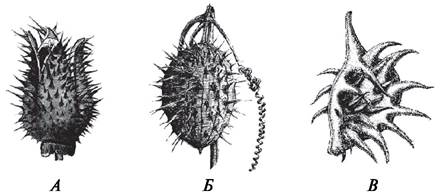
In addition to hairs, outgrowths known as emergences (from Latin emergentis — rising out) develop On the surface of stems and fruits in certain plants (Fig. 16). Not only epidermal cells but also the subepidermal cell layers participate in their formation. Emergences are often relatively large structures resembling prickles. However, unlike thorns, they are arranged on the SHOOT without a definite order and can be stripped off along with the epidermis. Examples of emergences include the prickles of roses, raspberries, and blackberries, as well as the spines on the fruits of thornapples (Datura), wild cucumbers (Echinocystis), many umbellifers, and horse chestnuts. Emergences primarily serve a protective function. Thus, the aforementioned Structural Features of the epidermis help protect young plant organs from unfavorable environmental factors.
Fig. 16. Emergences on the fruit walls of various plants: A — jimsonweed (Datura stramonium); B — wild cucumber (Echinocystis lobata); C — field buttercup (Ranunculus arvensis)
3.3.2. Concept of stomatal complexes: structure and functioning of stomata
A critical function of the epidermis is the active regulation of transpiration and gas exchange. This function is carried out through the stomatal apparatus, or stomatal complex, which consists of a stoma and functionally associated neighboring (subsidiary) cells. A stoma is a structure formed by two guard cells and a stomatal pore between them, which functions as an intercellular space (see Fig. 14). Stomatal guard cells may lie at the same level as the main epidermal cells, or they may project above them or be sunken significantly deeper (for example, sunken stomata in pine needles).
The guard cells of dicotyledonous plants are most commonly Kidney-shaped. The concave walls facing the pore (ventral or inner walls) are significantly thicker than the convex (dorsal or outer) walls. Furthermore, cuticular ledges form on them, giving the stomatal pore the appearance of a narrow channel expanded in a funnel-like manner at both ends. The outer expansion before the entrance to the pore is called the front vestibule (or outer stomatal cavity), and the inner expansion behind the pore is called the back vestibule (or inner stomatal cavity). Beneath the guard cells lies a substomatal or air chamber, where water vapor, carbon dioxide, and oxygen accumulate. Guard cells contain chloroplasts but lack plasmodesmata, which to a certain extent indicates their relative isolation from the remaining epidermal cells.
In monocotyledons, especially grasses, the guard cells are elongated, and thickenings are localized on the walls adjacent to both the stomatal pore and the subsidiary cells (see Fig. 14).
Stomata operate automatically, driven by a series of morphological and physiological factors.
The width of the stomatal pore, and consequently The rate of water evaporation, is regulated by changes in turgor pressure within the guard cells. When a plant has an abundant water supply, monovalent cations—primarily potassium—are actively transported into the guard cells from surrounding cells and the guard cells' own Organelles (i.e., against the concentration gradient and requiring energy). This leads to an increase in osmotic pressure within the stomata, causing them to swell and exert greater pressure on The cell wall as they absorb water from adjacent cells. The MICROSTRUCTURE OF THE guard cell walls plays a decisive role in altering cell shape (movement) and opening the stomatal pore. Cellulose microfibrils within the cell wall are arranged such that the wall facing the pore is less elastic, while certain fibers form hoop-like structures around the guard cells. As water is absorbed and turgor increases, these hoops prevent the cells from expanding radially while allowing them to elongate. A second structural constraint is located at the ends of the guard cells where they connect with one another. The rising turgor pressure forces the outer (dorsal), more elastic walls to push outward. In doing so, radial micelles pull the inner (ventral) walls of the stomatal cells along with this movement. As a result, the stomatal pore opens, and water transpiration takes place. Stomatal closure involves the passive efflux of potassium ions down their concentration gradient. Osmotic pressure drops, water leaves the guard cells, vacuole volume decreases, the stretched thin cell walls collapse, and the stomatal pore closes. The energy required for the operation of the stomatal apparatus comes from the Hydrolysis of reserve CARBOHYDRATES accumulated in the guard cells during Photosynthesis.
Different plant species have evolved their own specific rhythms for stomatal operation. In most plants, stomata are open during the day and closed at night. They typically close during periods of extreme heat. When the plant tissue is saturated with water (for example, following prolonged rainfall), the guard cells swell and are compressed by the surrounding epidermal cells, causing the stomatal pore to close as well.
The number and distribution of stomata vary depending on plant species and environmental conditions. Their density ranges from several tens to several thousand per 1 mm2 of epidermal surface. The total area of stomatal pores accounts for no more than 1–2% of the leaf surface area. However, transpiration through open stomatal pores is equivalent to the evaporation from approximately 70% of an equivalent open water surface. When stomata are closed, transpiration drops sharply, and water evaporation occurs solely through the cell walls and cuticle. Thus, the epidermis regulates gas exchange and transpiration with remarkable efficiency.
3.3.3. Development of Stomatal Complexes and Their Types
Stomatal complexes develop from protodermal cells. In angiosperms, the mother cell or precursor cell (meristemoid) of the guard cells forms as a result of an unequal division of a protodermal cell, representing the smaller of the two resulting daughter cells. This smaller cell subsequently divides to produce two guard cells. The intercellular substance between them swells and dissolves, forming the stomatal pore. Simultaneously, peristomatal (subsidiary) cells are formed. Thus, a stomatal apparatus, or stomatal complex, is created. It consists of the stoma and subsidiary cells. In rare cases, subsidiary cells are absent or indistinguishable from typical epidermal cells. The Study of stomatal apparatuses is known as stomatography (from the Latin stoma, meaning Mouth).
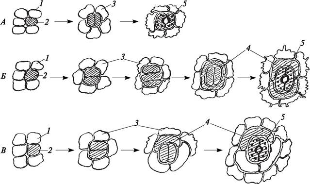
Based on their ontogenetic development in plants, three types of stomatal apparatuses are distinguished: perigenous, mesogenous, and mesoperigenous (The terms "perigenous" and "mesogenous" were first proposed by R. Florin and later widely popularized by D. Pant in 1965, who also introduced the term "mesoperigenous"). During The formation of a perigenous stomatal apparatus, the meristemoid divides to produce only a pair of guard cells. Subsidiary cells, if present, develop from separate meristemoids, meaning they do not share a common Lineage with the guard cells. The mature stoma is surrounded by ordinary epidermal cells (as seen in iris and pelargonium). In The Development of a mesogenous apparatus, the meristemoid gives rise to both the guard cells and all associated subsidiary cells (such as in grasses, Caryophyllaceae, and Brassicaceae), indicating a common origin. If the meristemoid gives rise to the guard cells and at least one subsidiary cell, while other surrounding cells originate from different meristematic initials, it is termed a mesoperigenous apparatus (Fig. 17).
Fig. 17. Diagram of stomatal complex ontogeny:
A—perigenous; B—mesogenous; C—mesoperigenous: 1—protodermal cells that give rise to epidermal cells; 2—protodermal cells that give rise to stomatal complexes (guard cells and subsidiary cells); 3—epidermal cells; 4—subsidiary cells; 5—stoma
As early as the late 19th century, many botanists (I. P. Borodin, 1888; J. Vesque, 1889, etc.) believed that the mode of Development of the stomatal apparatus was an entirely constant trait that could be applied in plant systematics. As I. P. Borodin wrote: "The particular method of stomatal initiation—whether without subsidiary cells, or with the formation of one, two, or none—proves to be highly consistent across entire families and ranks among the Anatomical Features useful for Taxonomy."
Through his studies of stomatal complex development, J. Vesque identified six ontogenetic stomatal types—three perigenous and three mesogenous—though it turned out that only four types remain distinguishable at maturity: ranunculaceous, cruciferous, rubiaceous, and caryophyllaceous. These types differ in the number of subsidiary cells and their arrangement relative to the long axis of the stomatal pore. Their names are derived from the plant families in which J. Vesque considered these types to be most characteristic. The primary categories of stomatal complexes established by J. Vesque gained widespread recognition, particularly in pharmacognosy, serving as a key criterion for identifying the species identity (authenticity) of herbal medicinal raw materials.
Further research into stomatal complexes revealed that the types established by J. Vesque occur in representatives of many other families, rendering the terms he proposed somewhat conventional.
It is now firmly established that regardless of how stomatal complexes develop during ontogeny, they can exhibit morphological similarity. Because it was discovered that identical types of stomatal complexes can occur across different families, it became necessary to introduce a new terminology devoid of ontogenetic implications and unlinked to specific taxon names.
Such a morphological Classification of stomatal apparatuses in dicotyledonous plants was proposed by English botanists C. Metcalfe and L. Chalk in 1950. They distinguished four (classical) types of stomatal complexes: anomocytic, diacytic, paracytic, and anisocytic (Fig. 18). Structurally, these correspond to the types identified by J. Vesque. Subsequently, a fifth type—actinocytic—was also defined.
Fig. 18. Morphological types of stomatal complexes: A—anomocytic; B—diacytic; C—paracytic; D—anisocytic: 1—stoma; 2—chloroplasts; 3—epidermal cells; 4—subsidiary cells
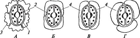
The anomocytic (from the Greek anomos, irregular, and kytos, cell), or irregular-celled type is found in various groups of higher plants, with the exception of horsetails. In this case, subsidiary cells are absent or indistinguishable from the other epidermal cells. The anomocytic stomatal complex is characteristic of primitive angiosperms belonging to the Ranunculaceae and Geraniaceae families.
The diacytic (from the Greek dis, two, and kytos, cell), or cross-celled type features two subsidiary cells positioned perpendicularly to the long axis of the stoma. This type of stomatal complex is found in Representatives of the Lamiaceae (mint, sage, etc.) and Caryophyllaceae (carnation, chickweed) families. It has also been discovered in ferns (Nephrolepis).
The paracytic (from the Greek para, beside, and kytos, cell), or parallel-celled type is characterized by two subsidiary cells arranged parallel to the long axis of the stoma. It is typical of species from the Rubiaceae (bedstraw, woodruff) and Chenopodiaceae families, as well as grasses, and is also found in horsetails, ferns, and Gnetophytes.
The anisocytic (from the Greek anisos, unequal, and kytos, cell), or unequal-celled type is found exclusively in flowering plants. The guard cells are surrounded by three subsidiary cells, one of which is markedly larger or smaller than the others. This type is most common in plants of the Brassicaceae (cabbage) and Crassulaceae (stonecrop, houseleek) families.
The actinocytic (from the Greek aktis, ray, and kytos, cell) stomatal complex is one in which the guard cells are surrounded by a varying number of radially arranged subsidiary cells.
The classification of stomatal complexes proposed for dicotyledonous angiosperms by C. Metcalfe and L. Chalk gained worldwide renown and formed the foundation for the modern classification of stomatal types.
The structure of stomatal complexes has been investigated in plants beyond angiosperms as well. Between 1931 and 1933, R. Florin published studies on gymnosperm stomata, and in 1970, W. Van Cotthem presented a classification of stomatal complexes in pteridophytes. During the latter half of the 20th century, considerable attention was devoted to studying the stomatal complexes of monocots, and discussions arose regarding the classification of stomata across various higher plant taxa. According to M. A. Baranova, 14 morphological types of stomatal complexes are currently known in higher plants.
In 1973, a new classification was introduced by E. Fryns-Claessens and W. Van Cotthem, which combined features of both morphological and ontogenetic classifications, resulting in the identification of 26 distinct stomatal complex types.
Since various authors approach the classification of stomatal complexes from different Perspectives, a single universally accepted classification of stomatal complexes does not yet actually exist. Nevertheless, when describing various families of angiosperms, the type of stomatal complex is indicated as one of the key diagnostic features.
If the aerial organs of a plant are covered by the epidermis, the primary protective tissue of the ROOT is the rhizodermis (from the Greek rhysa—root and derma—skin) or epiblem, which produces root hairs. It originates from the dermatogen—the outer cells of the root apical meristem near the root cap, and covers the young root tips. Functionally, this is one of the most vital tissues, as it is through it that water and mineral uptake from the soil takes place, i.e., root Nutrition of the plant is carried out. In its structure, the epiblem is similar to the epidermis, but it also has its own specific features associated with its location and primary function. The epiblem lacks stomata, but forms specialized outgrowths known as root hairs. Potentially, every cell of the epiblem is capable of forming a root Hair. However, this function is performed only by certain specialized cells called trichoblasts (Fig. 19).
Fig. 19. Primary protective tissue of the root (rhizodermis, epiblem): 1—rhizodermis; 2—epiblem; 3—root hairs; 4—atrichoblasts; 5—trichoblasts
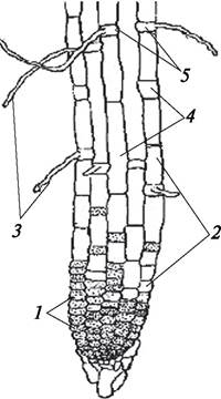
Epiblem cells are thin-walled, have a Primary Structure, contain a large vacuole and more viscous cytoplasm than epidermal cells. Due to the active uptake of aqueous mineral salt solutions, the root hairs of the epiblem are rich in Mitochondria. Chloroplasts are absent in root hairs, and there is no cuticle either. Root hairs are typically unicellular and, unlike epidermal trichomes, are not separated from the trichoblast by a cell wall. They reach 1–2 mm in length and have a short lifespan. After 10–15 days, the root hairs wither, and new ones emerge in an acropetal direction. Root hairs significantly increase the absorptive surface area of the root.
3.3.4. Secondary Protective Tissue
Primary protective tissue exists for a limited period of time and only in actively growing organs. In herbaceous plants, the epidermis persists throughout their life. In the stems of perennial woody plants (trees and shrubs), as well as in the roots of gymnosperms and dicotyledons characterized by secondary thickening, the primary protective tissues are replaced by a secondary tissue—the periderm (from the Greek peri—around, near, and derma—skin). Typically, the periderm forms on annual shoots by the end of the growing season, but sometimes it forms In the second (heather, some rhododendrons) or third year of the plant's life (bearberry). The modes of periderm initiation vary (Fig. 20). It originates from a specialized secondary meristematic tissue—the phellogen, or cork cambium. The stem phellogen most commonly arises from subepidermal cells of the ground parenchyma, which dedifferentiate and regain meristematic activity. In roots, as well as in the stems of certain plants (raspberry, briar rose, fireweed), the phellogen frequently develops from the pericycle.
Fig. 20. Modes of periderm initiation: A—in the epidermis and partially beneath it in pear (Pyrus communis); B—in the subepidermal layer in plum (Prunus domestica); C—in the inner layer of the primary cortex in red raspberry (Rubus odoratus): 1—epidermis with cuticle; 2—phellogen; 3—phelloderm; 4—phelleme; 5—primary cortex of the stem
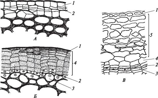
The phellogen can initiate either as separate patches that subsequently merge, or as a continuous ring around the entire circumference of the axial organ. Its cells are tabular in shape. They divide periclinally, parallel to the organ surface, successively cutting off phelloderm cells (or cork skin) inwardly, and phelleme cells (or cork) outwardly. Since with each division of the phellogen cells one of the daughter cells retains its meristematic properties while the second differentiates into a permanent tissue cell (phelleme or phelloderm), a long-functioning layer of phellogen is maintained between the phelleme and the phelloderm (Fig. 21). Phellogen cells produce more phelleme cells than phelloderm cells. Consequently, the phelloderm most often consists of 1–3 layers of living parenchymal cells containing reserve nutrients and performing a trophic function in relation to the phellogen. The phelleme layer is thicker. Its cells are tightly packed together, strictly tabular in shape, lacking intercellular spaces, and arranged in regular radial rows. Young phelleme cells are living; however, subsequently—often even before cell growth is complete—suberin is deposited on the primary wall from the inside, sometimes alternating with wax. The Plasma Membrane of the cell forms a secondary cell wall (cellulosic), which is laid down over the suberin layer. Because there are no pits in the walls of phelleme cells, the protoplast of the suberized cells dies. Their cavities become filled with air or dark-colored resinous or tanning substances, which is why the green color of shoots turns brown. In birch, the cell walls of the phelleme are impregnated with a special amorphous substance—betulin, which gives the branches their white color and also possesses antiseptic properties. Cork cells are impermeable to water and gases, effectively protecting the internal Tissues of the plant from desiccation, Temperature fluctuations, and pathogens. The periderm is especially important for plants living under seasonal conditions. On the trunks of certain trees, such as the cork oak and Amur cork tree, a thick layer of cork develops, reaching several centimeters in thickness. Thus, the periderm is a complex tissue comprising both living (phellogen, phelloderm) and dying cells (phelleme). During periderm formation, the epidermis dies and sloughs off.
Fig. 21. Diagram of periderm formation: A—parenchyma cell regaining meristematic function; B—phellogen cell formed from a dedifferentiated parenchyma cell; C—division of a phellogen cell producing phellogen derivative cells (1) and phelleme cells (2); D—enlargement of phellogen (1) and phelleme (2) cells; E—onset of phelleme cell suberization (2) and division of the phellogen cell producing a new phellogen derivative cell (1) and a phelloderm cell (3); F—growth of phellogen (1) and phelloderm (3) cells to typical dimensions, further thickening of the phelleme cell wall (2); G—formation of a new phellogen cell (1) and phelleme cell (2a), phelleme cell (2), phelloderm cell (3); H—periderm (4)

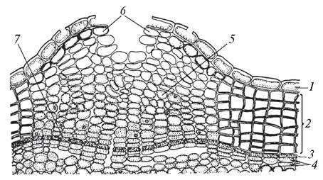
Possessing the aforementioned properties, the periderm effectively isolates internal tissues from the environment. At the same time, living tissues require gas exchange and the removal of excess moisture. Therefore, during periderm formation, lenticels begin to form first—specialized structures that provide aeration for internal tissues (Fig. 22). The phellogen primarily initiates beneath stomata (though not under every one) where gas exchange and transpiration processes are active. The cells resulting from The activity of the phellogen have an isodiametric-like shape and are loosely arranged due to the presence of large intercellular spaces. The aggregate of these cells is termed complementary or filling tissue. Through the intercellular spaces of this filling tissue, unregulated (unlike stomata) gas exchange and transpiration take place. Due to intense gas exchange and transpiration during the summer period, the cells of the filling tissue do not become suberized. The phellogen underlying the filling tissue also contains narrow intercellular spaces, thus it does not hinder gas exchange. With the onset of the cold season, the phellogen lays down a closing layer beneath the filling tissue, consisting of smaller, suberized, and tightly packed cells, and the lenticels "close". In spring, with the reactivation of the phellogen, this layer is ruptured by the pressure of new cells, and the lenticels resume function. Lenticels typically protrude above the periderm and exhibit various shapes characteristic of different plant species. In aspen and poplar, lenticels are rhomboid; in elderberry, they are rounded; in birch, they are linearly elongated along the circumference of the trunk, appearing as black dashes on a white Background. Lenticels are clearly visible on potato tubers, as well as on apple and pear fruits. Lenticels are typically absent in the periderm of roots.
Fig. 22. Periderm with a lenticel in red elderberry (Sambucus racemosa): 1—epidermal remnants; 2—phelleme; 3—phellogen; 4—phelloderm; 5—lenticel complementary tissue; 6—lenticel rupture; 7—closing layer
3.3.5. Tertiary Protective Tissue
In most woody plants, the periderm of axial organs is eventually replaced by rhytidome, or bark, which is a tertiary protective tissue (Fig. 23). It forms as a result of multiple successive initiations of new periderm layers in deeper tissues of the primary cortex. Consequently, the tissues enclosed between the periderms are deprived of water and nutrient supply; metabolic end products (resins, tannins, calcium oxalates, alkaloids, etc.) are deposited in them, and sclereids are formed. Over time, these tissues die, forming a robust protective complex. From the inside, the bark layer grows annually, while on the surface it cracks, breaks down, and is shed. Gas Exchange in this tissue also occurs via lenticels, which form in the periderm beneath rhytidome fissures. Rhytidome is classified into ring and scaly types. Ring rhytidome forms when periderms develop in a more or less continuous layer. As a result, the bark easily peels off the trunk like a stocking or splits into long strips (grapevine, clematis, honeysuckle, strawberry tree, cypress, cherry). However, scaly rhytidome is more common, during the formation of which new periderms arise as separate overlapping fragments. Such rhytidome develops in pine (Fig. 24), apple, and birch (Fig. 25). As the stem increases in thickness, this type of bark cracks and sheds in the form of scales.
Fig. 24. Scaly bark (rhytidome) of Scots pine (Pinus sylvestris)
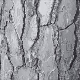
Fig. 23. Structure diagram of the rhytidome (bark) of pedunculate oak (Quercus robur): 1(a–c)—periderms successively developing in the parenchyma of the stem primary cortex; 2—sclereids; 3—sclerenchymatous fibers; 4—calcium oxalate druses in parenchyma cells; 5—rhytidome
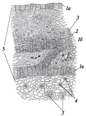
Fig. 25. Rhytidome (at the base) and periderm on the trunk of silver birch (Betula pendula)
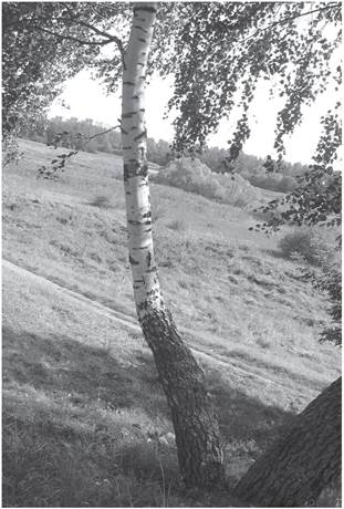
Rhytidome formation occurs at different ages across various plant species: in grapevines, in the second year of life; in apples and pears, in the seventh to eighth year; in pine, at 8–10 years; in oak, at 25–30 years; in hornbeam, after half a century. The rhytidome provides protection for trees against temperature fluctuations, sunscald, ground fires, and damage by microorganisms, animals, etc. In some woody species (cork oak, Amur cork tree), rhytidome does not form, and the protective function throughout the tree's life is performed by a powerfully developed periderm.
The epidermis, periderm, and rhytidome are classified as complex tissues because they consist of different cell types.
Last update: 07/08/2026
Editorial and Educational Adaptation: This material has been compiled based on the primary/original source text. The project team performed an editorial review, corrected technical inaccuracies, structured sections, and adapted the content for an educational format.
What was processed:
- elimination of formatting defects (OCR errors, structural breaks, corrupted characters);
- editorial organization of content;
- standardization of terminology in accordance with academic sources;
- verification of factual statements against the original source text.
All mentions of the author, publication year, and origin of the primary text have been preserved in accordance with the source.