BIOLOGY Volume 2 - A Guide to General Biology - 2004
17. COORDINATION AND REGULATION IN ANIMALS
17.1. The Nervous System
17.1.2. Synapses
A synapse is a specialized functional junction between two excitable Cells that serves to transmit excitation. In the case of Neurons, it is typically located between a fine branching terminal of one Cell's axon and the dendrite or perikaryon of another. There is no physical contact between the cells in this case—they remain separated by a small space known as the synaptic cleft. The number of synapses is usually very large, forming a vast surface area for information transmission. For example, there may be over 1,000 synapses on the dendrites and cell body of a single spinal motor neuron, and up to 10,000 on certain brain neurons (Fig. 17.8).
Class="center">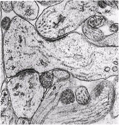
Fig. 17.8. Transmission electron micrograph of motor neuron synapses. The synaptic clefts can be seen between the dark, thickened synaptic membranes.
A special type of synapse with a similar function but a different Structure links motor neuron terminals to Muscle fibers. These are called neuromuscular junctions and will be discussed later in this section.
Structure of a Synapse
The structure of a typical synapse is shown in Figs. 17.9 and 17.10. It consists of a synaptic terminal in the form of a bulbous Swelling at the end of the axon, located in close proximity to the dendrite membrane. The Cytoplasm of this swelling contains numerous Mitochondria (supplying energy) and small synaptic vesicles, 50 nm in diameter. The latter contain a substance called a neurotransmitter, which serves to transmit the impulse across the synaptic cleft, which is approximately 20 nm wide. The Cell membranes facing the cleft are thickened and referred to as the presynaptic membrane on the axon and the postsynaptic membrane on the dendrite. The presynaptic membrane is modified to rapidly attach synaptic vesicles and release their contents into the synaptic cleft (exocytosis). The postsynaptic membrane contains large protein molecules that function as receptors for Neurotransmitters, as well as numerous channels (pores) that are closed at rest but can open to allow specific ions to enter the cell (Fig. 17.11).

Fig. 17.9. Structure of a synapse.

Fig. 17.10. Electron micrograph of a synapse.

Fig. 17.11. Mechanism of chemical transmission in a nerve synapse; A to E represent the temporal sequence of events.
The neurotransmitter is either synthesized in the perikaryon and transported to the presynaptic terminal of the neuron, or synthesized directly within the terminal itself. In both cases, this requires Enzymes assembled on Ribosomes in the neuron's cell body. Within the synaptic terminal, the neurotransmitter is "packaged" into vesicles and stored there until release. In the vertebrate Nervous system, There are two primary substances of this type—acetylcholine (ACh) and noradrenaline—although other neurotransmitters exist, which we will examine at the end of this section. Acetylcholine is an acetic acid ester of Choline. It was the first neurotransmitter isolated by scientists (in 1920). Noradrenaline is described in Section 17.6.5. Neurons whose communication is mediated by acetylcholine are called cholinergic, while those using noradrenaline for synaptic transmission are called adrenergic. Noradrenaline is released by sympathetic nerves, whereas Acetylcholine is released by almost all other nerves (except for certain ones in the brain).
Mechanism of Synaptic Transmission
We will examine this process using cholinergic neurons as an example. The arrival of nerve impulses at the synaptic terminal depolarizes the presynaptic membrane, causing calcium channels to open and thereby increasing its permeability to Calcium Ions (Ca2+). Calcium ions rush into the cell, leading to the fusion of synaptic vesicles with the presynaptic membrane and the release of their contents into the synaptic cleft (exocytosis). The vesicles then return to the cytoplasm, where they are refilled with neurotransmitter. Each vesicle contains approximately 3,000 acetylcholine molecules.
Acetylcholine diffuses across the synaptic cleft—causing a conduction delay of about 0.5 ms—and binds to a specific receptor (protein) on the postsynaptic membrane that "recognizes" the molecular STRUCTURE OF THE neurotransmitter. This causes the receptor region to change its conformation, leading to the opening of an associated ion channel in the postsynaptic membrane. Note that while these channels opened in response to depolarization during the propagation of a Nerve Impulse along the axon, in the postsynaptic membrane they open in response to the neurotransmitter binding to the receptor protein.
The influx of sodium ions into the dendrite through the postsynaptic membrane causes its depolarization (Fig. 17.4, A). If the excitation threshold is reached, an Action Potential is generated in the neuron, and the nerve impulse propagates further. Having altered the permeability of the postsynaptic membrane, acetylcholine is almost instantly removed from the synaptic cleft by the action of the enzyme acetylcholinesterase, sometimes simply called cholinesterase. This enzyme is localized on the postsynaptic membrane and hydrolyzes acetylcholine into choline and an acetate residue. As a result, the Ion Channels close and the synapse returns to its "resting state." Choline is reabsorbed by the synaptic terminal and converted back into acetylcholine within the synaptic vesicles (Fig. 17.11). Certain nerve gases, insecticides, and other poisons inhibit acetylcholinesterase, thereby disrupting nerve conduction, as discussed in Section 4.4.3.
Other Neurotransmitters
Some neurotransmitters do not have an excitatory effect on neurons, but rather an inhibitory one, leading to hyperpolarization rather than depolarization of the postsynaptic membrane. This increases the negative charge inside the cell, making it more difficult to reach the threshold depolarization required to generate an all-or-none action potential.
There are three known pathways for removing a neurotransmitter from the synaptic cleft: 1) reabsorption by the presynaptic membrane; 2) diffusion from the cleft; 3) Enzymatic Hydrolysis.
Role of Synapses
EXCITATORY SYNAPSES, SPATIAL AND TEMPORAL SUMMATION. In excitatory synapses, the neurotransmitter opens postsynaptic ion channels, allowing sodium ions to enter the cell while potassium ions leave it along their concentration gradients. This leads to the depolarization of the postsynaptic membrane, generating what is known as an excitatory postsynaptic potential (EPSP). Typically small in amplitude, an EPSP lasts longer than an action potential. The amplitude of an EPSP changes in a stepwise manner, suggesting a quantized rather than a graded release of the neurotransmitter. Apparently, each such "step" corresponds to the contents of a single synaptic vesicle. A single EPSP is generally insufficient to trigger the threshold depolarization required to generate an action potential and propagate the impulse along the postsynaptic neuron. However, a subsequent EPSP arriving within a short time interval has a much higher chance of achieving this. This phenomenon is known as synaptic facilitation. The depolarizing effects of multiple EPSPs add together in a process called summation. Several EPSPs occurring simultaneously at different synapses on the same neuron (usually originating from different neurons) can overlap to produce threshold depolarization, thereby generating a postsynaptic action potential. This is referred to as spatial summation (Fig. 17.12). Rapidly repeated release of neurotransmitter from several synaptic vesicles of the same synaptic terminal in response to an intense stimulus produces individual EPSPs that follow one another so closely in time that their effects also summate, triggering an action potential in the postsynaptic neuron. This is called temporal summation (Fig. 17.12). Thus, impulses in a single postsynaptic neuron can arise either as a result of weak stimulation from several connected presynaptic neurons or through repeated stimulation from a single presynaptic neuron.
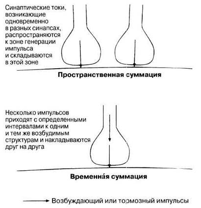
Fig. 17.12. Spatial and temporal summation.
INHIBITORY SYNAPSES. In inhibitory synapses, the release of a neurotransmitter increases the permeability of the postsynaptic membrane to chloride ions (Cl-) and potassium ions. As Cl- ions rush into the cell and K+ ions leave it along their respective concentration gradients, the membrane undergoes hyperpolarization, known as an inhibitory postsynaptic potential (IPSP). In other words, the interior of the cell becomes more negative (down to —90 mV). Clearly, this hinders threshold depolarization and, consequently, the generation of an action potential.
Neurotransmitters do not inherently possess exclusively excitatory or inhibitory properties. For example, acetylcholine exerts an excitatory effect at most neuromuscular junctions and interneuronal synapses, yet it inhibits The activity of the myocardium and the digestive tract musculature. The ultimate outcome depends on the structural CHARACTERISTICS OF THE postsynaptic membrane. The Organization of its receptor-pore complexes determines which ions will enter the cell in response to the neurotransmitter and, consequently, The Nature of the resulting postsynaptic potential.
Neuromuscular Junction (Figs. 17.13 and 17.14). Motor neurons are connected to Skeletal Muscle fibers by specialized synapses. Each striated muscle fiber features a specialized region known as the motor end plate. This region is approached by a branched, unmyelinated axon terminal with synaptic varicosities, which are embedded in shallow depressions of the muscle fiber's outer membrane (Fig. 17.13, A), called the sarcolemma. Each such depression is further elaborated with numerous deep folds (Fig. 17.13, B). Upon stimulation, the synaptic terminals release acetylcholine via the mechanism described above. Structural Changes in the receptors within the folds of the sarcolemma (the postsynaptic membrane) increase its permeability to sodium and potassium ions, causing a localized depolarization termed the end-plate potential (EPP). This depolarization is entirely sufficient to generate an action potential that propagates along the sarcolemma and deep into the muscle fiber via the transverse tubule system (T-system) (Section 18.4.4), ultimately triggering Muscle contraction as described in Section 18.4.7. In most skeletal Muscles, each fiber possesses only a single end plate.

Fig. 17.13. Structure of the neuromuscular junction. A. Light Microscopy view. B. Electron microscopy view. ACh — acetylcholine.
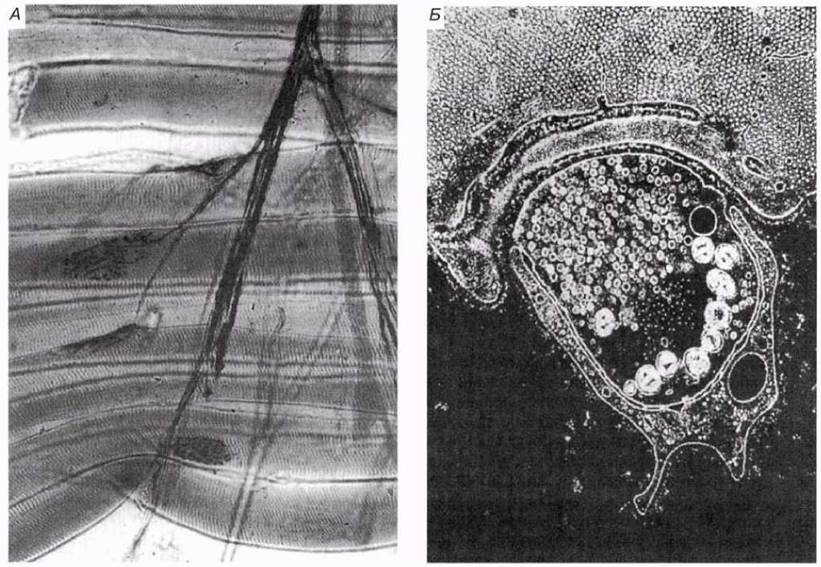
Fig. 17.14. Structure of the neuromuscular junction as observed under a Light Microscope (A) and a transmission Electron microscope (B). The nerve terminal (center) is closely associated with the muscle fiber. Small vesicles containing neurotransmitter are concentrated inside the nerve terminal near the muscle fiber.
Functions of Synapses
Given that chemical synapses entail a synaptic delay of approximately 0.5 ms, one might assume this drawback is outweighed by compensating advantages. What are these advantages? They can be summarized as follows.
1. Unidirectionality of transmission. Because neurotransmitter release is restricted to the presynaptic membrane and its receptors are localized exclusively on the postsynaptic membrane, nerve impulses can travel along this pathway in only one direction. This ensures the reliable operation of The Nervous System, guaranteeing that signals are "delivered to their destination."
2. Amplification. Each nerve impulse triggers the release of a sufficient quantity of acetylcholine at the neuromuscular junction to ensure that the depolarization of the postsynaptic membrane causes the muscle fiber to contract. Thus, nerve impulses arriving at the neuromuscular junction, regardless of how weak they may be, can elicit an effector response, thereby increasing the sensitivity of the system.
3. Adaptation and fatigue. With continuous stimulation, The amount of released neurotransmitter gradually decreases until its supply is completely exhausted; the synapse is then said to be fatigued. Further signal transmission becomes possible only after a recovery period. The adaptive significance of fatigue is that it prevents damage to the effector resulting from hyperstimulation. The same purpose is served by adaptation at the receptor level, which is discussed in Section 17.4.2.
4. Integration, convergence, and spatial summation. Due to The properties of its receptors, a postsynaptic neuron can simultaneously receive signals from A large number of excitatory and inhibitory presynaptic neurons. This phenomenon is termed convergence. The resulting spatial summation of all stimuli (a simple example is shown in Fig. 17.12) enables the postsynaptic neuron to act as an integration center for signals from various sources and to produce coordinated responses.
5. Facilitation. Observed in A number of synapses, this phenomenon means that following each stimulus, the synapse becomes more sensitive to the next one. Consequently, an action potential may eventually be triggered even by a weak signal that was previously insufficient to elicit such a response. Facilitation is not equivalent to temporal summation, as it is associated with chemical changes in the presynaptic membrane (enhanced neurotransmitter release) rather than simple electrical summation of postsynaptic membrane potentials.
6. Signal discrimination (filtering) and temporal summation. Temporal summation in synapses makes it possible to filter out weak Background stimuli, preventing them from entering the Central nervous system. For instance, the receptors of the eyes, ears, and Skin constantly receive environmental signals of no functional importance to the nervous system—so-called background noise. Only changes in signal intensity that lead to an increased impulse frequency are significant, ensuring proper transmission across the synapse and an appropriate response.
7. Inhibition. Information transfer across synapses and neuromuscular junctions can be inhibited by specific blocking agents that act on the postsynaptic membrane (see below). Presynaptic inhibition is also possible. It occurs at synaptic terminals that make contact with the inhibitory synapses of other neurons. Stimulation of these inhibitory synapses reduces the number of synaptic vesicles that release neurotransmitter at the excitatory synapse. Such a system allows a given terminal to produce varying responses depending on the activity of surrounding inhibitory and excitatory synapses.
8. In the brain, synaptic transmission involves multiple neurotransmitters. This chemical complexity of the system allows for finer regulation of processes associated with Higher Nervous Activity.
Neurotransmitters and Other Substances Affecting Synaptic Transmission
Outside the brain, the primary neurotransmitters are acetylcholine and noradrenaline, whereas over 50 other substances with similar functions have been isolated from the brain itself, each specifically associated with particular Neural Pathways. Synapses are influenced by numerous other substances of both natural and synthetic origin. Their effects manifest at the level of the brain—and consequently behavior—as well as at other levels, notably the neuromuscular junction. Several such substances and their properties are described below and summarized in Table 17.3.
Table 17.3. Substances Affecting Synaptic Transmission
Substance |
Function/effect |
||
Acetylcholine (ACh) |
Various Regions of the nervous system |
Excitation or inhibition |
|
Glutamic acid |
Brain |
Excitation |
|
Postsynaptic membranes in the BRAIN AND SPINAL cord |
Inhibition Muscle relaxation |
||
Strychnine |
Brain and Spinal Cord (glycine receptor blockade) |
Glycine antagonist Muscle contraction without relaxation |
|
Gamma-aminobutyric acid (GABA) |
Brain |
Inhibition |
|
β-Endorphins Enkephalins |
Opiate receptors in brain postsynaptic membranes |
Analgesic action and stimulation of the reward system |
|
Morphine Heroin |
Opiate receptors in brain postsynaptic membranes |
Mimic the action of endorphins and enkephalins |
|
Noradrenaline |
Brain and SNS |
Excitation |
|
Dopamine |
Brain |
Excitation, control of complex motor activity, various emotional responses |
|
Serotonin |
Brain |
Excitation, mood control, hallucinations |
|
LSD (lysergic acid diethylamide) Mescaline |
Brain |
Mimic serotonin action? Induce hallucinations |
|
Amphetamine |
Brain and SNS |
Inhibits monoamine oxidase, increasing noradrenaline levels |
|
Cocaine |
Brain and SNS |
Similar to amphetamines (see above) Other effects |
|
Prozac |
Brain and SNS |
Blocks serotonin reuptake, enhancing its effect |
|
Nicotine |
Postsynaptic membranes of SNS and PNS |
Mimics ACh action at nicotinic receptors |
|
Muscarine |
Postsynaptic membranes of PNS |
Mimics ACh action at muscarinic receptors |
|
Caffeine |
Brain |
Stimulates dopaminergic transmission |
|
Atropine |
SNS and PNS |
Blocks ACh action at muscarinic receptors |
|
Curare |
SNS, PNS, neuromuscular junctions, postsynaptic membranes |
Blocks ACh action at nicotinic receptors |
|
Tetanus toxin |
Presynaptic membrane |
Blocks the release of inhibitory neurotransmitters |
|
Botulinum toxin |
Presynaptic membrane |
Blocks ACh release |
|
Organophosphate pesticides, certain nerve gases |
Postsynaptic membrane |
Inactivate acetylcholinesterase, preventing ACh breakdown |
|
Psychotropic substances are shown in bold. SNS — sympathetic nervous system; PNS — parasympathetic nervous system. |
|||
Amino Acids. The principal excitatory neurotransmitter in the brain is glutamate (glutamic acid). A number of substances mimic its action by binding to glutamate receptors. Some of these receptors serve as the primary target for the psychotropic drug phencyclidine (phenylcyclohexylpiperidine), which is now banned in many countries and known among illicit drug dealers in England as "angel dust."
The amino acid glycine functions as an inhibitory neurotransmitter, opening chloride channels in the postsynaptic membrane and leading to its hyperpolarization (increasing the negative charge inside the cell). Glycine plays a crucial role in the spinal cord, where it helps control skeletal muscle movement by promoting muscle relaxation (preventing stimulation). The Importance of glycine is clearly demonstrated by the action of strychnine, which blocks glycine receptors, thereby abolishing The Effect of glycine. In this case, even very mild stimulation triggers muscle contraction. A victim of strychnine poisoning suffocates because they are unable to relax the muscles involved in breathing.
γ-Aminobutyric acid (GABA) is the most important inhibitory neurotransmitter in the brain. It is involved in the control of muscle contractions, acting at the level of the Cerebellum (discussed in Section 17.2.4). A deficiency in GABA leads to the uncoordinated movements characteristic of Huntington's chorea (Section 25.7.5). Its activity is enhanced by anti-anxiety medications, notably Valium (diazepam).
OPIOIDS, ENDORPHINS, AND ENKEPHALINS. Opioids (opiates) refer to the Alkaloids derived from opium—the milky sap of the opium poppy—and closely related synthetic substances. These are classic narcotics that have been used for centuries as painkillers (analgesics). Among them, morphine and its synthetic derivative heroin are the most widely known. The brain contains Natural Peptides with a similar effect that are up to 200 times more potent than morphine. These bind to the so-called brain opiate receptors, discovered in 1973, and are termed endorphins (meaning endogenously synthesized morphine-like compounds, or "endogenous morphines"). While quite a few of these are known, the best-studied are short peptides called enkephalins, such as Met-enkephalin and β-endorphin. Enkephalin molecules consist of five amino acid residues. They alleviate pain and elevate mood, and disruptions in their activity are linked to various psychiatric disorders. Endorphins act on the postsynaptic membrane by suppressing the nerve conduction that normally leads to the sensation of pain. In other words, they are endogenous analgesics. When administered externally, they produce only a short-lived effect but have high addictive potential. A surge of β-endorphin has been documented in women during childbirth.
Opioids and endorphins elevate mood (induce euphoria) and may be linked to intrinsic "reward" pathways that reinforce certain types of behavior. Because physical exertion raises β-endorphin levels, this may help explain the "runner's high" and the associated feelings of euphoria.
The Study of such substances has provided fresh insights into various aspects of brain function. Specifically, the results obtained have helped explain the pain-relieving mechanisms of interventions as diverse as hypnosis, acupuncture, and faith healing. It is believed that the body contains many more as-yet-unknown substances of this type that await isolation, identification, and functional characterization.
MONOAMINES. Noradrenaline (norepinephrine), a hormone produced by the Adrenal Glands, also serves as a neurotransmitter in the sympathetic nervous system, preparing the body for rapid action (Section 17.2.3). It is also found in the brain, where it enhances attention and maintains wakefulness, thereby generally increasing responsiveness to novel stimuli. Stimulant tablets containing amphetamines raise noradrenaline levels in the brain by inhibiting the enzyme monoamine oxidase (MAO). Normally, MAO oxidizes noradrenaline reabsorbed by synaptic terminals, thereby preventing hyperstimulation. Another effect of amphetamines is The stimulation of dopamine release, which in turn activates the reward pathway (see below). Furthermore, by increasing noradrenaline levels in sympathetic synapses, they stimulate the sympathetic nervous system.
Certain antidepressants act as MAO inhibitors, and their mood-improving ability is thought to stem from prolonging the action of noradrenaline. Clinical depression is a psychiatric disorder characterized by depressed neural conduction in the brain. MAO inhibitors increase the activity of all monoamine neurotransmitters and can produce adverse side effects. A comparatively recent drug, Prozac, specifically blocks the reuptake of serotonin by presynaptic axons, thereby enhancing only its action (see below). This medication is now widely prescribed for the Treatment of depression.
Dopamine is structurally very similar to noradrenaline (Fig. 17.15). This neurotransmitter is associated with the brain's "dopamine system," which is involved in the voluntary control of complex muscular activity. A deficiency in dopamine leads to Parkinson's disease (parkinsonism). Additionally, dopamine is implicated in the generation of emotional responses in the Cerebral Cortex AND the Pathophysiology of Schizophrenia. Dopamine also stimulates the "pleasure center" in the Hypothalamus, and the release of this neurotransmitter is induced by amphetamines.

Fig. 17.15. Structural formulas demonstrating the chemical similarity between the neurotransmitters noradrenaline and dopamine (A), and between LSD and the natural neurotransmitter serotonin (B). Many psychotropic (behavior-altering) substances, notably LSD, bear a close molecular structural resemblance to natural neurotransmitters.
Serotonin is involved in mood regulation, and disruptions in its activity can lead to depression, euphoria, or mania. In the hypothalamus, it is also linked to the areas controlling Sleep, sensory perception, and thermoregulation. The Chemical Structure of serotonin (Fig. 17.15) shows a structural resemblance to LSD (lysergic acid diethylamide). LSD is as potent a hallucinogen as mescaline. This effect is believed to stem from their ability to mimic certain functions of serotonin. The structural similarity between psychotropic substances and neurotransmitters is a widespread phenomenon, suggesting that all these compounds are recognized by the same receptors. This allows external agents to enhance or block synaptic transmission in the brain, thereby altering its activity.
NICOTINE. Nicotine mimics the action of acetylcholine at specific postsynaptic receptors known as nicotinic receptors. These receptors are located in both the sympathetic and parasympathetic nervous systems (Section 17.2.3). Activation of these receptors leads to depolarization (excitation) of postsynaptic neurons or effectors. Nicotine triggers pronounced "sympathetic" constriction of Blood Vessels in the abdominal viscera (e.g., the intestines) and the limbs; simultaneously, it elicits parasympathetic effects such as enhanced digestive tract activity and, at times, a slowed Heart rate.
Nicotinic receptors are also found at neuromuscular junctions. Applying nicotine directly to a neuromuscular junction causes the muscle to contract—in other words, nicotine mimics the effect of acetylcholine. While nicotinic receptors are present in certain regions of the brain, their relationship to the psychotropic effects of nicotine remains unclear. Many of these receptors stimulate dopamine release and, consequently, the reward and pleasure pathways. However, the behavioral effects of nicotine are very weak compared to other psychoactive substances.
ATROPINE. Cholinergic receptors—those that bind acetylcholine—fall into two main categories: nicotinic receptors (see above) and muscarinic receptors. The latter derive their name from muscarine, a toxic alkaloid found in certain mushrooms, such as the fly agaric (Amanita muscaria), and are located in the parasympathetic nervous system, which regulates functions such as heart rate and Digestion. Atropine blocks the action of acetylcholine at muscarinic receptors. Because acetylcholine normally slows The Heart rate and stimulates intestinal peristalsis, atropine produces the opposite effect by blocking the receptors and preventing acetylcholine binding.
Atropine is extracted from the deadly nightshade plant, or belladonna (Atropa belladonna). The plant got its species name from the historical practice among noble ladies in the Middle Ages of using its extract as eye drops to dilate their pupils, which was considered an attractive physical trait. As it turned out, atropine and other belladonna alkaloids inhibit the parasympathetic stimulation of the circular Muscles of the iris, which normally constrict the pupil. Today, these substances are widely used in medicine. Atropine is used to dilate pupils during ophthalmic examinations, to suppress respiratory mucus secretion prior to general anesthesia, to reduce gastric acid secretion in patients with hyperacidity-related gastritis, and as an antidote to organophosphate pesticides and certain nerve agents that otherwise amplify the effects of acetylcholine.
CURARE. Curare (specifically, its constituent curarine alkaloids) specifically blocks nicotinic acetylcholine receptors, acting as an antagonist to both acetylcholine and nicotine. The effects of curare are most pronounced at neuromuscular junctions: muscles lose their ability to contract, and the entire body is rapidly paralyzed. The victim dies from an inability to breathe; however, artificial Respiration can save them if maintained until the effects of curare wear off. The paralyzing properties of this substance are utilized by certain South American indigenous peoples, who tip their blowgun darts with it so that even a lightly wounded animal cannot escape the hunter. Curare-like agents are used medically as muscle relaxants during surgical Procedures to facilitate the surgeon's work, with respiration maintained artificially throughout the operation.
CAFFEINE. Like nicotine, caffeine is a stimulant, though it is even weaker than nicotine in terms of potency. It is believed to promote the release of dopamine in the brain, thereby activating the psychological reward pathway.
COCAINE. Cocaine is a potent stimulant whose physiological effects closely resemble, though do not fully match, those of amphetamines. It blocks the reuptake of monoamines by presynaptic axons, leading to the hyperstimulation of dopamine and other monoaminergic systems, which produces a wide array of effects. Cocaine is an alkaloid found in the leaves of the coca shrub, which have traditionally been chewed by indigenous peoples of the high Andes to elevate mood and combat fatigue. Until around 1900, it was included as an ingredient in Coca-Cola before being prohibited. As a recreational drug, it is used to relieve fatigue, induce euphoria, and cope with stressful situations (its highly purified form, known as "crack," is currently widespread). The dangers of cocaine stem from the rapid development of psychological dependence, leading to abuse and a complex of negative consequences—ranging from depression and personality disintegration to cardiovascular and renal damage, and ultimately fatal overdoses. Nevertheless, the substance is still used medicinally as a local anesthetic in ophthalmic, nasal, oral (dental), and pharyngeal surgeries.
Last update: 06/08/2026
Editorial and Educational Adaptation: This material has been compiled based on the primary/original source text. The project team performed an editorial review, corrected technical inaccuracies, structured sections, and adapted the content for an educational format.
What was processed:
- elimination of formatting defects (OCR errors, structural breaks, corrupted characters);
- editorial organization of content;
- standardization of terminology in accordance with academic sources;
- verification of factual statements against the original source text.
All mentions of the author, publication year, and origin of the primary text have been preserved in accordance with the source.