LEHNINGER PRINCIPLES OF BIOCHEMISTRY - VOLUME 3. INFORMATION PATHWAYS - 2017
PART III. INFORMATION PATHWAYS
27. PROTEIN METABOLISM
27.3. Protein Transport and Degradation
Eukaryotic Cells are organized into distinct compartments and Organelles that perform specialized Functions requiring specific sets of Proteins and Enzymes. These proteins (with the exception of those encoded by mitochondrial and plastid genomes) are synthesized on Ribosomes in the Cytoplasm and must subsequently be sorted and delivered to their precise cellular destinations.
Only recently have we begun to understand this complex and remarkable process. The Initial Stages of the pathway for proteins destined for secretion, incorporation into Cell/30.html">The Plasma Membrane, or sequestration within Lysosomes typically occur in The Endoplasmic reticulum. Mitochondrial, chloroplast, and Nuclear Proteins utilize three distinct transport pathways, whereas cytosolic proteins simply remain where they are synthesized.
A crucial role in targeting proteins to their proper intracellular locations is played by a short amino acid stretch known as a signal sequence. Its function was first elucidated by Günter Blobel and his colleagues in the 1970s. The signal sequence directs a protein to its correct cellular compartment and, in many cases, is cleaved off during transport or upon arrival at its destination. For proteins destined for Mitochondria, METABOLISM/14.html">Chloroplasts, or the endoplasmic reticulum, the signal sequence is located at the N-terminus of the newly synthesized polypeptide. In numerous instances, the targeting capability of a specific signal sequence has been demonstrated experimentally by fusing it to another protein, which is thereby redirected to the usual Location OF THE first protein. The selective degradation of proteins that are no longer needed by The Cell is similarly guided by a specific set of molecular signals embedded within each protein's Structure.
Class="center">
In this final section, we examine the processes of protein transport and degradation, focusing on the molecular signals and regulatory mechanisms that play such a vital role in cellular metabolism (unless otherwise noted, the Discussion pertains to eukaryotic cells).
Posttranslational Modification of Many Eukaryotic Proteins Begins in the Endoplasmic Reticulum
Protein transport systems originating in the endoplasmic reticulum (ER) are arguably the best understood. Most lysosomal, membrane-bound, and secretory proteins feature an N-terminal signal sequence (Fig. 27-37) that marks them for translocation into the ER lumen; hundreds of such sequences have been identified. At the C-terminus of the signal sequence lies a protease Cleavage site, which allows the signal sequence to be removed from the rest of the protein once it enters the ER. Signal sequences typically range from 13 to 36 amino acid residues in length and share common properties: (1) they contain a stretch of 10 to 15 hydrophobic Amino Acids; (2) one or more positively charged residues, usually located between the N-terminus and the Hydrophobic core; and (3) a short, relatively polar sequence at the C-terminus, commonly containing amino acid residues
with short side chains (particularly Ala) near the cleavage site.
Fig. 27-37. N-terminal signal sequences of several eukaryotic proteins that target them to the ER. The hydrophobic core (highlighted in yellow) is preceded by one or more basic amino acid residues (blue). Note the presence of polar residues with short side chains immediately preceding the cleavage sites (to the left of the cleavage sites indicated by red arrows).

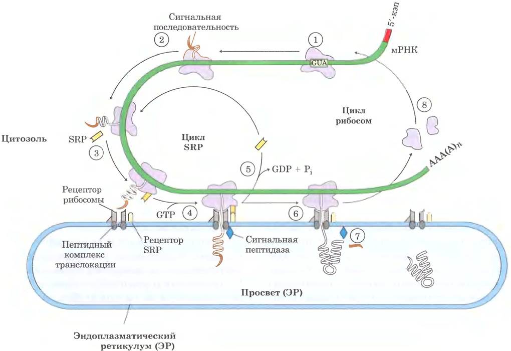
George Palade was the first to demonstrate that proteins bearing such signal sequences are synthesized on ER-associated ribosomes. The signal sequence helps guide the ribosome to the ER (steps ①–⑧ in Fig. 27-38). ① Targeting preparation begins at the initiation of Protein Synthesis on free ribosomes. ② The signal sequence is generated at the very beginning of synthesis because it resides at the N-terminus, which is synthesized first. ③ As the signal sequence emerges from the ribosome, it is bound by a large signal recognition particle (SRP); the SRP then binds GTP and arrests polypeptide elongation once the chain reaches approximately 70 amino acids in length and the signal sequence is fully exposed. ④ The GTP-bound SRP now directs the ribosome (still attached to the mRNA) and the nascent polypeptide to the GTP-bound SRP receptor on the cytoplasmic surface of the ER; the polypeptide is delivered to the ER protein translocation complex, which interacts directly with the ribosome. ⑤ The SRP dissociates from the ribosome, a process accompanied by GTP Hydrolysis in both the SRP and the SRP receptor. ⑥ Polypeptide elongation resumes with the assistance of an ATP-dependent translocation complex that feeds the growing polypeptide into the ER lumen until synthesis is complete. ⑦ The signal sequence is cleaved off by a signal peptidase within the ER lumen; ⑧ the ribosome dissociates and becomes available for another round of translation.

Fig. 27-38. Delivery of eukaryotic proteins bearing signal sequences to the endoplasmic reticulum. This process involves the SRP cycle, translocation, and Cleavage of the newly formed polypeptide (see text). The rod-shaped SRP complex contains a 300-nucleotide RNA (7SL RNA) and six distinct proteins (total molecular weight ~325,000). One SRP protein subunit binds directly to the signal sequence and arrests elongation by blocking access of aminoacyl-tRNA molecules and inhibiting peptidyl transferase. Another protein subunit binds and hydrolyzes GTP. The heterodimeric SRP receptor consists of an α subunit (Mr = 69,000) and a β subunit (Mr = 30,000), each of which binds and hydrolyzes multiple GTP molecules.
Glycosylation Plays a Pivotal Role in Protein Transport
Within the ER lumen, Newly synthesized proteins undergo various modifications. Following the removal of signal sequences, Polypeptide chains fold—often with The formation of Disulfide Bonds—and many proteins are glycosylated to become Glycoproteins. In numerous glycoproteins, attachment to the corresponding Oligosaccharides occurs via Asn residues. Such N-linked oligosaccharides are structurally diverse (Chapter 7 in Vol. 1), but the initial stage of protein glycosylation is invariant. First, a 14-residue core oligosaccharide is synthesized and then transferred from a dolichol phosphate donor molecule to specific Asn residues on the protein (Fig. 27-39).
Fig. 27-39. Synthesis of the oligosaccharide moiety of glycoproteins. The oligosaccharide is built up by the sequential addition of monosaccharide units. ①, ② The initial steps occur on the cytoplasmic surface of the ER. ③ Through a translocation event, the incomplete oligosaccharide is flipped across the membrane (mechanism not shown), and ④ completion of synthesis takes place within the ER lumen. Additional mannose and glucose residues for the growing oligosaccharide are supplied by dolichol phosphate derivatives. In the first step of N-linked glycoprotein formation ⑤, ⑥, the oligosaccharide is transferred from dolichol phosphate to an Asn residue of the protein within the ER lumen. Subsequently, depending on the specific protein, the oligosaccharide moiety is modified in various ways in the ER or the Golgi apparatus. However, the five sugar residues depicted on the beige Background (after step ⑦) are conserved in all N-linked oligosaccharides. ⑧ The released dolichol pyrophosphate is flipped back across the membrane so that the pyrophosphate group is exposed on the cytoplasmic surface of the ER, where ⑨ it is hydrolyzed to regenerate dolichol phosphate.

Transferase enzymes are located on the luminal surface of the ER and thus cannot catalyze the glycosylation of cytoplasmic proteins. Following membrane translocation, the oligosaccharides of different proteins are trimmed and modified in various ways, yet the pentasaccharide core of the original 14-residue oligosaccharide is conserved in all N-linked oligosaccharides. Certain Antibiotics block one or more steps of this pathway, providing valuable tools for investigating protein glycosylation. The best-characterized of these is the antibiotic tunicamycin, which mimics The structure of UDP-N-acetylglucosamine and blocks the first step (Fig. 27-39, step ①). Although some ER proteins undergo O-glycosylation, this modification typically takes place in the Golgi apparatus or the cytoplasm (for proteins that do not enter the ER).

Subsequently, the modified Proteins can be transported to various cellular compartments. Proteins travel from the ER to the Golgi apparatus via transport vesicles (Fig. 27-40). Within the Golgi apparatus, oligosaccharides are attached to certain proteins via O-glycosidic bonds, and N-linked oligosaccharides undergo further modifications. Through mechanisms not yet fully understood, the Golgi apparatus sorts proteins and dispatches them to their final destinations. The sorting of proteins destined for secretion, the plasma membrane, or lysosomes relies on structural features distinct from signal sequences, as the latter are cleaved off within the ER lumen.
Fig. 27-40. Protein transport to lysosomes, the plasma membrane, or the extracellular space. From the ER, proteins are directed in transport vesicles to the cis-face of the Golgi apparatus. Sorting predominantly occurs at the trans-face of the Golgi apparatus.

The sorting process for Hydrolases targeted to lysosomes is the best understood. When a hydrolase enters the Golgi apparatus, a yet-unidentified element of its three-dimensional structure (sometimes called a signal patch) is recognized by phosphotransferase, which phosphorylates specific mannose residues in the oligosaccharide portion of the glycoprotein (Fig. 27-41). The presence of one or more mannose-6-phosphate residues in the oligosaccharide chain of the hydrolase serves as a signal for its delivery to the lysosome. A receptor protein in the Golgi membrane recognizes mannose-6-phosphate and binds the thus-tagged hydrolase. Vesicles containing these receptor-hydrolase complexes bud off from the trans-Golgi network and are directed to sorting vesicles. Here, under conditions of low pH and through the action of a phosphatase that removes phosphate groups from the mannose-6-phosphate residues, the receptor-hydrolase complex dissociates. The receptor then returns to the Golgi apparatus, while vesicles containing the hydrolases bud off from the sorting vesicles and are directed to lysosomes. In cells treated with tunicamycin (Fig. 27-39, step ①), hydrolases destined for lysosomes are secreted instead, confirming the key role of the N-linked oligosaccharide moiety in delivering these enzymes to lysosomes.
Fig. 27-41. Phosphorylation of mannose residues in enzymes targeted to lysosomes. N-acetylglucosamine phosphotransferase recognizes as-yet-unknown Structural Features of hydrolases destined for lysosomes.

Protein delivery to Mitochondria and chloroplasts also relies on N-terminal signal sequences contained within these proteins. While mitochondria and chloroplasts possess their own DNA, the majority of their proteins are encoded in The Nucleus and, following synthesis, imported into the appropriate organelle. Unlike other targeted transport pathways, however, preparation for Protein transport into mitochondria and chloroplasts begins only after the precursor protein has been fully synthesized and released from the ribosome. Protein precursors destined for mitochondria and chloroplasts bind to cytosolic chaperones and are delivered to receptors on the outer surface of the organelle. Then, via specialized translocation systems, the protein is transported to its destination within the organelle, after which the signal sequence is cleaved off.
Nuclear protein signal sequences are not cleaved
Macromolecules must pass through nuclear pores to move between the nucleus and the cytoplasm. RNA molecules synthesized in the nucleus are exported to the cytoplasm. Ribosomal proteins synthesized on cytoplasmic ribosomes are imported into the nucleus and assembled into 60S and 40S ribosomal subunits in the nucleolus; these assembled subunits are then exported back to the cytoplasm. A wide variety of nuclear proteins (RNA and DNA polymerases, Histones, topoisomerases, Gene Expression regulatory proteins, etc.) are synthesized in the cytoplasm and imported into the nucleus. This pathway is regulated by a complex system of molecular signals and transport proteins whose mechanisms have become increasingly clear.
In most multicellular eukaryotes, the nuclear envelope breaks down during each Cell Division and re-forms upon its completion, at which point nuclear proteins dispersed in the cytoplasm are re-imported into the nucleus. Because of the necessity of returning to the nucleus, the signal sequence that directs a protein to the nucleus—the nuclear localization sequence (NLS)—is not cleaved after the protein reaches its destination. Unlike other signal sequences, an NLS can be located almost anywhere within the primary sequence of a protein. While NLS sequences can vary considerably, many consist of four to eight amino acid residues and contain a stretch rich in basic amino acid residues (Arg or Lys).
Nuclear transport is mediated by several proteins that shuttle between the cytoplasm and the nucleus (Fig. 27-42), including importins $\alpha$ and $\beta$ and the small GTPase Ran (Ras-related nuclear protein). The importin $\alpha/\beta$ heterodimer acts as a soluble receptor for nuclear-imported proteins, with the $\alpha$-subunit serving to bind NLS-containing proteins in the cytoplasm. The complex of the NLS-containing protein and importin approaches the nuclear pore and passes through it in an energy-requiring process. In the nucleus, importin $\beta$ binds to the GTPase Ran and releases the delivered protein. Importin $\alpha$ binds to Ran and CAS (cellular apoptosis susceptibility protein) and dissociates from the NLS-containing protein. Importins $\alpha$ and $\beta$, complexed with Ran and CAS, are then exported from the nucleus. In the cytoplasm, Ran hydrolyzes GTP, releasing the importins to begin a new transport cycle. Ran itself is transported back into the nucleus via the binding of Ran-GDP to nuclear transport factor 2 (NTF2). In the nucleus, Ran-bound GDP is exchanged for GTP through the action of the guanine nucleotide exchange factor RanGEF (see Box 12–2, Vol. 1).
Fig. 27-42. Nuclear protein transport. ① A protein bearing a nuclear localization sequence (NLS) binds to the importin $\alpha/\beta$ complex. ② The resulting complex binds to the nuclear pore and is translocated into the nucleus. ③ In the nucleus, the binding of Ran-GTP promotes the dissociation of importin $\beta$. ④ Importin $\alpha$ binds to Ran-GTP and CAS (cellular apoptosis susceptibility protein), releasing the nuclear protein. ⑤ Importins $\alpha$ and $\beta$ and CAS are exported from the nucleus and ready for a new transport cycle. Egress into the cytoplasm is accompanied by the hydrolysis of Ran-bound GTP. ⑥ Ran-GDP binds to NTF2 and returns to the nucleus. ⑦ In the nucleus, RanGEF facilitates the exchange of GDP for GTP, leaving Ran-GTP ready to transport the next complex of importins with an NLS-containing protein. b — scanning electron micrograph of the nuclear envelope surface, showing numerous nuclear pores.

Bacteria also use signal sequences for protein transport
In bacteria, proteins are targeted to the inner or outer membranes, the periplasmic space, or the extracellular medium. Bacterial proteins feature N-terminal signal sequences (Fig. 27-43) that bear a striking resemblance to the analogous signal sequences of eukaryotic proteins directed to the ER, mitochondria, and chloroplasts.
Fig. 27-43. Signal sequences directing bacterial proteins to various localizations. Basic amino acids near the N-terminus are highlighted in blue, and hydrophobic amino acids in the central region of the signal Peptides are shown in yellow. Signal sequence cleavage sites are indicated by red arrows. Note that the coat protein and DNA of phage fd assemble into phage particles at the inner membrane of the bacterial cell (see Fig. 1–6, Vol. 1). OmpA is an outer membrane protein; LamB is a receptor protein on the cell surface of bacteriophage $\lambda$.

In most cases, protein export from E. coli cells proceeds via the mechanism illustrated in Fig. 27-44. Following translation, proteins destined for export fold very slowly because this is hindered by the N-terminal signal sequence. The soluble chaperone SecB binds to the signal sequence or to other Regions of the partially folded protein. In this form, the protein is delivered to the inner membrane protein SecA. SecA functions as both a receptor and a translocating ATPase. Upon releasing SecB, the SecA-bound protein integrates into the membrane translocation complex—composed of SecY, SecE, and SecG—and traverses the membrane through the SecYEG complex, which is approximately 20 amino acid residues long. At each
stage, ATP hydrolysis catalyzed by SecA takes place.
Fig. 27-44. Model of protein export in bacteria. ① Upon completion of translation, the polypeptide binds in the cytoplasm to the chaperone SecB, which ② delivers it to the SecA protein associated with the translocation complex (SecYEG) in the bacterial cell membrane. ③ SecB is released, and SecA inserts into the membrane, pushing approximately 20 amino acid residues of the protein through the translocation complex. ④ ATP hydrolysis by SecA provides the energy for conformational changes that allow SecA to disengage from the membrane, leaving the polypeptide in the periplasmic space. ⑤ SecA binds another ATP molecule and pushes the next 20-amino-acid segment of the protein through the translocation complex. Stages ④ and ⑤ repeat until ⑥ the entire protein has crossed the membrane and been released into the periplasm. The Electrochemical Potential across the membrane (indicated by "+" and "-" signs) serves as an additional driving force facilitating protein translocation.

Thus, protein export across the membrane is mediated by the SecA protein localized on the inner surface of the membrane rather than by a protein of the periplasmic space. This arrangement may simply stem from the fact that the translocating ATPase resides where ATP is available. The transmembrane electrochemical potential may also assist in protein translocation (though much remains unclear in this regard).
While most bacterial proteins leave the cell via this pathway, some proteins are known to be exported through an alternative mechanism involving signal sequences and receptor proteins homologous to Components of the eukaryotic SRP and its receptor (Fig. 27-38).
Proteins enter cells via receptor-mediated endocytosis
Certain proteins enter cells from the surrounding environment; in eukaryotes, these include low-density Lipoproteins (LDLs), the iron carrier transferrin, Peptide Hormones, and circulating proteins destined for degradation. There are several Mechanisms of Protein entry into cells (Fig. 27-45). In one mechanism, proteins bind to receptors in membrane indentations known as coated pits, which contain a significantly higher concentration of endocytic receptors than other areas of the cell surface. On the cytoplasmic side, these pits are covered by a lattice of the protein clathrin, which forms closed polyhedral structures (Fig. 27-46). The clathrin lattice expands as the number of receptors bound to target proteins increases, until a fully membrane-enclosed vesicle buds off from the plasma membrane with the assistance of the large GTPase dynamin and is released into the cytoplasm. Clathrin is rapidly removed enzymatically, and the vesicle fuses with an endosome. ATPase activity within the endosomal membranes lowers the pH, facilitating the dissociation of receptor-target Protein Complexes. In a related process, caveolin induces the invagination of membrane domains containing lipid rafts associated with specific types of receptors (see Fig. 11–21, Vol. 1). The resulting vesicles then fuse with caveolin-containing internal structures called caveosomes, where internalized molecules are sorted and redirected to other PARTS OF THE cell, while caveolins recycle back to the membrane surface. Additionally, pathways exist that operate independently of clathrin or caveolin; some of these involve dynamin, while others do not.
Fig. 27-45. Endocytic pathways in eukaryotic cells. In clathrin- or caveolin-dependent pathways, vesicles pinch off from the plasma membrane with the help of the GTPase dynamin. Other pathways involve neither caveolin nor clathrin, and dynamin may or may not be required.

Next, the imported proteins and their receptors undergo various transformations, with their ultimate fate depending on the cell type and protein type. Transferrin and its receptor are ultimately recycled. Certain hormones, growth factors, and immune complexes are degraded along with their receptors following the induction of a corresponding cellular response. Low-density lipoproteins are degraded after the bound Cholesterol is delivered to its destination, but LDL receptors are used repeatedly (see Fig. 21-42 in Vol. 2).
Fig. 27-46. Clathrin. (a) Three light (L) chains (Mr = 35,000) and three heavy (H) chains (Mr = 180,000) form a clathrin unit (HL)3 with a three-pronged structure known as a triskelion. (b) Triskelions assembled into a polyhedral lattice structure. (c) Electron micrograph of a coated vesicle on the cytoplasmic surface of a fibroblast plasma membrane.

Protein degradation in all cells is carried out by specialized systems
Protein degradation prevents the accumulation of abnormal or unnecessary proteins and allows for the recycling of amino acids. The half-life of eukaryotic proteins ranges from 30 seconds to many days. The turnover of most proteins is quite rapid relative to the lifespan of the cell, although certain proteins (such as Hemoglobin) may persist for the entire lifetime of the cell (for example, erythrocytes live for about 110 days). Defective proteins that have been misfolded or damaged during function, as well as regulatory enzymes catalyzing key reactions in metabolic pathways, are rapidly degraded.
Defective proteins and those with the shortest half-lives are typically degraded in both bacterial and eukaryotic cells by selective ATP-dependent cytoplasmic systems. Another system, which functions in the lysosomes of vertebrates, releases amino acids from membrane
proteins, extracellular proteins, and long-lived proteins.
In E. coli, many proteins are degraded by the ATP-dependent Lon protease (named after the "long form" of proteins that appear in the cell only in the absence of this protease). Lon protease is activated in the presence of defective or short-lived proteins; the cleavage of each peptide bond consumes two ATP molecules. The details of this process have not yet been fully elucidated. Once the protein is cleaved into small inactive peptides, other ATP-independent proteases complete the process.
The ATP-dependent pathway of protein degradation in eukaryotic cells is somewhat different and relies on the action of the protein ubiquitin. Ubiquitin (76 amino acid residues) is one of the most highly conserved proteins, being virtually identical in organisms as diverse as Yeast and humans. Ubiquitin is covalently attached to proteins destined for degradation via the ATP-dependent pathway, involving three different enzymes (E1, E2, and E3 in Fig. 27-47).
Fig. 27-47. The three-stage process of ubiquitin attachment to a protein. The process proceeds via the formation of two different intermediate complexes of ubiquitin with enzymes. Ultimately, the free carboxyl group of the C-terminal Gly residue of ubiquitin is linked via an amide (isopeptide bond) to the ε-amino group of a Lys residue in the target protein. Repeating the cycle generates polyubiquitin—a covalent polymer of ubiquitin subunits that directs the tagged eukaryotic protein to the degradation site.

Ubiquitin-tagged proteins are degraded by a large protein complex known as the 26S proteasome (Mr = 2.5 × 10⁶) (Fig. 27-48).
Fig. 27-48. Three-dimensional STRUCTURE OF THE eukaryotic proteasome. The 26S proteasome is highly conserved across all eukaryotes. It consists of a central 20S particle and two regulatory 19S particles. (a) The central particle (PDB ID 1IRU) consists of four rings forming a barrel-shaped structure. The two inner rings contain seven different β subunits each (light blue), three of which possess proteolytic activity (dark blue). The two outer rings consist of seven different α subunits (gray). (b) The central barrel is capped at both ends by regulatory particles. The central region is colored as in part (a). The base and top of each regulatory particle are shown in different shades of pink. The regulatory particle unfolds ubiquitinated proteins (blue spiral) and threads them into the barrel.

The eukaryotic proteasome contains at least 32 distinct subunits present in two copies, with most of these subunits being highly conserved across many species, from yeast to humans. The proteasome consists of Two Types of complexes: a barrel-shaped central particle and regulatory particles at both of its ends. The central 20S particle is composed of four rings; the outer rings are formed from seven α subunits, and the inner rings from seven β subunits. Three of the seven subunits in each β ring possess proteolytic activity, and all of them differ in substrate Specificity. The rings of the central particle form a barrel-like structure where the degradation of target proteins takes place. The regulatory 19S particles at the ends of the barrel each contain 18 subunits, including those that recognize and bind ubiquitinated proteins. Six of the subunits are AAA+ ATPases (see Chapter 25), which presumably unfold ubiquitinated proteins and guide the unfolded Polypeptides into the central particle for destruction. In addition, the 19S particle removes ubiquitin from the proteins undergoing degradation. Most cells contain additional regulatory complexes capable of replacing the 19S particle. These alternative regulators do not hydrolyze ATP or bind ubiquitin, yet they are essential for the degradation of certain cellular proteins. Depending on intracellular conditions, the 26S proteasome can be assisted by various regulatory complexes.
We do not yet understand all the signals that induce ubiquitin attachment, but one simple signal has been discovered. The first amino acid remaining after the removal of the N-terminal Met residue or following other post-translational N-terminal proteolytic Processing determines the half-life of many proteins (Table 27-9). These N-terminal signals have remained unchanged over billions of years of evolution—they are identical in bacterial protein degradation systems and in the human ubiquitination system. More complex signals have also been discovered (see Fig. 12-46, Vol. 1).
Table 27-9. Relationship Between Protein Half-Life and Its N-Terminal Residue
N-Terminal Residue |
Half-Life* |
Stabilizing |
|
Met, Gly, Ala, Ser, Thr, Val |
> 20 h |
Destabilizing |
|
Ile, Gln |
~ 30 min |
Tyr, Glu |
~ 10 min |
Pro |
~ 7 min |
Leu, Phe, Asp, Lys |
~ 3 min |
Arg |
~ 2 min |
* Half-lives were determined using yeast β-galactosidase modified such that the enzyme possessed a different N-terminal residue in each experiment. The half-lives of various proteins and in different organisms may vary, but the general trend appears to be universal.
Ubiquitin-dependent proteolysis is equally critical for the Regulation of cellular processes and the destruction of defective proteins. Many proteins required only at a specific stage of The Introduction/5.html">Eukaryotic Cell cycle are rapidly degraded via the ubiquitin-dependent pathway once their function is complete. Ubiquitin-dependent cyclin destruction plays a crucial role in Cell Cycle regulation (Fig. 12-46 in Vol. 1). The E2 and E3 components involved in ubiquitination reactions (Fig. 27-47) represent two large Protein Families. E2 and E3 enzymes exhibit varying affinities for target proteins, thereby regulating distinct cellular processes. Some E2 and E3 enzymes are localized to strictly defined cellular compartments, reflecting their specialization.
It is unsurprising that impairments in the ubiquitination machinery lead to various diseases. The failure to degrade specific proteins (oncogene products) that activate cell division can lead to tumor development; conversely, excessively rapid degradation of proteins acting as Tumor Suppressors can have the same effect. Inefficient or overly rapid degradation of cellular proteins is thought to underlie Kidney disease, asthma, neurodegenerative disorders such as Parkinson's and Alzheimer's diseases (characterized by distinctive protein
structures in Neurons), cystic fibrosis (resulting from the excessively rapid degradation and malfunction of chloride channels; see Box 11-3 in Vol. 1), Liddle syndrome (in which renal sodium channels fail to be degraded, leading to excessive Na+ reabsorption and early-onset Hypertension), and many other disorders. Some of these conditions can potentially be treated using proteasome inhibitors. During metabolic shifts, protein degradation is just as vital for cell survival as protein synthesis. Much remains to be learned about these metabolic pathways. ■
Summary of Section 27.3 Protein Targeting and Degradation
■ Following synthesis, many proteins are directed to their specific cellular locations. One mechanism of targeted protein delivery relies on the recognition of a signal peptide, most commonly located at the N-terminus of the newly synthesized protein.
■ In eukaryotic cells, one class of signal sequences is recognized by a signal recognition particle (SRP), which binds the signal sequence as soon as it emerges from the ribosome and delivers the entire ribosome and unfinished polypeptide to the ER. Polypeptides bearing such signal sequences are translocated into the ER lumen during synthesis, where they undergo modification and are subsequently routed to the Golgi apparatus. From there, proteins are sorted and directed to lysosomes, the plasma membrane, or transport vesicles.
■ Eukaryotic proteins destined for mitochondria and chloroplasts, as well as bacterial secreted proteins, also feature an N-terminal signal sequence.
■ Proteins targeted for nuclear transport contain an internal signal sequence which, unlike other signal sequences, is not cleaved off after the protein reaches its destination.
■ Some eukaryotic cells import proteins via receptor-mediated endocytosis.
■ All cells eventually degrade proteins using specialized proteolytic systems. Defective proteins and those with short half-lives are typically degraded via an ATP-dependent pathway. In eukaryotic cells, proteins are first tagged by conjugation to the highly conserved protein ubiquitin. Ubiquitin-dependent proteolysis is carried out by proteasomes, which are likewise highly conserved. This mechanism plays a crucial role in The regulation of many cellular processes.
■ Many well-characterized antibiotics and toxins inhibit specific Stages of Protein Synthesis.
Last update: 06/08/2026
Editorial and Educational Adaptation: This material has been compiled based on the primary/original source text. The project team performed an editorial review, corrected technical inaccuracies, structured sections, and adapted the content for an educational format.
What was processed:
- elimination of formatting defects (OCR errors, structural breaks, corrupted characters);
- editorial organization of content;
- standardization of terminology in accordance with academic sources;
- verification of factual statements against the original source text.
All mentions of the author, publication year, and origin of the primary text have been preserved in accordance with the source.