Biological Membranes - A. N. Ogurtsov 2012
Structure and Functions of Biomembranes
Intracellular Protein Transport
Mechanisms of Protein Transport
Cellular Membrane Structures actively participate in Intracellular Protein Transport. Following Protein Synthesis on Ribosomes in the Cytosol of Eukaryotic Cells, these Proteins must be delivered to their designated functional Organelles (or outside The Cell).
There are three primary mechanisms by which the cell accomplishes this task (Figure 76).
1. Following synthesis and folding, the protein is delivered intact to the target organelle through specialized membrane pores. This type of delivery is known as gated transport.
2. Transmembrane Translocation of proteins, during which the polypeptide is first denatured, then the polypeptide chain is threaded across one or more membranes, and finally the functional protein refolds.
3. Vesicular trafficking of proteins, in which a vesicle containing the transported proteins buds off from a membrane.
Class="center">
Figure 76 - Mechanisms of intracellular protein transport
Gated transport, for instance, delivers all proteins into the Cell Nucleus through nuclear pores. Transmembrane translocation ensures the delivery of cytosol-synthesized proteins to Peroxisomes, Mitochondria, and METABOLISM/14.html">Chloroplasts. Vesicular trafficking mediates protein delivery to Lysosomes and the secretion of proteins from the cell.
Protein targeting is governed by specialized sorting signals within their Structure.
Immediately after Protein Synthesis on the ribosome in the cytosol, while still a mere polypeptide, the sorting signals consist of specific Amino acid sequences at the ends of the protein chain, known as targeting sequences or localization sequences.
For proteins synthesized on the membranes of the rough Endoplasmic reticulum, additional sorting signals (such as sugars or phosphate groups) can be appended by specialized Enzymes within the cisternae of the Golgi apparatus during Post-translational protein modification.
Such signals typically take the form of specific ligands recognized by receptor proteins. These receptor proteins, together with their associated cargo proteins, subsequently bind to the membrane translocation complexes of the corresponding compartment.
Protein targeting sequences, consisting of a chain of 3–80 Amino Acids, are likewise recognized by specialized receptors that deliver the protein to the appropriate translocation complexes. Once delivered to the target compartment, these targeting sequences are usually cleaved from the polypeptide chain by specialized enzymes.
Among the most thoroughly studied targeting sequences are signal Peptides (also known as signal sequences), which consist of chains of 5–15 predominantly hydrophobic amino acids. The presence of such a signal sequence in a nascent protein prompts the ribosome to bind to The endoplasmic reticulum, directing the synthesized polypeptide chain into the lumen of the reticulum rather than the cytosol.
Another example of a targeting sequence is the import signal for proteins destined for Transport from the cytosol to the mitochondrial matrix. This signal comprises a chain of 20–80 amino acids that form an amphipathic polar a-helix, featuring positively charged amino acids aligned on one side of the helix and hydrophobic amino acids on the other.
For protein targeting to the cell nucleus, a specific sequence of five positively charged amino acids has been identified.
Protein transport into peroxisomes is mediated by the peroxisomal targeting sequence Ser-Lys-Lys-COOH, a C-terminal tripeptide.
There are also retention signals which do not promote protein translocation; instead, they serve as a signal that a protein has already reached its final destination and should not be transported any further. For instance, proteins bearing the so-called KDEL sequence (Lys-Asp-Glu-Leu-COOH) at their C-terminus remain in the Endoplasmic reticulum and are prevented from leaving via vesicular transport.
An illustration of the foregoing is calreticulin, a calcium-binding protein of the smooth endoplasmic reticulum (calcium-binding protein of the endoplasmic reticulum - calreticulin), whose Primary Structure is shown in Figure 77.
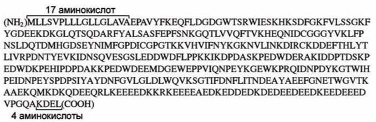
Figure 77 - Amino Acid Sequence of calreticulin
The first seventeen amino acids at the N-terminus of calreticulin function as a signal sequence that initiates protein translocation into the lumen of the endoplasmic reticulum, whereas the final four amino acids—the KDEL sequence—prevent the protein from escaping the reticulum. The Introduction/19.html">Primary structure of the functional protein is located between these two sorting signals.
Last update: 13/08/2026
Editorial and Educational Adaptation: This material has been compiled based on the primary/original source text. The project team performed an editorial review, corrected technical inaccuracies, structured sections, and adapted the content for an educational format.
What was processed:
- elimination of formatting defects (OCR errors, structural breaks, corrupted characters);
- editorial organization of content;
- standardization of terminology in accordance with academic sources;
- verification of factual statements against the original source text.
All mentions of the author, publication year, and origin of the primary text have been preserved in accordance with the source.