Medical Genetics - V. M. Zaporozhan 2005
Fundamentals of Oncogenetics
Targets of Gene Action Involved in Carcinogenesis
Tumor Suppressors
These Gene products inhibit Cell proliferation. They encode:
1. Proto-oncogene inhibitors — components of signaling pathways: NF1 (RAS protein inhibitor); p16 (cyclin D-Cdk4 complex inhibitor); pRb (METABOLISM/31.html">Transcription factor inhibitor).
2. Receptors for anti-mitogenic factors (TGF-βR).
3. Adaptor Proteins that transmit signals from these factors (SMAD2 and SMAD3).
4. Activator of the anti-mitogenic gene (SMAD4).
5. Genes that promote apoptosis (p53, E-cadherin, WT1 — on chromosome 11).
One of the most crucial tumor suppressor genes is the p53 gene (17p13.1), which plays a central role in triggering apoptosis in genetically altered Cells, particularly those that have undergone tumorigenic transformation. If a cell sustains DNA damage, errors in chromosome segregation during mitosis, microtubule disruption, or similar anomalies, the p53 gene is activated. It arrests the mitotic cycle at a specific checkpoint until the damage is repaired. If repair is impossible, the gene halts Cell Division (entering cellular senescence) or (if the damaged cell poses a potential threat to its microenvironment) initiates the apoptotic program. Due to these vital Functions, the p53 gene has been termed the “guardian of The Genome.”
Mutations in this gene can be inherited and lead to Li-Fraumeni syndrome, a rare familial syndrome characterized by multiple primary tumors. Carriers of the gene develop tumors in various Organs at an early age (sarcomas, osteosarcomas, breast Cancer, Brain Tumors, adrenal cortex carcinoma, colon cancer, leukemias). Fig. 8.3 illustrates the pedigree of a family with Li-Fraumeni syndrome. Gene penetrance is high: by the age of 70, malignant tumors develop in 90% of mutant gene carriers.
Somatic mutations of the p53 gene are detected in approximately 50% of all human tumors.
Mutations of the p53 gene play a significant role in mediating multiple drug resistance in malignant tumors.
Class="center">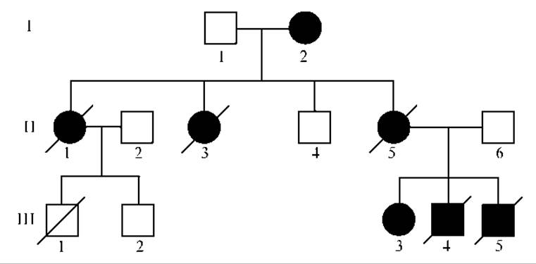
Fig. 8.3. Pedigree of a family with Li-Fraumeni syndrome:
I, 2 — bilateral breast cancer diagnosed at age 40; II, 1 — brain tumor at age 35; II, 3 — Sarcoma at age 19 and breast cancer at age 33; II, 5 — breast cancer at age 32; III, 3 — osteosarcoma at age 8; III, 4 — leukemia at age 2; III, 5 — sarcoma at age 3
Mutant tumor suppressor genes are recessive; therefore, altering the phenotype requires mutations in both alleles. They can be inherited and account for A large number of hereditary cancer syndromes (Table 8.3).
A classic example of a tumor caused by tumor suppressor gene mutation is retinoblastoma (Fig. 8.4). The retinoblastoma gene, RB1, is located on chromosome 13 (13q14.1-q14.2). In 60% of cases, the tumor is sporadic and affects one eye. In 40%
of cases, retinoblastoma is inherited, affecting both eyes. Tumor suppressor Gene Mutations are recessive. If a child inherits a mutant gene from one parent, they become heterozygous. The normal dominant gene is typically sufficient to maintain normal function. How, then, does homozygotization occur when a single recessive mutant suppressor gene is inherited?
Table 8.3. Hereditary cancers and oncogenetic syndromes (primarily caused by tumor suppressor gene mutations)
|
Disease |
Main tumor types |
Gene (locus) |
|
Hereditary breast cancer |
Early-onset breast carcinomas, often bilateral, ovarian tumors |
BRCA1 17q BRCA2 13q p53 (rare) 17p ATM (rare) 11q |
|
Hereditary familial polyposis of the colon (Lynch syndrome) |
Multiple colonic polyps, tendency toward malignant transformation |
APC 5q |
|
Hereditary non-polyposis Colorectal Cancer |
Multiple colon carcinomas, frequently combined with tumors of other localizations |
MSH2 2p MLH1 3p PMS1 (less frequent) 2q PMS2 (less frequent) 7p |
|
Li-Fraumeni syndrome |
Sarcomas, leukemias, breast and brain tumors, and other malignancies |
p53 17p |
|
von Hippel-Lindau syndrome |
Bilateral Renal Tumors, Central Nervous system lesions |
VHL 3p |
|
Retinoblastoma |
Bilateral retinal tumors, sarcomas |
RB1 13q |
|
Multiple endocrine neoplasia type 1 |
Involvement of the pituitary, Parathyroid glands, Pancreas, etc. |
MEN1 11q |
|
Multiple endocrine neoplasia type 2 |
Thyroid involvement, often associated with tumors of other endocrine organs |
RET (oncogene) 10q |
|
Gorlin syndrome |
Multiple basal cell carcinomas, less commonly brain tumors |
PTCH 9q |
|
Bilateral renal involvement |
WT1 11p |
|
|
Neurofibromatosis type 1 (Recklinghausen's disease) |
Neurofibrosarcomas, gliomas, pheochromocytomas, leukemia |
NF1 17q |
|
Neurofibromatosis type 2 |
Meningiomas, bilateral acoustic neuromas |
NF2 22q |
|
Familial melanoma |
Multiple melanomas |
CDK4 12q |
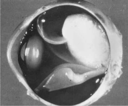
Fig. 8.4. Retinoblastoma (malignant tumor of the retina)
In 1971, Knudson proposed a hypothesis to explain this phenomenon. According to this hypothesis, tumor development requires the inactivation of both suppressor genes. In the hereditary form of the disease, the first mutation is inherited from the parents, while the second arises as a somatic mutation in the retina. Thus, in most heterozygotes, a somatic mutation is added to the germline mutation, leading to homozygotization — a process with an extremely high probability (90% in the case of retinoblastoma). This indicates that the presence of a single mutant suppressor gene increases the likelihood of a mutation in the remaining allele. In 10% of heterozygous carriers, tumors do not develop, yet they still transmit the mutant gene to 50% of their offspring. Consequently, retinoblastoma is inherited as an autosomal dominant trait with 90% penetrance. In sporadic retinoblastoma, both mutations occur in somatic cells.
Knudson's hypothesis became known as the two-hit mechanism of homozygotization.
Today, various mechanisms of homozygotization — both genetic and epigenetic — have been elucidated.
1. Genetic mechanisms:
— loss of an entire chromosome containing the normal dominant gene;
— deletion of the normal allele;
— point mutation inactivating the suppressor gene;
— mitotic recombination, which can lead to The entry of two chromatids with the retinoblastoma gene into cells.
2. Epigenetic mechanisms — methylation of suppressor gene promoters, leading to their inactivation.
Modern molecular genetic Research Methods have made it possible to identify all the described mechanisms of homozygotization.
Last update: 11/08/2026
Editorial and Educational Adaptation: This material has been compiled based on the primary/original source text. The project team performed an editorial review, corrected technical inaccuracies, structured sections, and adapted the content for an educational format.
What was processed:
- elimination of formatting defects (OCR errors, structural breaks, corrupted characters);
- editorial organization of content;
- standardization of terminology in accordance with academic sources;
- verification of factual statements against the original source text.
All mentions of the author, publication year, and origin of the primary text have been preserved in accordance with the source.