Review of Medical Physiology - William F. Ganong 2002
Functions of the Nervous System
Central Regulation of Visceral Functions
Hypothalamus - Regulation of Posterior Pituitary Secretion
In most mammals, the Hormones of the posterior pituitary are Arginine vasopressin (AVP) and oxytocin. In the vasopressin molecule of hippopotamuses and most swine species, arginine is replaced by Lysine (lysine vasopressin). The posterior pituitary of certain pig species and marsupials contains a mixture of arginine and lysine vasopressin. Posterior Pituitary Hormones are nonapeptides featuring a disulfide ring at one end of the molecule (Fig. 14-10).
Biosynthesis, Intraneuronal Transport, and Secretion
Posterior pituitary hormones are synthesized in The Cell bodies of large Neurons within the supraoptic and paraventricular nuclei. They are transported along the axons of these neurons to nerve terminals located in the posterior pituitary, where they are released in response to electrical signals reaching the terminals. Some neurons synthesize oxytocin, whereas others synthesize vasopressin. Both nuclei contain Nerve Cells that store either oxytocin or vasopressin.
Oxytocin and vasopressin are classic neurohormones—that is, hormones released into the bloodstream by nerve cells. This type of neural regulation, compared with
others, is illustrated in Fig. 14-11. The term neurosecretion originally referred specifically to the secretion of hormones by nerve cells, though it can sometimes be misleading since all neurons are now known to produce certain chemical substances, namely Neurotransmitters (see Chapter 1).
Like other Peptide Hormones, posterior pituitary hormones are synthesized as large precursor molecules. Within the neuronal granules where synthesis takes place, both oxytocin and vasopressin are bound to a specific carrier protein known as a neurophysin: neurophysin I in the case of oxytocin, and neurophysin II in the case of vasopressin. Initially, neurophysins were thought to act as binding Polypeptides forming weak, labile bonds with vasopressin or oxytocin molecules. It is now understood that they are integral PARTS OF THE precursor molecule. The precursor of arginine vasopressin—prepropressophysin—consists of a 19-amino-acid leader sequence, arginine vasopressin, neurophysin II, and a C-terminal glycopeptide (Fig. 14-12). The preprooxyphysin molecule, which is the precursor for oxytocin, is smaller because it lacks the glycopeptide component.
Precursor molecules are synthesized in the Ribosomes of neuronal cell bodies. Their leader sequences direct them into The Endoplasmic reticulum. These compounds then accumulate within secretory granules in the Golgi complex and are transported via axoplasmic flow to the terminals in the posterior pituitary. These secretory granules, known as Herring bodies, are easily identified because they stain densely in histological sections. Cleavage of the precursor molecule occurs during axonal transport, with the result that the granules in the nerve terminals contain free oxytocin and vasopressin, their respective neurophysins, and the glycopeptide fragment derived from the vasopressin precursor. The physiological function of these co-secreted substances remains unclear.
Electrical Activity of Large Neurons
The neurons that synthesize oxytocin and vasopressin also generate and propagate action potentials. Upon reaching the nerve terminals, these potentials trigger hormone release via Ca2+-dependent exocytosis. At least in anesthetized rats, these neurons show little to no resting electrical activity or exhibit low-level, irregular potentials (0.1–3.0 spikes/s). However, their response to stimulation varies significantly (see Fig. 14-13). Nipple stimulation induces high-frequency synchronous firing in oxytocin neurons after a characteristic latency period, which leads to oxytocin release and subsequent milk ejection (see below). Conversely, stimulation of vasopressin neurons, such as during Hemorrhage, initially produces a sustained increase in firing followed by a prolonged phasic discharge pattern, characterized by alternating periods of high-frequency spiking and electrical silence (phasic bursting). These phasic bursts are generally asynchronous among different vasopressinergic neurons. This firing pattern ensures a sustained, heightened release of vasopressin, contrasting with the synchronous, relatively brief, high-frequency discharge seen in oxytocin-synthesizing neurons.
Class="center">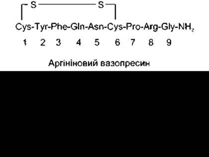
Fig. 14-10. Arginine vasopressin and oxytocin.
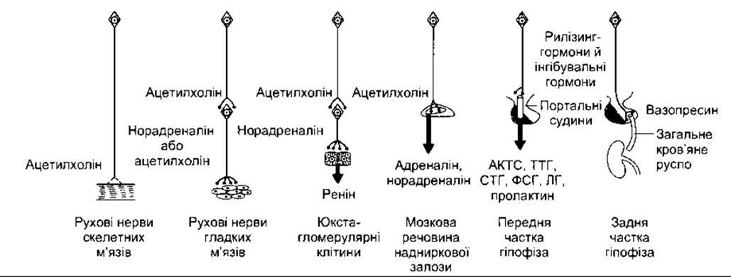
Fig. 14-11. Mechanisms of neural regulation. Two pathways on the far left: neurotransmitters act on Muscle nerve terminals. Two middle pathways: neurotransmitters regulate the secretion of Endocrine glands. Two pathways on the far right: neurons produce hormones released into the hypophysial portal system or the general bloodstream.
Vasopressin and Oxytocin in Other Regions
Vasopressinergic neurons are also located in the suprachiasmatic nuclei, and both vasopressin and oxytocin are found in nerve terminals whose axons project from the paraventricular nuclei to the Brainstem and Spinal Cord. These neurons appear to be involved in cardiovascular regulation. Additionally, vasopressin and oxytocin have been detected in the Gonads and the adrenal cortex, while oxytocin has also been found in the Thymus. The physiological function of these Peptides in these Organs remains unknown.
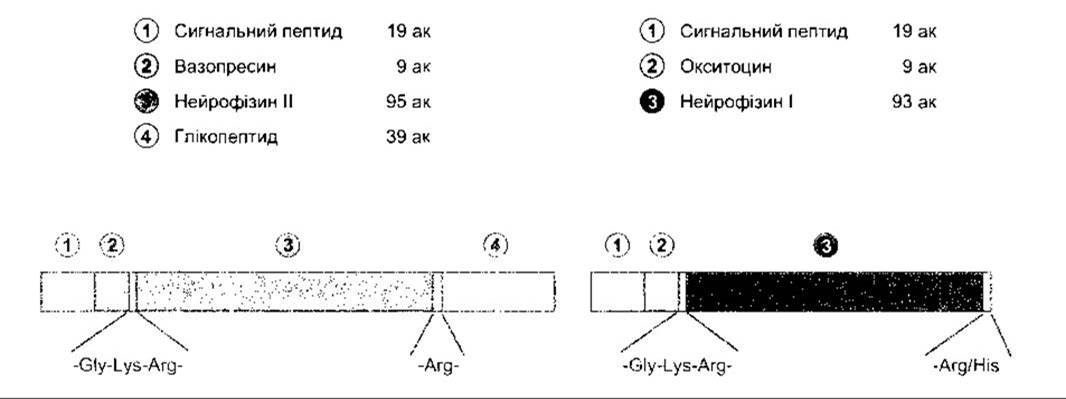
Fig. 14-12. Structure of bovine prepropressophysin (left) and preprooxyphysin (right). Glycine at position 10 in both peptides is required for the amidation of the glycine residue at position 9; aa, amino acid residues (reproduced with permission from Richter D: Molecular events in expression of vasopressin and oxytocin and their cognate receptors).
Regulation of Vasopressin Secretion: Osmotic Stimuli
Vasopressin is stored in the posterior pituitary and released into the bloodstream in response to neural impulses traveling along hormone-containing nerve fibers. Factors influencing its secretion are listed in Table 14-2. When the effective plasma osmotic pressure rises above the normal baseline (285 mOsm/kg), the firing rate of these neurons increases, leading to a rise in vasopressin secretion (Fig. 14-14). At 285 mOsm/kg, the plasma vasopressin concentration is at or near the sensitivity limit of current assay Methods, and it presumably decreases further when plasma osmolality falls below normal. Vasopressin secretion is regulated by osmoreceptors located in the anterior Hypothalamus. These receptors lie outside the Blood-Brain barrier and are believed to reside in the circumventricular organs, principally the organum vasculosum of the lamina terminalis (see Chapter 32). The osmotic threshold for thirst (see Fig. 14-8) is virtually identical to or slightly higher than the threshold for increased vasopressin secretion (see Fig. 14-14), though it remains unclear whether the exact same receptors mediate both responses.
Thus, The regulation of vasopressin secretion relies on a sensitive feedback mechanism that maintains plasma osmolality within strict limits. Even a 1% change in osmolality triggers a noticeable shift in vasopressin secretion rates. Consequently, the plasma osmolality of healthy individuals remains exceptionally close to 285 mOsm/L.
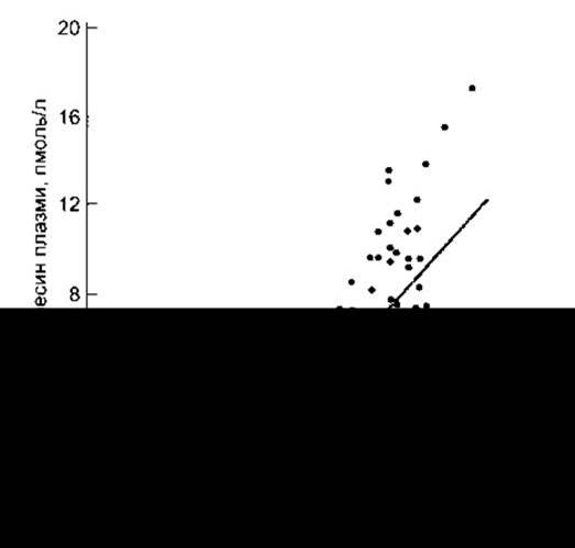
Fig. 14-14. Relationship between plasma osmolality and plasma vasopressin concentration in healthy humans during infusion of a hypertonic solution. MB, limit of measurement (reproduced with permission from Thompson CJ et al: The osmotic thresholds for thirst and vasopressin are similar in healthy humans. Clin Sci [Colch] 1986;71:651).
Table 12-2. Factors influencing vasopressin secretion
Factors increasing vasopressin secretion |
Factors decreasing vasopressin secretion |
Increased effective plasma osmotic pressure Decreased extracellular fluid volume Pain, emotion, "stress", exercise Nausea and vomiting Standing Clofibrate, carbamazepine Angiotensin II |
Decreased effective plasma osmotic pressure Increased extracellular fluid volume Alcohol |
Effect of Volume
Changes in ECF volume, as noted above, alter vasopressin secretion. Vasopressin secretion increases when ECF volume decreases and decreases when it increases (see Table 14-2). There is an inverse relationship between The rate of vasopressin secretion and the frequency of impulses in afferent neurons running from vascular stretch receptors in both low- and high-pressure areas of The Vascular System. Low-pressure receptors are located in the walls of the large Veins, the right and left atria, and the pulmonary vessels; high-pressure receptors are the carotid sinus and aortic arch receptors (see Chapter 31). The curve illustrating the exponential increase in plasma vasopressin concentration caused by a drop in blood pressure is shown in Fig. 14-15. However, low-pressure receptors primarily respond to vascular system filling. A minor reduction in blood volume lowers pressure in the large veins without decreasing arterial pressure, while also elevating plasma vasopressin levels.
Thus, low-pressure receptors act as critical initiators of the vascular filling-mediated effect on vasopressin secretion. Impulses from these receptors travel via the Vagus nerve to the Nucleus of the solitary tract (NST). From the NST, inhibitory impulses project to the caudal ventrolateral medulla (CVLM), which in turn gives rise to a direct excitatory pathway to the hypothalamus. Angiotensin II, acting on the circumventricular organs (see Chapter 32), enhances vasopressin secretion, thereby amplifying the response to hypovolemia and hypotension.
In cases of hypovolemia and hypotension caused, in particular, by blood loss, significant amounts of vasopressin are released, and during hypovolemia, the osmolality curve shifts to the left (Fig. 14-16); its slope also increases. The action of vasopressin results in Water retention and a decrease in plasma osmolality. This is accompanied by a drop in plasma Na+ concentration, leading to hyponatremia.
Other Factors Influencing Vasopressin Secretion
In addition to changes in osmotic pressure and ECF volume, factors affecting vasopressin secretion include pain, nausea, surgical stress, and certain emotions (see Table 14-2). Nausea is a particularly potent stimulus for vasopressin release. Alcohol suppresses vasopressin secretion.
Clinical Aspects
In various clinical scenarios, changes in fluid volume or other non-osmotic stimuli interfere with the osmotic regulation of vasopressin secretion. For instance, postoperative patients may exhibit elevated plasma vasopressin levels due to pain and hypovolemia, leading to decreased plasma osmolality and dilutional hyponatremia. In patients with vasopressin hypersecretion, high fluid intake can result in water intoxication.
In the syndrome of inappropriate antidiuretic hormone hypersecretion (SIADH), vasopressin not only causes dilutional hyponatremia but also—by reducing aldosterone secretion (see Chapter 20)—promotes renal salt wasting, provided water retention is sufficient to maintain ECF volume. This condition is observed in patients with cerebral ("cerebral salt wasting") or pulmonary pathologies ("pulmonary salt wasting"). Vasopressin hypersecretion in patients with pulmonary disorders, particularly Lung Cancer, may be partly due to the cessation of inhibitory impulses in vagal afferent fibers originating from stretch receptors in the atria and large veins. However, it is well established that many Lung Tumors and certain other neoplasms produce vasopressin. Patients with inappropriate vasopressin hypersecretion can be successfully treated with demeclocycline, an antibiotic that diminishes renal responsiveness to vasopressin.
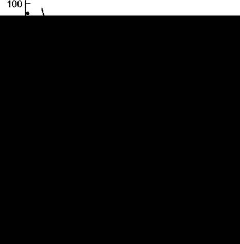
Fig. 14-15. Relationship between ARTERIAL BLOOD PRESSURE and plasma vasopressin concentration in healthy subjects in whom a gradual decrease in blood pressure was induced by administration of graded doses of the ganglionic blocker trimethaphan. The relationship curve is exponential rather than linear (adapted from Baylis PH: Osmoregulation and control of vasopressin secretion in healthy humans. Am J Physiol 1987; 253:R671).
Diabetes insipidus is a syndrome caused by vasopressin deficiency or unresponsiveness of the renal tubular epithelium to the hormone.
Causes of vasopressin deficiency include pathological processes in the supraoptic and paraventricular nuclei, the hypothalamohypophysial tract, or the posterior Pituitary Gland. Statistics show that 30% of clinical cases are caused by hypothalamic tumors (either primary or metastatic); 30% result from HEAD trauma; 30% are idiopathic; and the remainder are due to vascular damage, infections, systemic diseases such as sarcoidosis affecting the hypothalamus, or Mutations in the prepropressophysin Gene. Disturbances following surgical removal of the posterior pituitary may be transient if only the distal ends of the fibers originating from the supraoptic and paraventricular nuclei are damaged, as these fibers can regenerate, re-establish vascular connections, and restore vasopressin secretion. The symptoms of diabetes insipidus include The excretion of large volumes of dilute urine (polyuria) and excessive fluid intake (polydipsia), provided the thirst control mechanisms remain intact. Polydipsia in this case is appropriate. If the sensation of thirst is suppressed for any reason, water intake drops, leading to potentially fatal dehydration.
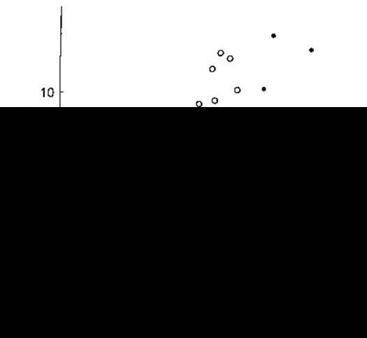
Fig. 14-16. Effect of hypovolemia and hypervolemia On the Relationship between plasma vasopressin concentration (ADH) and plasma osmolality (Posm). Seven blood samples were drawn at different times from ten healthy individuals in whom hypovolemia was induced by water deprivation, followed by hypervolemia via infusion of a hypertonic solution (black circles, solid line). Linear regression analysis revealed the relationships ADH = 0.52 (Posm - 283.5) during water deprivation and ADH = 0.38 (Posm - 285.6) during hypertonic infusion. MB — limit of detection (minimum detectable dose). Note the increase in the slope of the curve and its leftward shift during hypovolemia (courtesy of CJ Thompson).
Another cause of diabetes insipidus is the unresponsiveness of the renal tubular epithelium to vasopressin (nephrogenic diabetes insipidus). In one form of the disease, a congenital defect of the V2 receptor, resulting from various mutations in its gene, leads to impaired cAMP generation. This disorder is X-linked, as the V2 gene is located on the X chromosome. In another form, mutations in the autosomal aquaporin-2 gene result in The production of non-functional water channels. Interestingly, aquaporin-2 is normally present in urine, and in vasopressin-deficiency diabetes insipidus, injection of a vasopressin agonist causes a rapid increase in urinary aquaporin-2 concentration. However, no such increase is observed in nephrogenic diabetes insipidus.
The impact of concurrent anterior pituitary deficiency on the course of diabetes insipidus is discussed in Chapter 22.
Actions of Oxytocin
Oxytocin acts primarily on the mammary gland and Uterus, although it may also play a role in luteolysis (see Chapter 23). G protein-coupled serpentine oxytocin receptors have been identified in the human myometrium, with identical or similar receptors also found in mammary gland and ovarian Tissues. These receptors trigger an increase in intracellular Ca2+ concentration.
In mammals, oxytocin causes the contraction of myoepithelial cells—smooth muscle-like cells lining the ducts of the mammary gland. During Lactation, this contraction forces milk out of the glandular alveoli into the milk sinuses and onwards through the nipple (milk ejection). While numerous hormones interact to drive mammary gland growth and milk secretion (see Chapter 23), milk ejection in most species is regulated by oxytocin.
The Milk Ejection Reflex
Milk ejection is initiated via a neuroendocrine reflex pathway. The receptors are tactile nerve endings that are abundant in the mammary gland, particularly around the nipple. Impulses generated in these receptors travel via somatic tactile pathways to the supraoptic and paraventricular nuclei. Excitation of oxytocin-containing neurons prompts the posterior pituitary to release oxytocin (see Fig. 14-13). When an infant suckles, it stimulates tactile receptors, which in turn activate neurons in the supraoptic and paraventricular nuclei, releasing oxytocin that drives milk into the sinuses and ultimately into the hungry infant's Mouth. During lactation, stimulation of the reproductive organs as well as emotional stimuli can also trigger oxytocin release, occasionally causing milk leakage.
Oxytocin induces contractions of the uterine smooth muscle. Uterine sensitivity to oxytocin is enhanced by estrogen and inhibited by progesterone. The inhibitory effect of progesterone is mediated through the direct action of Steroids on uterine oxytocin receptors. In late Pregnancy, uterine sensitivity to oxytocin increases, accompanied by a marked upregulation of oxytocin receptors and their mRNA (see Chapter 23). Oxytocin secretion surges during labor. Following the dilation of the cervix, fetal descent through the birth canal initiates afferent nerve impulses that travel to the supraoptic and paraventricular nuclei, triggering the secretion of The amount of oxytocin required to intensify labor contractions (see Fig. 23-41). Baseline oxytocin levels are normal at the onset of labor. It is likely that the pronounced increase in oxytocin receptor density at this time, combined with normal oxytocin levels, initiates contractions and establishes a positive feedback loop. However, uterine oxytocin concentrations are also elevated during this period, suggesting that local oxytocin production may play a contributory role as well.
Oxytocin may exert similar effects on the non-pregnant uterus, facilitating sperm transport. The migration of sperm through the female reproductive tract to the fallopian tubes, where Fertilization predominantly takes place, depends not only on sperm motility but also, at least in some instances, on uterine contractions. Genital stimulation during sexual intercourse leads to oxytocin release, although it has not been definitively proven that oxytocin directly initiates the specific uterine movements that aid sperm transport. Oxytocin secretion is stimulated by stressful stimuli and inhibited by alcohol, much like vasopressin.
The level of circulating oxytocin in the male body increases during ejaculation, which may cause enhanced contraction of the smooth Muscles of the vas deferens and, consequently, the propulsion of sperm toward the Urethra.
Last update: 10/08/2026
Editorial and Educational Adaptation: This material has been compiled based on the primary/original source text. The project team performed an editorial review, corrected technical inaccuracies, structured sections, and adapted the content for an educational format.
What was processed:
- elimination of formatting defects (OCR errors, structural breaks, corrupted characters);
- editorial organization of content;
- standardization of terminology in accordance with academic sources;
- verification of factual statements against the original source text.
All mentions of the author, publication year, and origin of the primary text have been preserved in accordance with the source.