Review of Medical Physiology - William F. Ganong 2002
Functions of the Nervous System
Central Regulation of Visceral Functions
Hypothalamus - Regulation of Anterior Pituitary Secretion
Hormones of the Anterior Pituitary
The anterior pituitary secretes six hormones: adrenocorticotropic hormone (corticotropin, ACTH), thyroid-stimulating hormone (thyrotropin, TSH), Growth Hormone, follicle-stimulating hormone (FSH), luteinizing hormone (LH), and prolactin. The polypeptide ß-lipotropin (ß-LPH) is secreted along with ACTH, although its physiological role remains unknown. The actions of these hormones are illustrated in Fig. 14-17, and their synonyms and Abbreviations are listed in Table 22-1. The Functions of the hormones are described in detail in the chapters dedicated to the Endocrine System. The Hypothalamus plays an important stimulatory role in regulating the secretion of ACTH, ß-LPH, TSH, growth hormone, FSH, and LH. It also regulates prolactin secretion, although this action is inhibitory rather than stimulatory.
Nature of Hypothalamic Regulation
The secretory activity of the anterior pituitary is regulated by chemical agents delivered from the hypothalamus via the hypophyseal portal vessel system. These substances are called releasing factors and inhibitory factors. Recently, the collective term hypophysiotropic hormones has come into use. This new term is appropriate because these substances are secreted into the bloodstream and act at a distance from their site of production. They do not enter the general Circulation, but are found in high concentrations in the hypophyseal portal vessels.
Class="center">
Fig. 14-17. Hormones of the anterior pituitary. In females, FSH and R&H act sequentially to promote ovarian follicle growth, ovulation, and the formation and Maintenance of the corpus luteum. In males, FSH and R&H regulate testicular function (see Chapter 23). Prolactin stimulates Lactation.
Hypophysiotropic Hormones
To date, six hypothalamic releasing and inhibitory hormones have been identified (Fig. 14-18): corticotropin-releasing hormone (CRH); thyrotropin-releasing hormone (TRH); growth hormone-releasing hormone (GHRH); growth hormone-inhibiting hormone (GHIH, also known as Somatostatin); luteinizing hormone-releasing hormone (LHRH), also known as gonadotropin-releasing hormone (GnRH); and prolactin-inhibiting hormone (PIH). In addition, hypothalamic extracts exhibit prolactin-releasing activity. Therefore, it is believed that a prolactin-releasing hormone (PRH) exists; TRH, VIP, and several other Polypeptides found in the hypothalamus stimulate prolactin secretion, though it remains unclear whether one or several of these polypeptides constitute the physiological PRH. Recently, an orphan receptor was isolated from the anterior pituitary, and a 31-amino-acid polypeptide was identified from the hypothalamus during the search for its Ligand. This polypeptide stimulates prolactin secretion by acting on an anterior pituitary receptor, but further research is needed to determine whether it represents the physiological PRH. GnRH stimulates the secretion of both FSH and LH; thus, it is likely that a separate follicle-stimulating hormone-releasing hormone does not exist.
The structures of the six hypophysiotropic hormones are shown in Fig. 14-19. The Gene and preprohormone structures of TRH, GnRH, somatostatin, CRH, and GHRH are known. Prepro-TRH contains six copies of TRH (see Fig. 1-22). Individual preprohormones, In addition to hypophysiotropic Peptides, may contain other hormonally active peptides.

Fig. 14-18. Effect of hypophysiotropic hormones on anterior pituitary hormone secretion.

Fig. 14-19. Structure of human hypophysiotropic hormones. Preprosomatostatin is converted into a tetradecapeptide (somatostatin 14, [SS14], whose formula is shown above) as well as into a 28-amino-acid polypeptide (SS28).
The region where hypothalamic releasing and inhibitory hormones are released is the median eminence of the hypothalamus. It contains few nerve Cell bodies, but possesses numerous nerve terminals located directly adjacent to the capillary loops that give rise to the portal vessels.
The localization of neuronal cell bodies producing hypophysiotropic hormones, whose processes extend to the outer layer of the median eminence, is shown in Fig. 14-20. The distribution of Neurons synthesizing oxytocin and vasopressin is also depicted there. GnRH-producing neurons are located primarily in the medial preoptic area, somatostatin-producing neurons in the periventricular nuclei, TRH- and CRH-producing neurons in the medial PARTS OF THE paraventricular nuclei, and GHRH- and dopamine-producing neurons in the arcuate nuclei.
Most (if not all) hypophysiotropic hormones influence the secretion of more than one anterior pituitary hormone (see Fig. 14-18). The FSH-stimulating activity of GnRH was mentioned above. TRH stimulates the secretion of both prolactin and TSH. Somatostatin inhibits the secretion of both TSH and growth hormone. Normally, it does not inhibit the secretion of other Anterior Pituitary Hormones, but it suppresses abnormally high ACTH secretion in patients with Nelson's syndrome. CRH stimulates the secretion of ACTH and ß-LPH (see Chapter 22).
Receptors for many hypophysiotropic hormones are serpentine and G protein-coupled. Humans have two CRH receptors: CRHR-I and CRHR-II. The second differs from the first by the presence of a 29-amino-acid insertion in the first cytoplasmic loop of the receptor. The Physiological Role of CRHR-II remains unknown, although it is found in many Regions of the Brain. In addition, a CRH-binding protein circulates in the Blood and inactivates CRH. It is also detected in the Cytoplasm of anterior pituitary corticotropes, where it may play a role in receptor internalization. However, the precise physiological significance of this protein is not yet established. Other hypophysiotropic hormones have no known binding Proteins.
Administration of CRH into the cerebral ventricles causes hyperglycemia, increased Cardiac Output, suppressed reproductive function, anorexia, decreased gastrointestinal function, and other changes characteristic of the stress response. Nevertheless, it cannot yet be definitively stated that endogenously produced CRH mediates the changes that occur during stress.
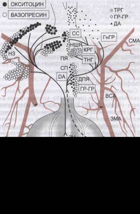
Fig. 14-20. Distribution of neuronal cell bodies releasing hypophysiotropic hormones (projection onto the ventral surface of the rat Hypothalamus and Pituitary). ICA — Internal Carotid Artery; MA — main artery; ARC — arcuate Nucleus; PCA — posterior cerebral artery; PP — posterior lobe; SOV — supraoptic nucleus; PVN — periventricular nucleus; IL — intermediate lobe; AL — anterior lobe; PaN — paraventricular nucleus; MCA — middle cerebral artery; ME — median eminence. Hormone names are enclosed in boxes (after L.W. Swanson and E.T. Cunningham Jr).
In other regions of the brain, the retina, and the Autonomic Nervous System, hypophysiotropic hormones act as Neurotransmitters (see Chapter 4). In addition, somatostatin is present in the pancreatic islets (see Chapter 19). TRH is produced by pancreatic tumor Cells, while somatostatin and TRH have also been detected in the gastrointestinal tract (see Chapter 26).
Significance and Clinical Aspects
The Study of the numerous neuroendocrine Regulatory Functions of the hypothalamus is important because it explains how they adapt to the demands of a constantly changing environment. The Nervous System receives information about Changes in the internal and external environments via Sensory Organs. This helps the body adapt through effector mechanisms that encompass not only somatic motor responses, but also alterations in hormone secretion rates. There are numerous Examples of neuroendocrine effector pathways. In birds and many mammals, an increase in daylight hours in the spring stimulates gonadotropin secretion, activates the Gonads, and triggers the breeding season. It has been observed that during the prolonged winter night, ovulation ceases in Eskimo women; regular menstrual cycles and sexual activity resume with the onset of spring. In warmer climates, menstruation occurs year-round, although its regularity is noticeably influenced by somatic and emotional stimuli. Documented phenomena include secondary Amenorrhea in young women leaving home ("dormitory amenorrhea"), cycle suppression caused by the fear of Pregnancy, and a tendency toward Menstrual cycle synchronization among close female friends in women's colleges. Thus, there is clear evidence of psychological factors influencing endocrine secretion.
Pathological processes arising in the hypothalamic region manifest as neurological disorders, endocrine dysfunctions, and metabolic abnormalities such as hyperphagia or hyperthermia. The relative frequency of signs and symptoms occurring in hypothalamic pathology is listed in Table 14-3. Hypothalamic pathology can also frequently account for many features of pituitary dysfunction, particularly those caused by deficiencies of specific hypophysiotropic hormones. In this regard, Kallmann syndrome deserves special attention, in which hypogonadism resulting from low circulating gonadotropin levels (hypogonadotropic hypogonadism) is combined with partial or complete loss of the SENSE OF SMELL (hyposmia or anosmia). Embryologically, GnRH-producing neurons develop in the Nasal cavity and migrate along the olfactory nerves and subsequently through the brain parenchyma to the hypothalamus. A disruption of this migration process due to a congenital anomaly of the olfactory pathways results in GnRH-producing neurons failing to reach the hypothalamus, leading to a failure of sexual maturation at Puberty. The syndrome occurs predominantly in males, and in most cases is caused by a mutation in the *KAL1* gene—an X-linked gene that presumably encodes an adhesion molecule required for the normal Development of the Olfactory nerve, along which GnRH-producing neurons migrate to the brain. A similar syndrome occurs in females, though it may result from other genetic defects.
Table 14-3. Symptoms and signs in 60 patients with hypothalamic pathology confirmed by post-mortem autopsy1
Symptoms and signs |
% of cases |
Endocrine and metabolic |
|
Precocious puberty |
40 |
Hypogonadism |
32 |
Diabetes insipidus |
35 |
Obesity |
25 |
Impaired thermoregulation |
22 |
Wasting |
18 |
Bulimia |
8 |
Anorexia |
7 |
Neurological |
|
Ocular symptoms |
78 |
Pyramidal and sensory disturbances |
75 |
Headache |
65 |
Extrapyramidal symptoms |
62 |
Vomiting |
40 |
Psychiatric disorders, rage attacks |
35 |
etc. |
|
Somnolence |
30 |
Seizures |
15 |
1 Data adapted from Bauer HG: Endocrine and other clinical manifestations of hypothalamic disease. J Clin Endocrinol 1954; 14:13. See also Kahana L et al: Endocrine manifestations of intracranial extrasellar lesions. J Clin Endocrinol 1962;22:304.
Last update: 10/08/2026
Editorial and Educational Adaptation: This material has been compiled based on the primary/original source text. The project team performed an editorial review, corrected technical inaccuracies, structured sections, and adapted the content for an educational format.
What was processed:
- elimination of formatting defects (OCR errors, structural breaks, corrupted characters);
- editorial organization of content;
- standardization of terminology in accordance with academic sources;
- verification of factual statements against the original source text.
All mentions of the author, publication year, and origin of the primary text have been preserved in accordance with the source.