Human Anatomy - Lecture Course - Kostylenko Yu.P. 2015
General Anatomy of the Oral Cavity Organs
Lecture Plan:
5.1 General anatomical layout of the Oral Cavity.
5.2 General Structure OF THE Organs within the oral cavity.
5.3 General Anatomy of the Teeth; concepts of articulation, occlusion, and bite.
5.4 Anomalies of bite, tooth development, and dental structure.
5.5 Dental charting and dental formulas.
5.6 MAIN STAGES OF tooth development.
5.1 General anatomical layout of the oral cavity
The oral cavity (cavitas oris; certain terms derive from the Greek word stoma, meaning Mouth, as in stomatology) is divided into two sections: the oral vestibule and the oral cavity proper. The oral vestibule is the space bounded externally by the Lips and Cheeks, and internally by the teeth and Gums. The vestibule opens to the exterior via the oral fissure.
The lips (labia oris) are composed of the fibers of the orbicularis oris Muscle, covered externally by Skin and internally by a mucous membrane. Between the skin and the mucous membrane along the free margin lies the intermediate red zone (vermilio), which differs from the skin by the absence of a cornified layer in its stratified squamous epithelium, and from the mucous membrane by the absence of Salivary Glands.
The upper lip is bounded by the Base of the Nose and the paired nasolabial sulci, while the lower lip is bounded by the mentolabial sulcus. At the corners of the mouth, the lips merge via labial commissures.
As the mucous membrane transitions from the upper lip to the alveolar process of the Maxilla, it forms a fold known as the superior labial frenulum. Similarly, the transition from the lower lip to the Mandible forms the inferior labial frenulum.
The cheeks (buccae) share a similar histological structure (lined internally by mucous membrane and covered externally by skin) and contain the buccinator muscle, laterally adjacent to the buccal fat pad, which is particularly prominent in infants and young children. Superiorly, the cheeks are bounded by the zygomatic arch, inferiorly by the base of the mandible, anteriorly by the nasolabial sulcus, and posteriorly by the masseter muscle. The lips are demarcated from the cheeks by the nasolabial and mentolabial sulci, which are faint in children and deepen with age. The skin of the cheeks and lips contains sweat and Sebaceous Glands, and in men, it is typically covered with Hair.
The mucous membrane of the cheeks and lips is thin, transparent, and represented by non-keratinized stratified squamous epithelium containing numerous minor salivary glands. Additionally, the buccal mucosa along the line of dental occlusion contains a small number of sebaceous glands.
The oral cavity proper (cavitas oris propria) extends from the teeth anteriorly and laterally to the entrance of the Pharynx posteriorly. Superiorly, it is bounded by the hard palate and the anterior portion of the soft palate; the floor is formed by the oral Diaphragm and occupied by the Tongue. When the mouth is closed, the dorsal surface of the tongue contacts the palate, causing the oral cavity to appear as a narrow slit-like space between them. The mucous membrane, passing onto the Inferior surface of the tongue's apex, forms the lingual frenulum along the midline. On either side of the frenulum, small papillae are visible, marking the openings of the excretory ducts for the submandibular and sublingual salivary glands. When the dental arches are closed, the oral vestibule communicates with the oral cavity proper through the interdental spaces and the retromolar space (posterior to the third molars)—the latter being of significant clinical importance for passing a feeding tube when the jaws are immobilized.
The palate (palatum) consists of two parts. Its anterior two-thirds have a bony foundation, the hard palate (palatum osseum), formed by the palatine processes of the maxillae and the horizontal plates of the palatine bones; the posterior third is the soft palate (palatum molle), a muscular structure with a fibrous framework.
During quiet nasal breathing, the soft palate hangs obliquely downward, separating the oral cavity from the pharynx.
Failure of the suture between the palatine processes of the right and left maxillary bones to fuse results in a pathological opening between the oral and nasal cavities, a congenital malformation known as a cleft palate (wolf's mouth). The mucous membrane covering the inferior surface of the hard palate is tightly fused to the periosteum via dense fibrous tissue.
The soft palate is a fold of mucous membrane enclosing Muscles, a fibrous sheet known as the palatine aponeurosis, and minor salivary glands. The soft palate comprises the uvula, the palatine velum, and the paired palatoglossal (anterior) and palatopharyngeal (posterior) arches. The tonsillar sinus, housing the palatine tonsil, is located between the anterior and posterior arches. The anterior edge of the soft palate attaches to the posterior margin of the hard palate, while its posterior section (the palatine velum) hangs freely downward and backward, featuring a median projection known as the uvula.
The opening connecting the oral cavity to the pharynx is called the fauces (isthmus faucium). It is bounded laterally by the arches, superiorly by the soft palate, and inferiorly by the dorsum of the tongue.
5.2 General structure of the organs within the oral cavity
The Organs of the oral cavity include the tongue, salivary glands, and teeth.
The tongue (lingua, glossa) is an unpaired muscular organ located on the floor of the oral cavity. It has a cone-like shape and is covered on its superior surface, sides, and partially on its inferior surface by mucous membrane.
The anatomical PARTS OF THE tongue include the apex (tip), body, ROOT, dorsum (superior surface), inferior surface, and two lateral margins. The mucous membrane of the tongue is covered with non-keratinized stratified squamous epithelium, which may accumulate cornified scales during certain Digestive System disorders, resulting in a grayish coating on the dorsal surface.
The mucous membrane of the tongue is equipped with specialized structures called papillae. These include filiform, conical, leaf-like (foliate) papillae (well-developed in children, but undergo atrophy in adults), fungiform, and vallate (circumvallate) papillae. The latter are arranged along the sulcus terminalis, which separates the body of the tongue from the root. Fungiform, foliate, and vallate papillae contain taste buds harboring receptors that evaluate the taste qualities of food. At the junction of the dorsum and root of the tongue lies the foramen caecum, a remnant of the embryonic thyroglossal duct. Failure of the thyroglossal duct to obliterate can lead to The formation of a median cervical cyst, which serves as an indication for surgical intervention. Posterior to the sulcus terminalis lies an accumulation of lymphoid follicles known as the lingual tonsil.
The Muscles of the tongue are paired and divided into intrinsic muscles (superior longitudinal, inferior longitudinal, transverse, and vertical), which form the bulk of the tongue, and extrinsic muscles (genioglossus, styloglossus, and hyoglossus), which connect the tongue to the Skeleton. The intrinsic muscles alter the shape of the tongue, whereas the extrinsic muscles facilitate its movement within the oral cavity.
Functions of the tongue: 1) it takes part in the intake and mastication of food within the oral cavity, as well as in the swallowing of the food bolus; 2) it plays a vital role in articulated speech production; 3) it performs sensory functions, including specific gustatory (taste) reception; 4) the lingual tonsil constitutes an integral component of the oral immunological barrier.
Developmental anomalies of the tongue:
1. absence of the tongue (aglossia);
2. underdevelopment of the tongue (hypoplasia);
...3. excessive enlargement of the tongue (macroglossia);
4. bifurcation of the tongue body;
5. duplication of the tongue;
6. restricted tongue mobility (due to a short lingual frenulum);
7. plicated (folded) tongue.
Salivary glands (glandulae salivatoriae, sialoaden) are classified into minor and major glands. The Location and number of minor salivary glands can be identified by punctiform openings on the mucosal surface using a magnifying Glass. Some of these glands are arranged in clusters within the mucosa of the oral vestibule—namely, labial, buccal, and molar glands—while others reside in the mucosa of the oral cavity proper. The latter include palatine glands, lingual glands, and minor sublingual glands, which are associated with the major sublingual salivary gland. The major salivary glands are situated outside the Oral Cavity and communicate with it via excretory ducts. There are three pairs of major salivary glands: 1 - sublingual glands, 2 - submandibular glands, and 3 - parotid glands.
The sublingual gland is a complex alveolar-tubular gland comprising both minor sublingual glands and a distinct aggregate of glandular units united by a common duct. Each sublingual gland occupies the space beneath the mucosa of the floor of the oral cavity, bounded medially by the mylohyoid, genioglossus, and hyoglossus muscles, laterally by the inner surface of the mandibular body, and inferiorly by the mylohyoid muscle.
The sublingual gland is elongated anteroposteriorly and flattened laterally, measuring approximately 3–4 cm in length, about 1 cm in thickness, and weighing roughly 5 g. When the gland possesses a common excretory duct, it either merges with the adjacent submandibular duct or opens via a shared orifice at the apex of the sublingual caruncle.
The submandibular gland is approximately three times larger than the aforementioned sublingual gland and is a complex alveolar-tubular structure. It has a flattened ovoid shape and lies within the submandibular triangle, occupying an area bounded superiorly by the inferior surface of the mylohyoid muscle and laterally by the inner surface of the mandibular body. Its excretory duct courses along the Medial surface of the sublingual gland, opening via an orifice at the apex of the sublingual caruncle.
The parotid gland is the largest of the salivary glands, with a weight ranging from 20 to 30 g. Histologically, it is a complex alveolar-tubular gland. Located within the parotid-masseteric region, the bulk of the gland occupies the retromandibular fossa, which is bounded superiorly by the cartilaginous part of the external acoustic meatus, anteriorly by the posterior border of the mandibular ramus and the masseter muscle, and posteriorly by the anterior border of the sternocleidomastoid muscle. Medially, the parotid gland abuts the Ligaments and Muscles originating from the styloid process of the Temporal bone, while a portion of the gland overlaps the posterior part of the masseter muscle.
The parotid gland has the shape of an irregular triangle, with its upper margin directed along the zygomatic arch and its lower angle situated beneath the inferior angle of the mandible, where it frequently contacts the posterior part of the submandibular gland. Of significant importance in surgical practice is the anatomical feature wherein the External Carotid Artery (with its numerous branches), the retromandibular vein, and the parotid plexus of the Facial Nerve pass directly through the substance of the parotid gland.
The excretory duct of the parotid gland, approximately 4 mm in diameter, emerges from its anterior aspect (about 1 cm below the zygomatic arch), crosses the superficial surface of the masseter muscle, reaches its anterior border, and then makes a sharp medial turn to pierce the buccal adipose pad and the buccinator muscle. It opens into the Vestibule of the oral cavity via an orifice on the buccal mucosa opposite the second maxillary molar. Additional parotid glands are frequently found along the course of the excretory duct.
Functions of the salivary glands. The Major and minor salivary glands, sharing a common developmental origin and central nervous regulation centers, function as a unified system aimed primarily at supplying the oral cavity with an adequate volume of fluid. Notably, they are capable of producing about 2 liters of saliva per day. Approximately 30% of this volume is contributed by the minor salivary glands, enabling them to compensate for the functional insufficiency of the major salivary glands under certain extreme conditions.
Furthermore, the salivary glands play a crucial role in establishing the immunological defense mechanisms of the oral cavity, as they serve as the source of secretory immunoglobulin A, which, in conjunction with salivary Lysozyme, forms an "antiseptic barrier" against pathogenic microflora.
5.3 General Anatomy of the Teeth; Concepts of Articulation, Occlusion, and Bite
In humans, the teeth form an integral part of the masticatory-speech apparatus, which is currently understood as a complex of interacting and interdependent organs involved in mastication, Respiration, vocalization, and speech. Human teeth belong to heterodont (differing in shape) and thecodont (embedded in bony sockets) systems and are characterized by a diphyodont type of dentition (undergoing two successive sets of teeth throughout life).
Teeth are calcified mucosal papillae adapted for the mechanical Processing of food. Phylogenetically, teeth are derived from fish scales that grew along the margin of the jaws and acquired new functions there. While tooth replacement occurs numerous times throughout life in lower vertebrates, it happens twice in humans. Accordingly, teeth are classified into: 1) temporary or deciduous teeth (dentes decidui), and 2) permanent teeth (dentes permanentes). Deciduous teeth differ from permanent ones in color (being somewhat whiter), smaller size, proportional crown-to-root dimensions, the presence of an enamel ridge on the vestibular surface, and a relatively larger pulp cavity.
As a result of evolution, the uniform conical teeth of fish—which served merely to retain prey—were replaced in mammals by specialized forms adapted for various Methods of food capture and processing, namely tearing (canines), cutting (incisors), crushing (premolars), and grinding (molars). The teeth are housed within the alveoli (sockets) of the alveolar processes of the maxilla and mandible. The tissue covering the alveolar processes is termed the gingiva (gums). Here, the mucosa is firmly fused to the periosteum via fibrous tissue; the gingival tissue is rich in Blood Vessels (making it relatively prone to bleeding) and has a sparse nerve supply.
Because the gingiva lacks a submucosal layer, its mucous membrane tightly adheres to the periosteum of the alveolar processes of the upper and lower jaws. This portion of the gingiva is referred to as the alveolar gingiva or attached gingiva. The section of the gingiva adjacent to the tooth surface is known as the marginal or free gingiva. The tissue occupying the interdental spaces between adjacent teeth is called the interdental papilla. Two gingival papillae are distinguished between any two adjacent teeth: a vestibular papilla facing the oral vestibule and an oral papilla located more toward the tongue. The shallow sulcus situated between the tooth surface and the free margin of the gingiva is termed the gingival sulcus (pocket).
Each tooth (dens) consists of: 1) the crown (corona dentis), 2) the neck (collum dentis), and 3) the root (radix dentis). The crown projects above the gingiva, the neck (a slightly constricted region) is embraced by the gingiva, and the root is anchored within the dental alveolus, terminating in an apex where a microscopic apical foramen can be discerned even with the naked eye. Blood Vessels and nerves enter the tooth through this foramen. Inside the crown lies the pulp cavity (cavitas dentis), which is subdivided into the coronal pulp chamber—the most expansive portion of the cavity—and the root portion, a tapered canal designated as the root canal. The canal opens at the root apex via the aforementioned apical foramen.
The crown of each tooth presents 5 surfaces: 1 - facing the oral vestibule, the vestibular surface (facies vestibularis), which in anterior teeth contacts the mucosal membrane of the lip (facies labialis), and in posterior teeth - the mucosal membrane of the cheek (facies buccalis); 2 - facing the oral cavity (facies oralis): directed toward the tongue in the lower teeth, the lingual surface (facies lingualis), and toward the palate in the upper teeth, the palatal surface (facies palatinus); 3 and 4 - contacting adjacent teeth of the same dental arch, the proximal surfaces (facies contactus s. approximales). The proximal surfaces of the teeth directed toward the center of the dental arch are designated as the mesial surface (facies mesialis, where mesos means middle). In anterior teeth, this surface is medial, whereas in posterior teeth, it is anterior. The proximal surfaces of the teeth directed away from the center of the dental arch are termed the distal surfaces (facies distalis). In anterior teeth, this surface is lateral, while in posterior teeth, it is posterior; 5 - the masticatory surface, or The surface of occlusion with the teeth of the opposing arch (facies occlusalis, from occlusivus meaning closing or shutting).
To describe the localization of pathological processes on a tooth, dental practitioners use terms corresponding to the aforementioned surfaces: vestibular, oral, medial, mesial, distal, occlusal, and apical (toward the apex radicis).
Three distinct features serve to determine whether a tooth belongs to the right or left side: 1) the root sign, 2) the crown angle sign, and 3) the crown curvature sign. The root sign implies that the longitudinal axis of the root is inclined distally, forming an angle with a line passing through the midpoint of the crown.
The crown angle sign indicates that the line of the masticatory margin of the tooth on the vestibular side forms a sharper angle upon transitioning to the mesial surface than it does upon transitioning to the distal surface.
The crown curvature sign denotes that the vestibular surface of the crown transitions into the mesial surface more abruptly than into the distal surface. Consequently, the mesial segment of the vestibular surface in the transverse direction appears more convex than the distal segment.
The dental cavity is filled with dental pulp (pulpa dentis), which is represented by loose Fibrous Connective Tissue rich in blood vessels and nerves.
The pulp comprises 3 distinct layers: 1) the peripheral layer; 2) the intermediate, or cambial, layer; and 3) the central layer.
The pulp of the root canals differs somewhat from the coronal pulp; in the former, Collagen fiber bundles predominate over cellular elements. In the region of the apical foramen, the pulp tissue transitions directly into the periodontal tissue.
The pulp provides trophic support to the hard dental Tissues (via the blood vessels and nerves running through it) and performs a plastic function—the formation of new dentin through its cellular elements.
The pulp is enclosed by hard dental tissues, which include: 1) dentin (dentinum, subst. eburnean), 2) enamel (enamelum, subst. adamantina), and 3) cementum (cementum, subst. оssea) (Fig. 5.1).
The bulk of the tooth surrounding the pulp cavity is formed by dentin, which consists of 28% organic substances, principally collagen, and 72% inorganic substances (calcium and magnesium phosphates, calcium fluoride).
Dentin is composed of a ground substance (collagen fibers and a cementing substance) permeated by a system of dentinal tubules.
Two layers of dentin are distinguished:
the outer mantle layer, and the inner circumpulpal layer.
The collagen fibers of dentin run in both radial and tangential directions. This reciprocal intersection of fibers imparts exceptional strength to the tooth. Within the dentinal tubules lie the processes of odontoblasts, which terminate in the peripheral layer of dentin. The portion of dentin facing the pulp is termed "predentin" or the "dentinogenetic layer," where the formation and apposition of dentin take place (secondary/replacement dentin).
Class="center">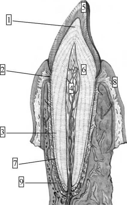
Fig. 1.5 Tooth structure (schematic diagram):
1 - tooth crown; 2 - neck of the tooth; 3 - tooth root; 4 - pulp; 5 - enamel; 6 - dentin; 7 - cementum; 8 - gingiva (gums); 9 - periodontium.
The dentin forming the crown is covered by dental enamel, whereas the root dentin is covered by dental cementum. The coronal enamel and root cementum meet in the cervical region of the tooth.
Enamel externally covers the crown, reaching a thickness of 3.5 mm at the cusp apex. Enamel contains a negligible amount of organic matter (about 3–4%) and inorganic salts (96–97%). Among the Inorganic Components, calcium phosphates and carbonates predominate, with calcium fluoride accounting for about 4%. Enamel is built of enamel prisms 3–5 µm in thickness. Each prism consists of a delicate fibrillar network housing hydroxyapatite crystals.
Cementum covers the root, and in its chemical composition, it closely approximates Bone tissue: it contains 30% organic matter and 70% inorganic matter, with calcium phosphate and carbonate salts predominating.
Between the cementum-covered root of the tooth, embedded deep within the alveolus, and the alveolar wall lies the alveolar periosteum, which lines the inner walls of the alveoli, is tightly integrated with the gingiva, and plays a significant role in stabilizing the teeth. The periosteum is a Cytology/practical/45.html">Dense connective tissue composed of bundles of fibers whose ends continue directly—as so-called Sharpey's fibers—on one side into the root cementum and on the other into the bony walls of the alveolus. This dense connective tissue is termed the periodontium (periodontium) or the root membrane.
The periosteal fibers run as ligamentous bundles arranged along the lines of functional stress exerted on the tooth. Some bundles extend radially from the tooth root into the alveolar wall, preventing tooth oscillation. Other bundles run tangentially (along the lines of contact), preventing the Rotation of the tooth around its axis. The thickness of the periodontium is 0.2–0.25 mm in the upper jaw and 0.15–0.22 mm in the lower jaw.
The periodontium performs several vital functions. First, it anchors the tooth within the alveolus; second, it provides trophic support to the tooth (as vessels and nerves pass through it to reach the tooth); third, it allows for a degree of physiological tooth mobility (Shock absorption/amortization), which is essential for complete dental occlusion. Finally, the periodontium prevents the spread of inflammatory processes and participates in tooth eruption and development.
All the tissues surrounding the neck and root of the tooth, including the gingiva, the alveolus, and the region of the alveolar process of the jaw that forms it, are regarded as an integrated anatomical and functional system termed the periodontium (parodontium) or amphodontium.
Any positions and Movements of the mandible relative to the maxilla performed by means of the masticatory muscles are called articulation. The positioning of the dental arches when they close is called occlusion. There are four MAIN TYPES OF occlusion: central, anterior, and two lateral—right and left. Central occlusion is characterized by the closure of the teeth with the maximum number of contacting points. The facial midline coincides with the line passing between the central incisors. The articular heads are located on the slope of the articular tubercle at its base. At the same time, a simultaneous and uniform contraction of the masseter and temporal muscles on both sides is observed.
Anterior occlusion involves the forward protrusion of the mandible. This is achieved by the bilateral contraction of the lateral pterygoid muscles. The facial midline, as in central occlusion, coincides with the line passing between the central incisors. The articular heads in anterior occlusion are shifted forward and located at the apex of the articular tubercles.
Lateral occlusion occurs when the mandible moves to the right—right occlusion, or to the left—left occlusion. When the mandible shifts to the right, the articular HEAD on the shift side remains at the base of the articular tubercle, rotating slightly. Meanwhile, on the left side, the articular head is located at the apex of the articular tubercle. Right lateral occlusion is accompanied by the contraction of the lateral pterygoid muscle of the opposite side (the left one) and, conversely, left lateral occlusion is accompanied by the contraction of the homonymous muscle on the right side.
The Relationship of the dental arches in central occlusion is called bite (mordex). Both physiological and pathological bites are possible. With physiological bites, chewing, speech, and facial profile are unimpaired, whereas pathological bites exhibit various disruptions.
There are four types of physiological bite: orthognathia, progenia, biognathia (biprognathism), and edge-to-edge bite (straight bite).
In orthognathia (orthos - straight, gnathio - jaw), There is a slight overlapping of the lower jaw teeth by the upper jaw incisors.
Progenia (pro - forward, genio - chin) is characterized by a reverse relationship.
Biognathia is typically characterized by the forward inclination of the upper and lower teeth, with the lower ones overlapping the upper ones.
In an edge-to-edge bite, the cutting edges of the upper and lower incisors meet each other.
5.4 Malocclusions, Developmental Anomalies, and Dental Structure
Anomalous types of bites:
1. Deep bite—absence of contact between the upper and lower incisors as a result of dentoalveolar or gnathic disorders. In a deep, traumatic bite, the cutting edges of the incisors rest against the mucous membrane of the gingival margin or alveolar process.
2. Open bite—characterized by the presence of a horizontal gap in the anterior or lateral segments of the dental arches when the teeth are closed in central occlusion; contacts are preserved only on the distal posterior teeth. There are unilateral and bilateral, symmetrical and asymmetrical open bites (most commonly caused by oral habits).
3. Crossbite (syn.: lateral bite)—a bite in which the buccal cusps of the upper posterior teeth fit into the longitudinal grooves of the lower ones or slide past them from the lingual side, meaning that the closure of the dental arches in the transverse direction is disrupted.
4. Pathological progenia—significant forward protrusion of the mandibular teeth.
5. Pathological prognathism—significant forward protrusion of the maxillary teeth.
Main dental developmental anomalies:
Dental developmental anomalies manifest as disorders in The Development of teeth, dental arches, or jaws.
I. Anomalies in the number of teeth:
1. Primary adentia—absence of teeth, which can be complete or partial; observed in both primary and permanent dentition. Secondary adentia occurs after a tooth extraction.
2. Tooth retention—delayed eruption of a fully formed tooth, THE POSITION OF which within the jaw is detected radiographically.
3. Supernumerary teeth—teeth located outside the dental arch, or sometimes within the arch without disrupting its shape.
II. Anomalies in the shape and size of tooth crowns—enlargement of the size of all teeth in the arch ("gigantism"). The presence of small tooth crowns leads to large spaces between the teeth. The gap between the central incisors is called a diastema, and between the other teeth, tremas.
III. Anomalies in the position of individual teeth: palatal, lingual, vestibular, distal positioning, tooth rotation, etc.
IV. Anomalies in the development of hard tooth tissues manifest as hypoplasia.
Enamel hypoplasia is a developmental defect of the tooth enamel manifested by chalky spots, pits, or grooves without compromising the structural integrity of the enamel. The presence of enamel hypoplasia indicates that METABOLISM in the growing Organism was severely disrupted during tooth formation. The development of hypoplasia in deciduous teeth is associated with the prenatal and neonatal periods, whereas in permanent teeth, it dates back to early childhood. This condition typically arises following Rickets or severe infectious diseases experienced in childhood.
5.5 Concept of Dental Formulas
As mentioned above, an adult human has 32 teeth, which include incisors (4 in the upper and lower jaws), canines (2 in each), premolars (4 in each), and molars (6 in each) (Fig. 5.2).

Fig. 5.2 Permanent teeth of an adult human, right side. A - upper; B - lower:
1 - medial incisor, 2 - lateral incisor, 3 - canine; 4 - first premolar; 5 - second premolar; 6 - first molar; 7 - second molar; 8 - third molar.
In both anatomy and clinical medicine, THE CONCEPT OF a dental formula is used to represent the arrangement of teeth and their belonging to a specific group.
The complete, or clinical, dental formula is constructed such that the teeth of each jaw quadrant are recorded using Arabic numerals, and for an adult, it appears as follows:

Deciduous teeth in the complete formula are designated by Roman numerals:

Group, or anatomical, dental formulas show the number of teeth in each group per jaw half. The group dental formulas for an adult and a child with deciduous teeth are as follows: for an adult — 2.1.2.3, for a child — 2.1.0.2. Group dental formulas can also be denoted using the initial letters of the Latin names of the teeth (I — incisors, C — canines, P — premolars, M — molars). Permanent teeth are designated by uppercase letters, while temporary teeth are indicated by lowercase letters. For an adult — I2 C1 P2 M3; for children with deciduous teeth — i2 c1 m2.
Currently, clinical practice widely utilizes a formula convenient for computer processing and recommended by the WHO, in which each jaw segment is designated by numbers 1–4 for the permanent dentition and 5–8 for the deciduous dentition, followed by the sequence number of the respective tooth, for example: 21, 22, 47, 48, 84.
5.6 Main Stages of Tooth Development
Teeth are derivatives of the embryonic oral mucosa. The enamel organ develops from the mucosal epithelium, whereas the dentin, pulp, cementum, and periodontium originate from the mucosal mesenchyme. Tooth development proceeds in three stages: the first involves the formation of tooth germs; the second involves the differentiation of tooth germs; and the third involves tooth formation.
Stage I. At the 4th to 6th week of development, epithelial thickenings — dental laminae — appear on the upper and lower surfaces of the oral cavity and grow into the mesenchyme. On the vestibular surface of the dental laminae, 10 flask-shaped evaginations appear, giving rise to the enamel organs of the deciduous teeth. By the 10th week of embryonic development, mesenchyme grows into the enamel organs, forming the dental papillae. By the end of the 3rd month, the enamel organs detach from the dental laminae via a neck, and the dental follicle forms around the enamel organ.
Stage II. The enamel organ differentiates into layers, with the pulp forming centrally and a layer of inner enamel Cells forming peripherally, which will subsequently give rise to the enamel (Fig. 5.3). The dental papilla enlarges, and several rows of odontoblasts (dentin-forming cells) appear on its surface. The tooth germs separate from the dental lamina, and bone septa form around them, creating the walls of the dental alveoli.

Fig. 5.3 Tooth germ of a deciduous incisor in a three-month human embryo:
1 — embryonic connective tissue;
2 — dental follicle;
3 — outer epithelium of the enamel organ;
4 — stellate reticulum (pulp) of the enamel organ;
5 — inner epithelium of the enamel organ;
6 — dental papilla.
Stage III. In the 4th month, dental tissues develop, including dentin, enamel, and pulp. Enamel first appears on the tips of the dental papillae in the region of the chewing cusps and then spreads down the lateral surfaces of the teeth.
The Development of the tooth root takes place during the postembryonic period. Following the Formation of the tooth crown, the upper section of the enamel organ regresses, while the lower section transforms into the epithelial root sheath, which grows into the mesenchyme and gives rise to the root dentin. The root cementum and periodontium are formed by the action of cementoblasts and Cells of the dental follicle.
Calcification of deciduous tooth crowns begins during the second half of the prenatal period. After birth, crown calcification is completed, immediately followed by the mineralization of the tooth roots (Fig. 5.4).
Permanent teeth likewise originate from the dental laminae. By the 5th month of development, posterior to the primordia of the deciduous teeth, the enamel organs of the incisors, canines, and premolars (successional teeth) are formed. Simultaneously, the dental laminae extend posteriorly, where the enamel organs of the permanent molars (accessional teeth) bud off along their margins. Subsequent stages of permanent tooth formation resemble those described for deciduous teeth, with the primordia of the permanent teeth residing together with the deciduous tooth within a common bony alveolus.

Fig. 5.4. A fragment of the lower jaw from a 24-week human fetus.
Photograph:
1 - molar primordia; 2 - canine primordium; 3 - deposition of hard tissues. Patterns of tooth eruption:
1. Strict sequential order.
2. Symmetry (pairing).
3. Mandibular teeth erupt earlier than their maxillary counterparts.
The timing of tooth eruption is presented in Table 1.
Permanent teeth begin to erupt in humans in a specific sequence, starting with the first molars (ages 5–8) and concluding with the third molars (ages 18–30).
Last update: 08/08/2026
Editorial and Educational Adaptation: This material has been compiled based on the primary/original source text. The project team performed an editorial review, corrected technical inaccuracies, structured sections, and adapted the content for an educational format.
What was processed:
- elimination of formatting defects (OCR errors, structural breaks, corrupted characters);
- editorial organization of content;
- standardization of terminology in accordance with academic sources;
- verification of factual statements against the original source text.
All mentions of the author, publication year, and origin of the primary text have been preserved in accordance with the source.