Human Anatomy and Physiology - N. I. Fedyukovich 2003
Bones and Articulations
Skeleton of the Head
The Skeleton of the HEAD is represented by bones that, tightly joined by sutures, protect the Brain and Sensory Organs from mechanical impacts. It provides support for the face and the initial PARTS OF THE respiratory and digestive systems.
The Skull (cranium) is divided into two parts: the neurocranium (braincase) and the viscerocranium (facial skeleton). The BONES OF THE neurocranium form the cranial cavity for the brain and, partially, cavities for the sensory organs. The bones of the viscerocranium make up the bony framework of the face and the skeleton of the initial parts of the respiratory and digestive systems. Some skull bones contain air-filled cavities (sinuses) that communicate with the Nasal cavity. This Bone Structure significantly reduces the weight of the skull while maintaining its necessary strength. The neurocranium consists of eight bones: two paired bones—the temporal and parietal—and four unpaired bones—the frontal, ethmoid, sphenoid, and occipital. Some bones of the viscerocranium form the skeleton of the masticatory apparatus: the paired Maxilla and the unpaired Mandible. Other facial bones are smaller. These are paired bones: the palatine, nasal, lacrimal, zygomatic, and Inferior nasal concha, while the unpaired ones include the Vomer and the Hyoid bone. They contribute to the cavities of the facial skeleton and determine its configuration.
Bones of the neurocranium. The Frontal bone (os frontale) is located anterior to the paired parietal bones and participates in The formation of the anterior part of the cranial vault and the anterior cranial fossa (Fig. 21).
Class="center">
Fig. 21. Frontal bone:
1 — squamous part (squama); 2 — frontal eminence; 3 — temporal line; 4 — zygomatic process; 5 — supraorbital margin; 6 — supraorbital foramen; 7 — nasal part; 8 — glabella; 9 — superciliary arch
The frontal bone consists of the frontal squama, orbital parts, and a nasal part. The frontal squama participates in forming the cranial vault. On the convex external surface of the frontal bone, there are paired elevations—the frontal eminences, and below them, the superciliary arches. The flat surface between the superciliary arches is called the glabella. Inferiorly, the superciliary arches transition into the supraorbital margin, which terminates laterally as the zygomatic process and articulates with the Zygomatic bone. The right and left orbital parts are separated by a deep ethmoidal notch, which houses the cribriform plate of the Ethmoid bone.
The orbital parts of the frontal bone, with their inferior orbital surface, participate in forming the superior wall of the Orbit, and with their internal surface, in forming the anterior cranial fossa. In the lateral aspects of the orbital surface, There is a shallow lacrimal fossa, and medially, the trochlear fossa.
The nasal part is located between the orbital parts and bounds the ethmoidal notch anteriorly and laterally. On the nasal part, there is the nasal spine, which participates in forming the nasal septum, with the openings into the frontal sinus located on its sides.
The Parietal bone (os parietale) is a paired plate that forms the middle part of the cranial vault. It has convex (external) and concave (internal) surfaces (Figs. 22, 23), four margins, and four angles.
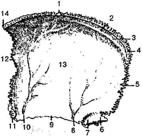
Fig. 22. Right parietal bone (internal surface):
1 — sagittal margin; 2 — parietal foramen; 3 — groove for superior sagittal sinus; 4 — occipital angle; 5 — occipital margin; 6 — mastoid angle; 7 — groove for sigmoid sinus; 8—10 — grooves for middle meningeal artery; 11 — sphenoidal angle; 12 — frontal margin; 13 — internal surface; 14 — frontal angle
The superior (sagittal) margin articulates with the contralateral parietal bone, while the anterior (frontal) and posterior (occipital) margins articulate with the frontal and occipital bones, respectively. The inferior margin of the parietal bone is overlapped by the squama of the Temporal bone. The relief of the internal surface of the parietal bone is determined by the adjacent dura mater and its vessels. Thus, the groove for the superior sagittal sinus runs along the superior margin of the parietal bone, and Branches of the meningeal Arteries are visible on the internal surface.

Fig. 23. Left parietal bone (external surface):
1 — sagittal margin; 2 — parietal foramen; 3 — occipital angle; 4 — occipital margin; 5 — superior temporal line; 6 — mastoid angle; 7 — squamous margin; 8 — sphenoidal angle; 9 — inferior temporal line; 10 — frontal margin; 11 — parietal eminence; 12 — external surface; 13 — frontal angle
The Occipital bone (os occipitale) consists of a basilar part, two lateral parts, and the occipital squama (Figs. 24, 25).
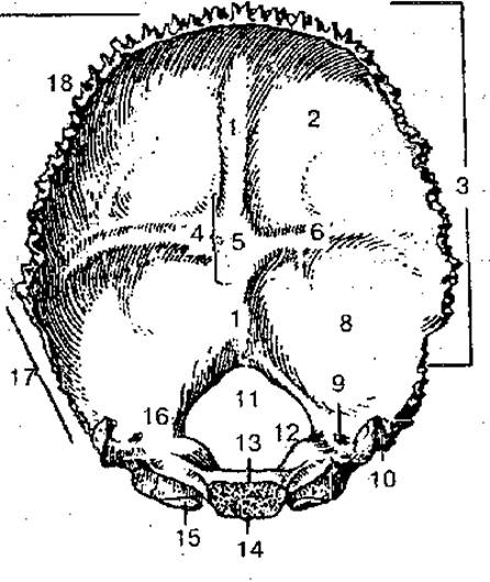
Fig. 24. Occipital bone (internal surface):
1 — groove for superior sagittal sinus; 2 — cerebral fossa; 3 — occipital squama; 4 — cruciform eminence; 5 — internal occipital protuberance; 6 — groove for transverse sinus; 7 — internal occipital crest; 8 — cerebellar fossa; 9 — condylar canal; 10 — jugular process; 11 — foramen magnum; 12 — jugular tubercle; 13 — basilar part; 14 — pharyngeal tubercle; 15 — occipital condyle; 16 — lateral part; 17 — mastoid margin; 18 — lambdoid margin
These parts surround the foramen magnum, through which the cranial cavity communicates with the vertebral canal. Anterior to the foramen magnum lies the basilar part of the occipital bone, which fuses with the body of the Sphenoid bone to form a slightly sloping surface—the clivus.

Fig. 25. Occipital bone (posterior and inferior view):
1 — occipital squama; 2 — jugular tubercle; 3 — clivus; 4 — foramen magnum; 5 — condylar canal; 6 — occipital condyle; 7 — condylar fossa; 8 — inferior nuchal line; 9 — external occipital crest; 10 — superior nuchal line; 11 — highest nuchal line; 12 — occipital plane; 13 — external occipital protuberance
On the Inferior surface of the lateral parts is the occipital condyle, which serves for articulation with the first cervical vertebra. The basilar and lateral parts, as well as the lower portions of the occipital squama, participate in the Formation of the skull base (posterior fossa), where the Cerebellum and other brain structures are located.
The occipital squama participates in the formation of the cranial vault. In the center of its internal surface is the cruciform eminence, which forms the internal occipital protuberance. Slightly below it lies the internal occipital crest. Extending upward from the occipital eminence to the junction with the parietal bones is the groove for the superior sagittal sinus, laterally is the groove for the transverse sinus, and downward is the groove for the sigmoid sinus. On the external surface of the occipital squama, the external occipital protuberance, external occipital crest, and other structures are prominent. The serrated margin of the squama articulates with the parietal and temporal bones via the lambdoid suture.
The ethmoid bone (os ethmoidale), along with other bones, participates in the formation of the anterior part of the neurocranial base, the walls of the orbits, and the nasal cavity of the viscerocranium.
The bone consists of the cribriform plate, from which the perpendicular plate projects downward, participating in the formation of the nasal septum. On both sides of the perpendicular plate are the ethmoidal labyrinths, consisting of air Cells. There are three groups of ethmoidal air cells that communicate with the nasal cavity: anterior, middle, and posterior. Hanging down from the ethmoid bone into the nasal cavity are the superior and middle nasal conchae, which form three nasal meatuses in each half of the nasal cavity. Laterally, the ethmoidal labyrinths are covered by the thin orbital plate, which participates in forming the medial wall of the orbit.
The sphenoid bone (os sphenoidale) is located between the frontal and occipital bones and lies in the center of the skull base (Fig. 26).
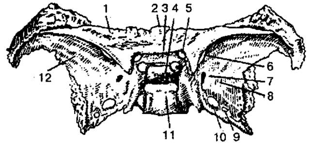
Fig. 26. Sphenoid bone (superior view):
1 — lesser wing (left); 2 — body; 3 — prechiasmatic groove; 4 — hypophyseal fossa; 5 — optic canal; 6 — superior orbital fissure; 7 — foramen rotundum; 8, 12 — greater wings; 9 — foramen ovale; 10 — foramen spinosum; 11 — dorsum sellae
In shape, this bone resembles a butterfly. It consists of a body and three paired processes: the greater and lesser wings, and the pterygoid processes. On the superior surface of the body of the bone is a depression (sella turcica) that houses the master endocrine gland, the Pituitary Gland. Within the body of the sphenoid bone is a sinus that communicates with the nasal cavity. Extending laterally from the anterosuperior surface of the sphenoid bone are the two lesser wings; at the base of each lies the large opening of the optic canal, through which the Optic nerve passes into the orbit. Located between the lesser and greater wings is the superior orbital fissure, through which the oculomotor, trochlear, abducens, and ophthalmic nerves (the first branch of the Trigeminal nerve) pass from the cranial cavity into the orbit.
The greater wings present orbital, temporal, maxillary, and cerebral surfaces. At the Base of the greater wing are the foramen rotundum, foramen ovale, and foramen spinosum, through which the branches of the trigeminal nerve and Blood Vessels pass. On the cerebral surface, digital impressions, gyral impressions, and arterial grooves are clearly visible.
The pterygoid process projects downward from the body of the sphenoid bone on each side and consists of lateral and medial plates, with the pterygoid fossa located between them on the posterior surface. The medial plate helps form the nasal cavity, while the lateral plate forms the infratemporal fossa. At the base of the pterygoid process runs the pterygoid canal of the same name, through which Vessels and nerves pass.
The temporal bone (os temporale) is a paired bone (Figs. 27, 28) that forms part of the skull base and the lateral part of the cranial vault; it articulates anteriorly with the sphenoid, posteriorly with the occipital, and superiorly with the parietal bones. The temporal bone houses the organs of Hearing and Equilibrium, and its canals transmit vessels and nerves. The temporal bone is divided into squamous, petrous, and tympanic parts. It forms a joint with the mandible and, together with the zygomatic bone, forms the zygomatic arch.

Fig. 27. Right temporal bone (internal surface):
1 — superior border of the petrous part; 2 — groove for the superior petrosal sinus; 3 — groove for the sigmoid sinus; 4 — internal process; 5 — jugular notch; 6 — styloid process; 7 — internal acoustic opening; 8 — internal acoustic meatus; 9 — groove for the inferior petrosal sinus; 10 — posterior border of the petrous part; 11 — posterior surface of the petrous part
On the internal surface of the squamous part, there are digital impressions and gyral impressions, and the groove for the middle meningeal artery is visible.
On the external convex surface of the squamous part, slightly above and anterior to the external acoustic opening, originates the horizontally oriented zygomatic process. At the base of the latter lies the mandibular fossa, with which the condylar process of the mandible articulates.

Fig. 28. Right temporal bone (external surface):
1 — tympanosquamous fissure; 2 — petrotympanic fissure; 3 — styloid process; 4 — tympanomastoid fissure
The petrous part (pyramid) of the temporal bone has a triangular shape. It presents anterior, posterior, and inferior surfaces. The anterior surface of the petrous part participates in the formation of the middle cranial fossa, the posterior surface in the posterior cranial fossa, and the inferior surface forms part of the external base of the skull. On the anterior surface near the apex of the petrous part is the trigeminal impression, which houses the trigeminal ganglion. On the posterior surface of the petrous part is the internal acoustic opening, which leads into the internal acoustic meatus. At the bottom of the latter, there are several openings for the facial and vestibulocochlear Cranial Nerves, as well as the Vessels of the Vestibulocochlear Organ. On the posterior surface of the petrous part are the external opening of the vestibular aqueduct, the cochlear canaliculus, and the groove for the sigmoid sinus. Projecting from the inferior surface of the petrous part is the long styloid process, near which lies the stylomastoid foramen, through which the Facial Nerve exits. In the center of the inferior surface of the petrous part, a round opening is prominent, which marks the beginning of the carotid canal. Through this canal, the Internal Carotid Artery enters the cranial cavity. Posterior to the external opening of the carotid canal, the jugular fossa is visible, which continues as the jugular notch at the posterior border of the petrous part.
The jugular notches of the temporal and occipital bones, when articulated on the intact skull, form the jugular foramen, through which the Internal jugular vein and three cranial nerves pass: the glossopharyngeal, vagus, and accessory nerves.
Within the substance of the petrous part lie the tympanic cavity and the bony labyrinth. The tympanic cavity communicates with the air Cells of the mastoid process, as well as with the nasopharyngeal cavity via the auditory tube.
The carotid and facial canals, as well as the canaliculus for the chorda tympani, the tympanic canaliculus, the mastoid canaliculus, and the caroticotympanic canaliculi, pass through the petrous part of the temporal bone, housing vessels, nerves, and the tensor tympani Muscle.
Bones of the facial skeleton. These are represented by paired bones (maxilla, palatine, zygomatic, nasal, lacrimal, and inferior nasal concha) and unpaired bones (mandible, hyoid bone, and vomer).
The maxilla consists of a body and four processes: frontal, zygomatic, palatine, and alveolar.
The body has four surfaces: anterior, infratemporal, orbital, and nasal. Within the body of the maxilla, there is a fairly large maxillary (antrum of Highmore) sinus (Figs. 29, 30).

Fig. 29. Maxilla (lateral view):
1 — orbital surface; 2 — infraorbital groove; 3 — zygomatic process; 4 — alveolar foramina; 5 — infratemporal surface; 6 — anterior surface; 7 — canine fossa; 8 — anterior nasal spine; 9 — body of maxilla; 10 — nasal notch; 11 — infraorbital canal; 12 — infraorbital foramen; 13 — zygomaticomaxillary suture; 14 — frontal process; 15 — lacrimal border; 16 — infraorbital margin
The maxilla contributes to the formation of the nasal cavity, orbit, Oral Cavity, infratemporal and pterygopalatine fossae. The alveolar process contains sockets for the eight upper Teeth.
The Palatine bone (os palatinum) is paired and consists of two bony plates — perpendicular and horizontal, which form part of the nasal cavity wall and the hard palate.
The zygomatic bone (os zygomaticum) is paired and has lateral, temporal, and orbital surfaces, as well as frontal and temporal processes. The size of this bone determines the width and shape of the face.

Fig. 30. Left maxilla (medial view):
1 — frontal process; 2 — nasal surface; 3 — anterior nasal spine; 4 — pterygopalatine groove; 5 — maxillary (antrum of Highmore) sinus
The Lacrimal bone (os lacrimale) is paired, participates in the formation of the medial wall of the orbit, and bounds the lacrimal fossa.
The inferior nasal concha (concha nasalis inferior) is a paired bone. It articulates with the maxilla and palatine bone along one border, while its other parts project into the nasal cavity, bounding the inferior nasal meatus.
The mandible (mandibula) is the only mobile bone in the human skull, consisting of a body and two rami (Fig. 31).
On the external surface of the body is the mental protuberance, flanked on each side by a mental tubercle and a mental foramen. Along the superior border of the mandibular body are the dental alveoli, separated by interalveolar septa. This border is called the alveolar part. Superiorly, each ramus ends in an anterior coronoid process and a posterior condylar (articular) process. On the Medial surface of the ramus, there is a foramen leading into the mandibular canal.

Fig. 31. Mandible:
1 — head of mandible; 2 — pterygoid fovea; 3 — neck of mandible; 4, 5 — rami of mandible; 6 — angle of mandible; 7 — mandibular canal; 8 — temporal crest; 9 — mandibular foramen; 10 — coronoid process; 11 — mandibular notch; 12 — condylar process
The hyoid bone (os hyoideum) consists of a body, a pair of greater horns, and a pair of lesser horns, and is located in the neck region between the mandible and the Larynx.
The vomer, articulating with the ethmoid bone, participates in forming the nasal septum and separates the paired posterior nasal apertures — the choanae.
The bones of the skull are joined together by continuous joints — synarthroses. The latter include bony fusions — synostoses; cartilaginous joints — synchondroses; and fibrous joints — syndesmoses. The predominant type of fibrous joints in the skull are sutures. Depending on their shape, sutures are classified as serrate, squamous, or plane. For example, serrate sutures include the coronal (between the frontal and parietal bones), sagittal (along the midline between the paired parietal bones), and lambdoid (between the occipital squama and parietal bones) sutures.
The squamous part of the temporal bone and the parietal bone on the lateral surface of the cranial vault are joined by the squamous suture. The facial bones are joined by plane sutures.
A cartilaginous joint exists between the occipital and sphenoid bones only until about 20 years of age, after which it ossifies (synostosis).
The temporomandibular joint (articulatio temporomandibularis) is a paired, complex, ellipsoidal joint. It is formed by the head of the condylar process of the mandible and the mandibular fossa of the temporal bone. Inside the joint, an articular disc divides the joint cavity into two separate compartments: upper and lower. The two temporomandibular joints function together (movement in one joint induces movement in the other) and are considered a single combined joint. Movements permitted at the joint include depression and elevation of the mandible, lateral movements (to the right and left), as well as protrusion and retraction.
The Skull as a whole. The Skeleton of the Skull (Figs. 32, 33) is conventionally divided into the calvaria, or skullcap, and the base.

Fig. 32. Human skull (anterior view, after R. D. Sinelnikov):
1 — coronal suture; 2 — parietal bone; 3 — orbital part of the frontal bone; 4 — orbital surface of the greater wing of the sphenoid bone; 5 — zygomatic bone; 6 — inferior nasal concha; 7 — maxilla; 8 — mental protuberance of the mandible; 9 — nasal cavity; 10 — vomer; 11 — perpendicular plate of the ethmoid bone; 12 — orbital surface of the maxilla; 13 — inferior orbital fissure; 14 — lacrimal bone; 15 — orbital plate of the ethmoid bone; 16 — superior orbital fissure; 17 — squamous part of the temporal bone; 18 — zygomatic process of the frontal bone; 19 — optic canal; 20 — Nasal bone; 21 — frontal eminence
The cranial vault is formed by the squamous parts of the frontal, temporal, and occipital bones, and by the parietal bones. The cranial base consists of the frontal, ethmoid, sphenoid, temporal, and occipital bones. A distinction is made between the internal and external cranial bases.

Fig. 33. Human skull (lateral view, after R. D. Sinelnikov):
1 — parietal bone; 2— coronal suture; 3 — frontal eminence; 4 — temporal surface of the greater wing of the sphenoid bone; 5 — orbital plate of the ethmoid bone; 6 — lacrimal bone; 7 — nasal bone; 8 — temporal fossa; 9 — anterior nasal spine; 10 — body of the maxilla; 11 — mandible; 12 — zygomatic bone; 13 — zygomatic arch; 14 — styloid process; 15 — condylar process of the mandible; 16 — mastoid process; 17 — external acoustic meatus; 18 — lambdoid suture; 19 — occipital squama; 20 — superior temporal line; 21 — squamous part of the temporal bone
The internal cranial base (basis cranii interna) has three cranial fossae: anterior, middle, and posterior (Fig. 34). The anterior cranial fossa contains the lobes of the cerebral hemispheres, the middle contains the temporal lobes of the cerebral hemispheres, and the posterior houses the cerebellum and parts of the Brainstem: the cerebral peduncles and the Medulla Oblongata.
The anterior cranial fossa is formed by the orbital part of the frontal bone, the ethmoid bone (cribriform plate), and the lesser wings of the sphenoid bone, and communicates with the nasal cavity through the foramina in the cribriform plate. These foramina serve as a passage for the olfactory nerves (I pair).
The walls of the middle cranial fossa are formed by the body and greater wings of the sphenoid bone, the anterior surface of the petrous pyramids, and the squamous part of the temporal bones. The middle cranial fossa communicates with the orbit and the pterygopalatine fossa. From this fossa, the optic nerve (II pair), ophthalmic artery, and vein pass into the orbital cavity through the optic canal. The oculomotor (III pair), trochlear (IV pair), abducens (VI pair), and ophthalmic (the first branch of the trigeminal nerve (V pair)) nerves pass into the orbit through the superior orbital fissure. Slightly posterior to the superior orbital fissure lies the foramen rotundum, through which the maxillary nerve (the second branch of the V pair) passes, while the mandibular nerve (the third branch of the V pair) exits the skull through the foramen ovale. The pituitary gland, an endocrine gland, is located in the hypophyseal fossa of the sella turcica.

Fig. 34. Internal cranial base:
1 — orbital part of the frontal bone; 2 — crista galli; 3 — cribriform plate; 4 — optic canal; 5 — hypophyseal fossa; 6— dorsum sellae; 7 — foramen rotundum; 8 — foramen ovale; 9 — foramen lacerum; 10 — foramen spinosum; 11 — internal acoustic opening; 12 — jugular foramen; 13 — hypoglossal canal; 14 — lambdoid suture; 15 — clivus; 16 — groove for transverse sinus; 17 — internal occipital protuberance; 18 — foramen magnum; 19 — occipital squama; 20 — groove for sigmoid sinus; 21 — petrous part (pyramid) of the temporal bone; 22 — squamous part of the temporal bone; 23 — greater wing of the sphenoid bone; 24 — lesser wing of the sphenoid bone
The occipital bone and the posterior surfaces of the petrous parts of the temporal bones participate in the formation of the posterior cranial fossa.
The clivus is located between the dorsum sellae and the foramen magnum.
The internal acoustic opening (right and left) opens into the posterior cranial fossa, giving passage to the Vestibulocochlear nerve (VIII pair), while the facial nerve (VII pair) exits from the facial canal. The glossopharyngeal (IX pair), vagus (X pair), and accessory (XI pair) nerves exit through the jugular foramen of the skull base. The Hypoglossal nerve (XII pair) passes through the hypoglossal canal. In addition to the nerves, the internal jugular vein, which is continuous with the sigmoid sinus, exits the cranial cavity through the jugular foramen. The foramen magnum connects the cavity of the posterior cranial fossa with the vertebral canal, at which level the medulla oblongata transitions into the Spinal Cord.
The external cranial base (basis cranii externa) is covered anteriorly by the facial bones (where the bony palate, bounded anteriorly by the alveolar process of the maxilla and the teeth, is distinguished), while its posterior part is formed by the external surfaces of the sphenoid, occipital, and temporal bones (Fig. 35).
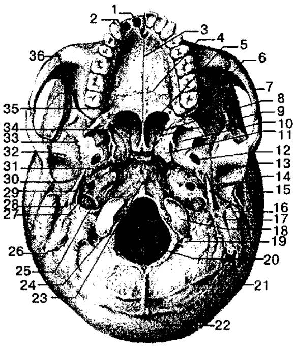
Fig. 35. External cranial base:
1 — palatine process of the maxilla; 2 — incisive foramen; 3 — median palatine suture; 4 — transverse palatine suture; 5 — choana; 6 — inferior orbital fissure; 7 — zygomatic arch; 8 — ala of the vomer; 9 — pterygoid fossa; 10 — lateral plate of the pterygoid process; 11 — pterygoid process; 12 — foramen ovale; 13 — mandibular fossa; 14 — styloid process; 15 — external acoustic meatus; 16 — mastoid process; 17 — mastoid notch; 18 — occipital condyle; 19 — condylar fossa; 20 — foramen magnum; 21 — inferior nuchal line; 22 — external occipital protuberance; 23 — pharyngeal tubercle; 24 — condylar canal; 25 — jugular foramen; 26 — occipitomastoid suture; 27— external opening of the carotid canal; 28 — stylomastoid foramen; 29 — foramen lacerum; 30 — petrotympanic fissure; 31 — foramen spinosum; 32 — articular tubercle; 33 — sphenosquamous suture; 34 — pterygoid hamulus; 35 — greater palatine foramen; 36 — zygomaticomaxillary suture
This region contains numerous foramina through which vessels and nerves pass, providing Blood supply to the brain. The central part of the external cranial base is occupied by the foramen magnum, flanked by the occipital condyles. The latter articulate with the first cervical vertebra. The exit from the nasal cavity is represented by paired openings (choanae) leading into the nasal cavity. In addition, the external surface of the skull base features the pterygoid processes of the sphenoid bone, the external opening of the carotid canal, the styloid process, the stylomastoid foramen, the mastoid process, the musculotubal canal, the jugular foramen, and other structures.
In the facial skeleton, the central features are the nasal cavity, the orbits, the oral cavity, and the infratemporal and pterygopalatine fossae.
The nasal cavity (cavitas nasi) is the initial part of the respiratory tract and contains the Organ of Smell. It has a single anterior inlet, the piriform aperture, and two posterior outlets, the choanae.
The nasal cavity is divided into two halves by a bony septum. The nasal cavity has superior, inferior, and lateral walls. The superior wall is formed by the nasal bones, the ethmoid bone, the nasal part of the frontal bone, and the body of the sphenoid bone. The inferior wall is represented by the palatine processes of the maxillae and the horizontal plates of the palatine bones. The lateral wall consists of the frontal process of the maxilla, the lacrimal bone, the ethmoidal labyrinth, the perpendicular plate of the palatine bone, and the medial plate of the pterygoid process of the sphenoid bone.
The nasal conchae divide the lateral part of the cavity into three nasal meatuses: superior, middle, and inferior. The sphenoid sinuses and posterior ethmoidal cells open into the superior nasal meatus; the maxillary and frontal sinuses, as well as the ethmoidal cells, open into the middle nasal meatus; and the nasolacrimal duct, which begins in the orbit, opens into the inferior nasal meatus.
The orbit (orbita) is a paired cavity shaped like a four-sided pyramid with rounded edges, its apex directed posteriorly and medially. The optic canal passes through this region. The orbital cavity contains the Eyeball with its Muscles, the lacrimal gland, and other structures. It has an opening and four walls: superior, inferior, medial, and lateral. The superior wall is formed by the orbital part of the frontal bone and the lesser wings of the sphenoid bone; the inferior wall by the zygomatic bone and the maxilla; the medial wall by the frontal process of the maxilla, the lacrimal bone, the orbital plate of the ethmoid bone, the body of the sphenoid bone, and a part of the frontal bone; the lateral wall by the zygomatic bone and the greater wing of the sphenoid bone. Between the lateral and inferior walls lies the inferior orbital fissure, which opens into the pterygopalatine and infratemporal fossae. The superior orbital fissure and the optic foramen open into the middle cranial fossa; the nasolacrimal canal connects with the nasal cavity.
The oral cavity (cavitas oris) is formed by the bony (hard) palate, the palatine processes of the right and left maxillae, and the horizontal plates of the palatine bones; the lateral and anterior walls are formed by the alveolar processes of the maxillae, which together make up the superior alveolar arch. The bony palate serves as the hard (bony) foundation of the superior wall of the oral cavity. The superior and inferior alveolar arches, along with the teeth and the body of the mandible, constitute the skeleton of the anterior and lateral walls of the oral cavity.
The infratemporal fossa is located posterior to the maxilla, medial to the zygomatic bone and zygomatic arch, and lateral to the pterygoid process of the sphenoid bone, forming part of the external base of the neurocranium.
The pterygopalatine fossa is located between the bones of the neurocranium and viscerocranium, and has four walls: anterior, superior, posterior, and medial. The anterior wall is formed by the maxillary tuberosity, the superior wall by a part of the body and the base of the greater wing of the sphenoid bone, the posterior wall by the base of the pterygoid process of the sphenoid bone, and the medial wall by the perpendicular plate of the palatine bone. Various canals and foramina open into the pterygopalatine fossa, through which it communicates with adjacent cavities.
Age-related Features of the skull. The newborn skull has several significant characteristics. Due to the rapid growth of the brain, its neurocranium is 8 times larger than the viscerocranium, whereas in adults, it is only twice as large. A characteristic feature of the newborn is the presence of fontanelles: anterior, posterior, and paired lateral (sphenoidal and mastoid). The anterior fontanelle is the largest and has a quadrangular shape. The lateral fontanelles, if present, close by the second or third month, while the anterior fontanelle closes during the second year of the child's life.
Cranial sutures form by 3–5 years of age, and skull growth is completed by 25–30 years of life.
Another feature is the presence of cartilaginous layers between the bones of the skull base. Additionally (the third feature), in newborns, the air sinuses, tuberosities, and processes are undeveloped, teeth are absent, and the jaws are poorly developed. In old age, changes occur in the facial skeleton As a result of tooth loss: the alveolar processes of the jaws decrease in size, the facial region of the skull becomes somewhat shortened, and the cranial bones become thinner and more fragile.
Last update: 08/08/2026
Editorial and Educational Adaptation: This material has been compiled based on the primary/original source text. The project team performed an editorial review, corrected technical inaccuracies, structured sections, and adapted the content for an educational format.
What was processed:
- elimination of formatting defects (OCR errors, structural breaks, corrupted characters);
- editorial organization of content;
- standardization of terminology in accordance with academic sources;
- verification of factual statements against the original source text.
All mentions of the author, publication year, and origin of the primary text have been preserved in accordance with the source.