Human Anatomy and Physiology - N. I. Fedyukovich 2003
Bones and Joints
Skeleton of the Trunk
The Skeleton of the Trunk (Fig. 11) consists of THE Vertebral Column and The thoracic cage, and is part of the Axial Skeleton.
Class="center">
Fig. 11. Human skeleton (anterior view):
1 — Skull; 2 — vertebral column; 3 — clavicle; 4 — rib; 5 — Sternum; 6 — humerus; 7 — radius; 8 — ulna; 9 — Carpal Bones; 10 — Metacarpal bones; 11 — Phalanges of the hand; 12 — ilium; 13 — sacrum; 14 — pubis; 15 — ischium; 16 — Femur; 17 — Patella; 18 — Tibia; 19 — Fibula; 20 — Tarsal Bones; 21 — Metatarsal Bones; 22 — phalanges of the FOOT
Vertebrae in different Regions of the vertebral column share not only common features and Structure, but also possess distinctive characteristics associated with the upright posture of humans.
A vertebra consists of a body (corpus vertebrae) and an arch (arcus vertebrae), which encloses to form the vertebral foramen (foramen vertebrale). When all the vertebrae are joined, they form the vertebral canal (canalis vertebralis), which houses the Spinal Cord. Arising from the vertebral arch are two superior and two inferior articular processes, as well as the right and left transverse processes. Posteriorly, along the midline, projects the spinous process. At the junction of the arch and the body of the vertebra are the superior and inferior vertebral notches, which, when the vertebrae articulate, form the intervertebral foramen (foramen intervertebrale). Blood Vessels and a spinal nerve pass through this opening.
Cervical vertebrae (vertebrae cervicales) differ from the vertebrae of other regions (Fig. 12). Their bodies are relatively small and elliptical in shape. Their primary distinguishing feature is the presence of the transverse foramen (foramen transversarium). The first two vertebrae are involved in HEAD movement and articulate with the skull (which distinguishes them from other cervical vertebrae).

Fig. 12. Cervical vertebra:
1 — superior articular process; 2 — vertebral arch; 3 — vertebral foramen; 4 — spinous process; 5 — lamina of the vertebral arch; 6 — inferior articular process; 7 — posterior tubercle; 8 — groove for the spinal nerve; 9 — transverse foramen; 10 — anterior tubercle; 11 — vertebral body; 12 — uncus of the body; 13 — transverse process
Under METABOLISM/18.html">The Influence of increasing load, the bodies of the cervical vertebrae increase in size from the III to the VII vertebra. The spinous processes of the cervical vertebrae are bifid, except for the VII, which is significantly longer than the others and easily palpable under the Skin. The anterior tubercle of the VI cervical vertebra is more prominent than in the other vertebrae. The carotid artery passes close to it, which is why it is called the carotid tubercle. To temporarily stop bleeding, the carotid artery can be compressed against this tubercle.
Thoracic vertebrae (vertebrae thoracicae) are larger than cervical ones (Fig. 13). Their vertebral foramen is somewhat smaller than that of the cervical vertebrae, and on the lateral surfaces of the body, there are superior and inferior costal facets required for articulation with the heads of the Ribs. The height of the thoracic vertebral bodies (from I to XII) gradually increases. The spinous processes are somewhat longer, directed posteriorly and downward, overlapping each other like shingles, which limits the mobility of this region of the spine (especially extension).

Fig. 13. Thoracic vertebrae:
1 — pedicle of the vertebral arch; 2 — superior vertebral notch; 3, 7 — transverse process; 4 — superior articular process; 5, 9 — superior costal facet; 6 — vertebral canal; 8 — spinous process; 10 — transverse costal facet; 11 — inferior articular process; 12 — inferior vertebral notch; 13, 14 — inferior costal facet; 15 — vertebral body
Lumbar vertebrae (vertebrae lumbales) have a more massive body than other vertebrae (Fig. 14).

Fig. 14. Lumbar vertebra (superior view):
1 — spinous process; 2 — superior articular process; 3 — costal process; 4 — vertebral arch; 5 — vertebral foramen; 6 — pedicle of the vertebral arch; 7 — vertebral body; 8 — accessory process; 9 — mammillary process
The body of a lumbar vertebra is Kidney-shaped, and its transverse diameter is greater than its anteroposterior diameter. The body of the V lumbar vertebra is the largest in both height and width. The spinous processes are massive and project almost horizontally backward, while the articular processes are oriented sagittally. This allows for significant mobility in the lumbar region of the spine. The vertebral foramen, which is larger than in other regions, is triangular with rounded corners.
The sacral vertebrae (vertebrae sacrales) fuse together to form a single bone, the sacrum (os sacrum). The sacrum (Fig. 15) is triangular in shape, with its base articulating with the V lumbar vertebra, and its apex directed downward and forward.
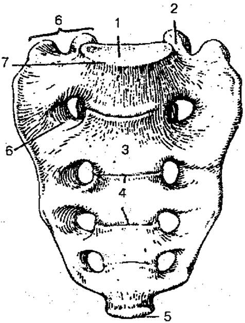
Fig. 15. Sacrum (anterior view):
1 — Base of the sacrum; 2 — superior articular process; 3 — anterior surface of the sacrum; 4 — transverse lines; 5 — apex of the sacrum; 6 — anterior sacral foramina; 7 — promontory; 8 — lateral part
On the concave anterior pelvic surface, there are four transverse lines, which represent the fusion sites of the sacral vertebral bodies. On the convex (dorsal) surface, longitudinal sacral crests (median, intermediate, and lateral) are well-defined. On both sides of the sacral surfaces, there are four pairs of sacral foramina, through which the Branches of the Spinal Nerves exit the sacral canal. The massive lateral parts feature an auricular surface, which is designed for articulation with the corresponding articular surfaces of the hip bones. The junction of the sacrum with the V lumbar vertebra forms a prominent anterior projection called the promontory (promontorium). The apex of the sacrum articulates with the coccyx.
The coccyx (os coccygis) consists of 1—5 (usually 4) fused vestigial vertebrae, vertebrae coccygeae (Fig. 16). It is triangular in shape, curved anteriorly, with its base directed upward and forward, and its apex pointing downward and forward. Distinct vertebral features are observed only in the first coccygeal vertebra; the remaining ones are significantly smaller and rounded.

Fig. 16. Coccyx (posterior view)
1 - coccyx; 2 - coccygeal horn
The rib (costa), of which there are 12 pairs, consists of a long posterior bony part and a short anterior cartilaginous part (costal Cartilage). The upper seven pairs of ribs (I—VII) connect directly to the sternum via their costal cartilages and are called true ribs. The cartilages of the VIII, IX, and X pairs of ribs do not articulate with the sternum but join the cartilage of the rib immediately above; these are called false ribs. Ribs XI and XII have short cartilaginous portions that end in the Muscles of the abdominal wall. They are more mobile and are referred to as floating ribs.
A rib consists of a head, body, and neck. In the upper 10 pairs of ribs, a tubercle is located between the neck and the body. Each rib has an inner and outer surface, as well as superior and inferior borders. Along the inferior border of the inner surface runs the costal groove, which accommodates the intercostal vessels and nerve. On the outer surface, at the junction of the neck and body, lies the tubercle of the rib, whose articular facet articulates with the transverse process of the corresponding vertebra.
Ribs vary in shape and size (Figs. 17, 18). The shortest are the two uppermost and two lowermost ribs. The first rib lies horizontally; its superior surface features a small tubercle for the attachment of the scalenus anterior Muscle and two grooves: an anterior one for the subclavian vein and a posterior one for the Subclavian Artery.

Fig. 17. The First and Second ribs (superior view): 1 — articular facet of the head of the rib; 2 — head of the rib; 3 — tubercle of the rib; 4 — body of the rib; 5 — articular facet of the tubercle of the rib; 6 — neck of the rib
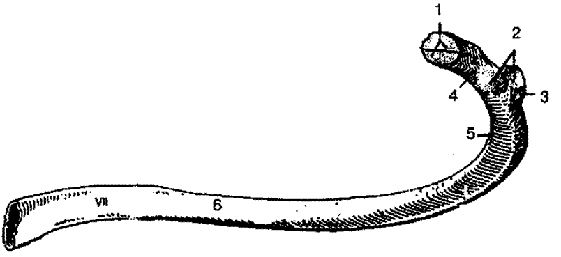
Fig. 18. The seventh rib (inner surface):
1 — articular facet of the head of the rib; 2 — articular facet of the tubercle of the rib; 3 — tubercle of the rib; 4 — neck of the rib; 5 — angle of the rib; 6 — body of the rib
The sternum is an elongated, flat bone consisting of three parts: the manubrium, the body, and the xiphoid process. In adults, these parts fuse into a single bone. The superior border of the manubrium features the jugular notch and paired clavicular notches. Costal notches are located along the lateral margins of the sternal body.
The xiphoid process varies in shape and size and may sometimes be bifid.
The vertebral column (columna vertebralis) provides structural support, connects various parts of The Human Body, and protects the spinal cord and the spinal nerve roots emerging from it. The human vertebral column consists of 33—34 vertebrae. The lowest 6—9 vertebrae fuse to form the sacrum and the coccyx (Fig. 19).
The spine is divided into five regions: the cervical (consisting of 7 vertebrae), thoracic (12), lumbar (5), sacral (5), and coccygeal (2—5 vertebrae).
The human vertebral column is characterized by several natural curvatures. An anteriorly convex curvature is called lordosis (cervical and lumbar), while a posteriorly convex curvature is called Kyphosis (thoracic and sacral). The prominent VII cervical vertebra marks the transition from cervical lordosis to thoracic kyphosis. At the junction of the lumbar lordosis and sacral kyphosis, the anteriorly projecting sacral promontory is formed. These spinal curvatures (lordoses and kyphoses) act as Shock absorbers during walking, running, and jumping. Asymmetrical development of muscle mass can lead to a pathological lateral curvature known as Scoliosis.

Fig. 19. Vertebral column:
1 — cervical vertebrae; 2 — thoracic vertebrae; 3 — lumbar vertebrae; 4 — sacrum; 5 — coccyx
The rib cage (compages thoracis) is formed by the thoracic spine, ribs, sternum, and their articulations. It encloses the thoracic cavity, which houses vital Organs: The Heart, Lungs, major blood vessels, Trachea, Esophagus, and nerves (Fig. 20).

Fig. 20. Skeleton of the thoracic cage (anterior view):
1 — superior thoracic aperture; 2 — jugular notch; 3 — ribs (1—12); 4 — first rib; 5, 16 — second rib; 6 — manubrium of sternum; 7 — body of sternum; 8 — xiphisternal joint; 9 — xiphoid process; 10 — floating ribs (11—12); 11— false ribs (8—12); 12 — thoracic vertebra; 13 — inferior thoracic aperture; 14 — sternum; 15 — true ribs (1—7); 17 — clavicular notch
The shape of the rib cage depends on sex, body build, physical development, and age.
The thoracic cage features superior and inferior apertures. The superior aperture is bounded posteriorly by the body of the I thoracic vertebra, laterally by the first ribs, and anteriorly by the manubrium of the sternum. Through it, the apex of the lung projects into the neck, and the esophagus, trachea, vessels, and nerves pass; the inferior aperture is bounded by the body of the XII thoracic vertebra, the XI and XII ribs, the costal arches, and the xiphoid process, and is closed by the Diaphragm.
The human thoracic cage is somewhat flattened, with its anteroposterior dimension being significantly smaller than its transverse dimension. The shape of the thoracic cage can be influenced by Rickets, respiratory diseases, and other conditions.
Last update: 08/08/2026
Editorial and Educational Adaptation: This material has been compiled based on the primary/original source text. The project team performed an editorial review, corrected technical inaccuracies, structured sections, and adapted the content for an educational format.
What was processed:
- elimination of formatting defects (OCR errors, structural breaks, corrupted characters);
- editorial organization of content;
- standardization of terminology in accordance with academic sources;
- verification of factual statements against the original source text.
All mentions of the author, publication year, and origin of the primary text have been preserved in accordance with the source.