THEORETICAL FOUNDATIONS OF FISH FARMING - I.M. Sherman - 2011
3. THEORETICAL FOUNDATIONS OF FISH BREEDING TECHNOLOGICAL PROCESSES
3.2 Fundamentals of Effective Fish Reproduction
The formation of replacement and brood stocks is an undeniably crucial baseline component of fish breeding technological processes, which in turn requires a solid understanding of the fundamentals of efficient fish reproduction.
According to various authors, the world's ichthyofauna comprises between 16,000 and 22,000 fish species, reflecting their immense diversity shaped by the struggle for survival over a long phylogenetic history. The adaptation of species to environmental conditions throughout their history has followed various directions, leading to the ecological differentiation of fish, which is based on THE PRINCIPLE OF habitat partitioning.
In this regard, G.V. Nikolsky notes that fish species, by analogy with other living organisms, inhabit their inherent habitats. Building on the proposed concept of fish habitats, this largely characterizes a species in terms of its relationship with ecological parameters
of the environment and significantly determines its biology of reproduction. Accordingly, based on the level of ADAPTATION TO A specific habitat, the following ecological groups are conventionally distinguished: marine, freshwater, diadromous, and brackish-Water.
Given that knowledge of fish ecological groups is of great importance when addressing issues related to natural reproduction, it is advisable to consider this information before proceeding directly to the core topic focused on studying efficient reproduction and, consequently, fish breeding.
Marine fish. They live in saline seawater throughout their entire life. Within this ecological group, a distinction is made between pelagic fish, which inhabit the water Column, and benthic fish, which live near the bottom in bottom-water layers. Marine fish are also subdivided into oceanic fish, inhabiting the open and surface layers of the ocean; neritic fish, inhabiting coastal marine waters; and deep-sea or abyssal fish. The latter are further subdivided into bathypelagic and benthic.
Freshwater fish. Fish that live in fresh water throughout their entire life and are generally not found in brackish water. Freshwater fish are divided into rheophilic species, adapted to living in running water, and limnophilic species, adapted to living in standing water. In turn, both rheophilic and limnophilic fish are subdivided into pelagic (inhabiting the water column), benthopelagic, and benthic.
Diadromous fish. Fish that spend part of their life in fresh water and part in salt water. Their reproduction process involves transitioning from saline seawater to fresh river water, or vice versa from fresh to salt water. Depending on their feeding grounds, they are subdivided into trophic-marine and trophic-freshwater.
Brackish-water fish. Fish that inhabit desalinated areas of seas, estuaries, and inland seas characterized by reduced salinity due to freshwater river systems. They are subdivided into semi-diadromous fish, which enter the lower reaches of rivers to spawn while feeding in brackish water, and true brackish-water fish, which permanently inhabit desalinated waters.
A characteristic feature of the vast majority of representatives from the discussed groups is that upon reaching sexual maturity, they undertake more or less pronounced spawning Migrations, during which sexual development is completed, and the ripening of Gametes in the Gonads takes place prior to spawning.
Marine fish permanently live and reproduce in the sea. Freshwater fish inhabiting rivers and freshwater lakes are characterized by weakly expressed migrations and spawn directly near their permanent habitats. The overwhelming majority of diadromous fish live in the sea and ascend rivers to spawn, many of them traveling hundreds of kilometers while overcoming currents, waterfalls, and rapids. Semi-diadromous fish localize in desalinated areas of the sea and enter rivers for reproduction without ascending as far as diadromous species.
In the pre-spawning period, the Ovaries of females contain reproductive products corresponding to the completed stage IV of maturity. As they approach the spawning grounds, under the Influence of the pituitary gonadotropic hormone, they undergo changes: The process of oogenesis is completed, and ovulation occurs.
The ovulation process is quite complex; ensuring its normal progression requires a complex of biotic and abiotic factors whose parameters must correspond to the species-specific traits of the fish. Among the environmental factors essential for ensuring ovulation, water Temperature, chemical regime, presence or absence of current, and the presence of males are of exceptional importance; for many fish species, a specific spawning substrate is also required.
In connection with spawning, of clear interest are the studies by N.L. Gerbilsky, who established the existence of a specific neurohumoral regulation system for the spawning process. Moreover, if after the onset of spawning temperatures, some of the mentioned environmental factors are unsatisfactory or do not meet requirements, ovulation does not occur. Such a mechanism has an adaptive significance for fish: the ovaries are cleared of older-generation oocytes, and a new development cycle begins.
Under normal environmental conditions corresponding to the species-Specific features of the fish, ovulation takes place, followed by the mating act known as spawning, with the spawning site referred to as the spawning ground.
Spawning sites, taking into account species-specific traits, meet the optimal requirements for the embryonic and early post-embryonic periods of fish life. By analogy with fish habitats, There is a differentiation of fish based on the principle of their relationship to spawning substrates.
In other words, to deposit their eggs, fish select areas whose conditions match their biological characteristics during the periods of insemination, Fertilization, embryonic development, and early post-Embryogenesis, which should be viewed in terms of species adaptation during phylogeny.
Depending on the conditions of reproduction, development, and primarily the habitat where spawning takes place, G.V. Nikolsky distinguishes the following ecological groups of fish: lithophiles, phytophiles, psammophiles, pelagophiles, and ostracophiles.
Lithophiles. They reproduce by depositing eggs on stony substrates, usually in rivers in the current, or on the bottom of oligotrophic lakes and coastal marine areas where, as a rule, though not always, favorable respiration conditions exist. This group includes: sturgeon, beluga, stellate sturgeon, sterlet, Atlantic salmon, chum salmon, chinook salmon, pink salmon, and black-sea roach.
Phytophiles. They reproduce by depositing eggs on plant substrates in slow-flowing or standing water on vegetating or decaying plants. In this case, respiration conditions vary greatly. This group includes: bream, common carp, pike, perch, roach, crucian carp, and catfish.
Psammophiles. They reproduce by depositing eggs on sand, sometimes attaching them to eroded plant roots. Egg membranes are often encrusted with sand grains, and development usually occurs under favorable respiration conditions. This group may include gudgeons and certain loach species.
Pelagophiles. They reproduce by releasing eggs into the water column. Eggs and free embryos remain suspended in the water column, moving freely under METABOLISM/18.html">The Influence of currents, waves, and wind phenomena. Development, as a rule, takes place under favorable respiration conditions. This group may include many species of herrings, cods, flounders, sabrecarp, grass carp, black carp, silver carp, and bighead carp.
Ostracophiles. They reproduce by depositing eggs into the mantle or gill cavity of Mollusks, and sometimes under the carapace of crabs and other animals. Eggs typically develop under suboptimal respiratory conditions. Bitterlings belong to this group.
Obviously, the considered groups of fish do not encompass the entire diversity of ichthyofauna. Along with the aforementioned ecological groups and their constituent species, There are also viviparous fish in which embryogenesis occurs fully or partially within the maternal Organism. Within the ichthyofauna, many species occupy an intermediate position in terms of their reproduction patterns, belonging to various groups. Depending on environmental conditions, vimba bream, kutum, or ide may spawn on stony substrates or vegetation, whereas pike-perch and smelt spawn on sand or vegetation.
Knowledge of ecological groups in relation to reproductive features is of exceptional importance within the chain of Problems associated with fish farming, as it dictates The Need for a scientifically grounded biological approach to aquaculture technology.
In ichthyology in general, and in fish farming in particular, there is THE CONCEPT OF "spawning temperature." This is due to the fact that while a combination of factors is necessary for fish spawning, water temperature acts as the triggering signal that initiates the spawning mechanism. However, each fish species requires a precisely defined water temperature range for spawning to occur, which must coincide with the availability of suitable spawning grounds. Furthermore, the duration of spawning, or the spawning season, is closely linked to water temperature dynamics. When observing a spawning season, a specialist is typically guided by temperature fluctuation parameters or ranges, as well as the thermal limits at which a particular species begins or finishes spawning. Hence, it is evident that the duration of spawning or the spawning season depends on the rate at which the temperature changes during spawning, or the number of days required for the water temperature to reach the threshold marking the start or end of spawning. Under artificial propagation conditions, by regulating water temperature, it is possible to directly influence the duration of individual technological cycles based on the specific features of given facilities.
The aforementioned principle is general in nature and explains the relationship between spawning duration and water temperature. Depending on the dynamics of the thermal regime, the duration of spawning or the spawning season can be sharply reduced or significantly prolonged. In addition, the type of spawning has a direct impact on spawning duration. Fractional spawners typically have a longer spawning period than fish that release all their eggs at once. The age Structure OF THE stock also plays a considerable role; characteristically, the wider the age range of individuals representing the spawning population, the longer the spawning period. When the spawning population comprises distinct ecological groups, this feature can also contribute to extended spawning. Other things being equal, sharp fluctuations in water temperature that exceed the spawning limits negatively affect spawning success. The final and logical Conclusion of spawning is the contact of eggs with sperm, fertilization, and the formation of a zygote.
These ecological reproduction features must be taken into account when designing and adapting fish farming technologies, backed by adequate specialized training in these matters.
These ecological reproduction features must be taken into account when designing and adapting fish farming technologies, backed by adequate specialized training in these matters.
For the ichthyofauna of temperate and high latitudes, where seasonality is relatively pronounced and dictates regular shifts in the thermal regimes of water bodies, two biological groups of fish naturally exist. It is well known that the spawning times of various fish species vary considerably, yet they are adapted to specific seasons of the year, making it possible to distinguish the following groups of fish:
- Spring-summer spawners, which spawn during the spring-summer period (pike, perch, pike-perch, roach, bream, common carp, sturgeon, beluga, vimba bream, stellate sturgeon);
- Autumn-winter spawners, which spawn during the autumn-winter period (Atlantic salmon, chum salmon, chinook salmon, coho salmon, trout, burbot, whitefishes).
For fish spawning in the spring-summer period, the spawning season typically extends from March to August, whereas for autumn-winter spawners, it lasts from September to January. Moreover, fish of the same species and even the same population may have different calendar dates for spawning.
It has repeatedly been noted that distinct groups of migratory fish belonging to the same species exhibit different timings for entering rivers to spawn. In this regard, L. S. Berg concluded that some salmon and sturgeon species objectively possess winter and spring races. Spring-run individuals undertake spawning migrations from the sea into rivers, where they spawn in the same year and return to the sea to feed. Winter-run individuals undertake spawning migrations from the sea into rivers, where they overwinter, spawning only the following year, after which they return to the sea to feed. Furthermore, differentiation exists in spawning sites between spring and winter forms. The spawning grounds of winter fish are located significantly further upstream than those of spring fish. Such intra-specific specialization regarding the Location of spawning grounds is adaptive in nature, ensuring a certain Stability of the species across its range. The species gains The ability to utilize spawning grounds in different sections of a river over considerable distances, which enhances reproductive efficiency, increases the Prospects for the survival and thriving of the species, helps avoid potential unfavorable conditions that periodically arise in individual spawning grounds, and promotes better progeny survival.
Different biological groups within a population are a consequence of the Features of the species' range, but a characteristic feature is a specific plasticity, whereby
if appropriate conditions are absent, intrapopulation differentiation does not manifest itself.
Differentiation within a population should be regarded as an adaptive mechanism to the conditions of reproduction and development. Intrapopulation differentiation is nothing other than an adaptation to the conditions of the range, allowing for the fullest utilization of its potential. Information on the differentiation of population composition is of more than theoretical interest; relevant knowledge in this field enables the qualified resolution of many practical tasks related to regulating the catch of valuable anadromous fish, siting fish hatcheries, and intentionally planning aquaculture technology. In artificial fish propagation, working with different biological groups of the same species at the required quantitative ratio assumes exceptional significance. Only in this way is it possible to preserve the genetic and ecological structure of the population and ensure population growth, which is particularly crucial for the artificial propagation of sturgeons and salmonids.
A rather important factor determining an individual's sex is the behavioral pattern of females and males. In the simplest case, reproductive behavior involves the formation of spawning aggregations and the joint release of gametes at spawning grounds. More complex behavior is driven by "courtship rituals" and the pursuit of females by males (carp, pike). Even more diverse behavior is associated with the preparation of spawning sites by males, the Construction of a unique "nest," and the attraction of females (tilapias). During the reproductive period, males exhibit aggressiveness driven by display behaviors, courtship, and territory defense.
Following the release of reproductive products, many species exhibit some form of parental care: fanning the eggs during embryogenesis, incubating eggs in the Oral Cavity, and tending to the offspring during early postembryogenesis. Such behavior in males and females results from the activation of innate behavioral stereotypes under the influence of the hormonal system, one of the links of which is the gonads themselves.
In the 18th century, scientists practically lacked a clear understanding of how aquatic animals reproduce. Even the most prominent naturalists of that time were mistaken regarding The Role of spermatozoa in egg fertilization, considering them to be extraneous and non-essential components of Male Germ Cells.
The features of embryogenesis and early postembryogenesis are integral components of fish reproduction under natural conditions as well as in aquaculture. It is difficult to name any links in the technological cycle of fish breeding that are more or less significant for the process as a whole. All links are interconnected, sequential, and interdependent. Losses incurred at any stage of the technological cycle cannot be compensated for by subsequent efforts to optimize aquaculture technology. Nevertheless, it is well known that maximum losses in fish farming occur during embryogenesis and early postembryogenesis.
Following egg fertilization and zygote formation, the process of embryonic development begins immediately. The duration of embryogenesis in fish varies across a very wide range, from tens of hours to several months, determined by the species-Specific characteristics of various taxonomic groups. The duration of embryonic development can also undergo significant changes within a single species, depending—other things being equal—on water temperature. Meanwhile, for A number of fish species, it has been established that within the normal range, water temperature affects the duration of embryogenesis, while the number of degree-days remains practically constant. For instance, in trout, this figure is 410 and is virtually independent of the water temperature within which normal embryonic development is possible:
Class="center">Temperature, °C |
Duration of embryogenesis, days |
Number of degree-days |
2 |
205 |
2 х 205 = 410 |
5 |
82 |
5 х 82 = 410 |
10 |
41 |
10 х 41 = 410 |
From this example, it is evident that by altering water temperature during embryogenesis, one can influence its duration. This principle is regular and holds true under conditions close to the species' thermal requirements during embryonic development.
At the same time, one must account for the fact that each fish species is characterized by a specific range of spawning temperatures, which may vary within narrow (stenothermal) or wide (eurythermal) limits. In any case, and this is crucial for fish farming, optimal spawning temperatures exist.
Consequently, when considering the possibilities of thermal regulation in the hatchery propagation of certain fish species, it is necessary to take into account such concepts as the optimal thermal regime, its limits, and the species-specific requirements of the farmed organism.
It is known that due to dissolved salts, the freezing point of seawater is below zero, which does not prevent the eggs of several marine fish species from developing normally at sub-zero temperatures. Changes in the duration of the incubation period caused by a one-degree temperature fluctuation are much more pronounced at low temperatures than at high ones. However, in a number of cases, these changes are not proportional.
During embryogenesis, light exerts a fairly significant influence on developing eggs. In some cases, increased illumination has a stimulating effect, accelerating egg development. For instance, flounder eggs kept in shade develop one and a half to two days longer than those in the light at identical temperatures. Acceleration of egg development in the light and deceleration in the dark have been observed in stellate sturgeon.
Fish roe, whose embryogenesis naturally occurs in darkness, reacts diametrically opposite to light. For instance, trout deposit their eggs in spawning grounds featuring gravel and pebble substrates, and since embryogenesis takes place in areas shielded from sunlight, exposure to light leads to a four-to-five-day extension of the incubation period.
Intense illumination of roe and free embryos in salmon causes metabolic disruptions accompanied by massive mortality. Due to this species Specificity, the larvae of these fish have developed a negative reaction to light, or negative phototaxis, during ontogenesis; they actively shun the light and seek out shadowed areas. It is quite possible that this active withdrawal from illuminated zones serves, among other reasons, as a form of defense against predators.
Changes in environmental parameters within the amplitude matching a species' adaptive capacity trigger specific, adaptive morphological modifications in the developing embryo. Research has shown that a drop in water temperature or an increase in its mineralization during the embryo's segmentation period leads to an increased number of vertebrae in the caudal region, which can be viewed as a specific adaptive response driven by shifts in water density. Conversely, higher water temperatures and lower mineralization produce the opposite effect: the number of caudal segments—and consequently the number of vertebrae in adults—decreases in a number of herring and salmon species.
Throughout embryogenesis, the strength of egg membranes undergoes significant changes, which is clearly observable in sturgeon roe. In sturgeons, membrane strength not only varies during embryogenesis but is also species-specific. Beluga roe possesses the highest strength, as its development occurs in river stretches with a rather strong current, and beluga spawning grounds are located furthest upstream. It is followed in membrane strength only by the sturgeon, which spawns further downstream, while minimum strength is characteristic of stellate sturgeon roe, whose spawning grounds are situated the furthest downstream.
In fish inhabiting ephemeral water bodies or depositing their eggs in the upper intertidal zone, development may halt during periods of roe desiccation, inducing a diapause that can last up to three months, after which embryogenesis resumes upon contact with water.
Embryonic hatching from the membrane occurs due to a reduction in its strength, achieved through the action of a special enzyme secreted by hatching glands located on the embryo's HEAD. When the membrane ruptures mechanically, the hatching enzyme is not produced. The subsequent Development of the free embryo after hatching from the egg, much like beforehand, is a species-specific process of an adaptive nature.
In this regard, it is essential to consider The Theory of developmental stages in fish, which enhances the understanding and perception of the changes characteristic of embryogenesis and early postembryogenesis. A detailed Analysis of the Morphology and physiology of reproduction against the backdrop of ecology led V.V. Vasnetsov (citing G.V. Nikolsky) to establish that an individual's biological traits and interaction with the environment change throughout its life—a principle that forms The basis of the theory of developmental stages. Concerning this theory in the context of embryogenesis and early postembryogenesis, It is worth emphasizing that while each stage applies to the entire diversity of global ichthyofauna, the specific developmental conditions of each ecological group and species-specific features introduce a distinct individuality dictated by structural and physiological traits.
Based on the foregoing, it is advisable to outline The Scope of issues to be examined. The concept of embryogenesis lacks a standardized definition in specialized literature, which has allowed for the presentation of material without rigid temporal constraints. Practically all classical works and individual applied publications define embryogenesis or the embryonic developmental period as the time interval from egg fertilization until the transition of the pre-larva or free embryo to active feeding, which signifies the completion of embryogenesis and the attainment of the larval developmental stage. As for the meaning and timeframe of postembryogenesis, various opinions exist. Some specialized studies restrict themselves to early postembryogenesis, allowing for a focus on processes that do not transcend this phase. One widely accepted formulation defines early postembryogenesis as the period of fish development from the onset of active feeding to the formation of an individual possessing all the characteristic traits of the species—in other words, reaching the fry stage. Thus, it is evident that the timespan between the larval and fry stages can be viewed as early postembryogenesis, which forms part of the broader postembryogenesis encompassing the interval between the larval stage and the remainder of the fish's lifespan.
Returning to early postembryogenesis, we find that free embryos hatched from the egg can, depending on species traits, developmental temperature regimes, and other favorable environmental factors, feed on their yolk sac for a more or less prolonged period.
For spring-summer spawning fish with a relatively short embryogenic period, the so-called yolk-feeding phase is brief. Prolonged yolk feeding is characteristic of autumn-winter spawning fish, which have a comparatively long embryogenesis. The normal progression of embryogenesis and early postembryogenesis in both pre-larvae and larvae requires an adequate supply of dissolved oxygen in the water and the normal development of corresponding respiratory Organs. The respiratory adaptations of free embryos and larvae across various systematic fish groups are quite diverse. During ontogenesis, fish respiratory organs undergo substantial changes associated with shifts in lifestyle. In fish embryos and larvae, prior to The Development of definitive gills, oxygen uptake occurs via a Vascular System located on the yolk sac and fin fold. Furthermore, it has been noted that under favorable oxygen regimes, the embryonic and larval circulatory Respiratory system is less developed, whereas reduced oxygen levels in the water stimulate a more robust capillary network, thereby enhancing the organism's oxygen supply.
During subsequent development, accompanied by the resorption of the yolk sac, the respiratory vascular network also shrinks, but this phenomenon is compensated for by an expansion of the vascular network within the fin folds.
Prior to the development of definitive respiratory organs characteristic of adults, lungfish larvae possess larval (external) gills, and pseudo-branchiae or false gills may also be present. Anal appendages featuring well-developed Blood Vessels serve as an embryonic respiratory organ.
Examining the features of pre-larval and larval respiratory organs reveals a clear correlation with spawning sites. Larval respiratory organs are least developed in pelagophilic fish, whose development typically proceeds under favorable conditions against a Background of high dissolved oxygen levels. In contrast, the larval respiratory organs of phytophils achieve a high degree of perfection, represented by a network of blood vessels not only on the yolk sac but also in the fin folds and opercula. However, larval respiratory organs reach their ultimate development and perfection in lithophilic fish. The appearance of formed blood elements as oxygen carriers and a prerequisite for respiration occurs earlier in species developing in less favorable conditions (phytophils, lithophils, ostracophils) and typically takes place before hatching from the membrane. In embryos developing in favorable oxygen environments (pelagophils), formed blood elements appear much later.
Upon hatching from the egg, the pre-larva or free embryo is equipped with specialized functional systems that enable it to anchor in sites most favorable for early postembryogenesis. During this period, pelagic pre-larvae and larvae possess distinct bodily outgrowths that allow them to withstand currents and maintain their position in the water column.
Pre-larvae of phytophilic fish, whose eggs develop attached to plants in the littoral zone, frequently possess a “cement” organ enabling them to anchor to the substrate and remain in the upper water layers with optimal oxygen conditions.
This phenomenon is not characteristic of ostracophils, and such species are relatively few—for instance, the bitterling, whose embryos develop special outgrowths of the yolk sac that anchor the pre-larva to the mollusk's gill filaments. In psammophils, maintenance during early postembryogenesis is typically achieved through the enlargement of the pectoral fins, while certain gudgeons develop a sucker-like apparatus.
Certain peculiarities of embryogenesis and early postembryogenesis are linked to viviparity, which warrants a Brief Overview of the topic. The viviparous eelpout exhibits true viviparity; the yolk in eelpout embryos is extremely small, and the nutritive medium for embryonic development—embryotrophic fluid—is formed within the gonad cavity from degenerating oocytes and embryos. Cymatogaster of the embiotocid family is characterized by intra-uterine development so prolonged that the young are born sexually mature. In this species, the embryotroph is formed from sperm residues, degenerating oocytes, and embryos. Nutrient absorption occurs across The surface of the yolk bladder, while respiration takes place via highly developed fin rays. A rather sophisticated process of intra-uterine development is also found in viviparous fish such as platies, guppies, mollies, and other aquarium fish. The embryos of these fish feature an intensive development of an external vascular network that sustains respiration, while the follicles surrounding the embryo form a nutritive pseudo-Placenta. A high level of intra-uterine development is attained by embryos of certain sharks, which are born fully formed and may exceed 20 cm in length. Their embryogenesis occurs within specialized uteri equipped with nutritive structures resembling mammalian placentas.
The species diversity of global ichthyofauna, alongside the distinctiveness and wide Variability of quantitative and qualitative habitat parameters,
has driven differences in the features and nature of embryogenesis among individual systematic groups as species adapted to their environmental conditions.
Given that aquaculture objects include species whose embryonic and early postembryogenic development occurs in the external environment rather than within the maternal organism, optimizing environmental conditions grants us a realistic opportunity to manage embryogenesis and early postembryogenesis in fish farming.
Since the carp is the primary traditional and thoroughly studied subject of warm-water pond aquaculture, and special attention is paid to its breeding, its embryogenesis and early postembryogenesis must be examined in greater detail.
The carp is a typical phytophil, depositing its eggs on vegetation in standing or sluggishly flowing water at temperatures of 17°C and above—a trait shared with its ancestral form, the wild carp (sazan), although reports indicate that successful spawning is possible at significantly lower water temperatures.
The roe is typically yellow, though colorless or greenish-tinted eggs are also found; the average egg diameter is 1.5–1.8 mm with a narrow perivitelline space. In terms of Cytoplasm volume, it ranks among the leaders in cyprinid fish roe. The yolk bladder averages 1.2 mm in diameter. The egg membrane is adhesive. The duration of carp egg development until the hatching of free embryos or pre-larvae depends primarily on temperature conditions. It has been established that normal egg development through to hatching requires a specific sum of heat, which for carp amounts to 60–80 degree-hours.
Duration of carp egg development as a function of temperature (according to F.M. Sukhoverkhov and A.P. Sivertsev):
Water temperature, °С |
22 |
20 |
19 |
17 |
below 16 |
Incubation period, days |
2.5-3 |
3.5-4 |
4.5-5 |
7-7.5 |
more than 8 |
Modern concepts indicate that the embryonic development of the common carp consists of several stages (Fig. 3.1).
At Stage I (1, Fig.), the perivitelline space and blastodisc are formed. In an unfertilized egg, the membrane tightly adheres to the yolk. The onset of The First stage of ontogenesis is marked by the Formation of the zygote, which continues until Cleavage begins. A few minutes after fertilization in water, changes occur in the egg due to water penetration. This causes the membrane to detach from the yolk, forming the perivitelline space. The egg Swelling process at a temperature of 19°C takes approximately one hour.
10
Fig. 3.1 Stages of embryonic development in the carp (see text for explanations)
The diameter of the eggs increases on average by one-third. Concurrently, during the swelling period, the embryonic disc, or blastodisc, is formed.
Egg activation triggered by fertilization leads to profound metabolic changes. During the first hour after fertilization, when rapid Hydration of the eggs occurs, the relative dry matter content drops from 30–32% to 10–12% and remains at approximately this level until embryonic hatching. The content of Glycogen—the main energy source during blastodisc formation—decreases by half, while the level of adenosine triphosphate, which plays a central role in Energy Metabolism, drops almost threefold.
At Stage II (2–3, Fig.), Cleavage of the blastodisc takes place, progressing from two blastomeres to the blastula, with an increase in Cell number and a reduction in cell size. The egg passes through a series of developmental stages. Three hours post-fertilization, the cleavage stage begins, and the first furrow appears, dividing the blastodisc into two cells (blastomeres), followed by the four- and eight-cell stages. Six hours after fertilization, the large-cell morula stage is reached. Subsequently, the blastodisc cells undergo further cleavage, leading to the small-cell morula stage. A small cavity, or blastocoel, forms between the blastodisc and the yolk, marking the blastula stage. The blastula represents a distinct formation—the blastoderm—located at the animal pole of the yolk.
Overall, the cleavage process is accompanied by significant internal Energy Expenditure. During this period, ATP levels decrease by almost half.
In fish farming practice, at the 4-to-8-blastomere Stages of the second phase, egg quality is evaluated based on normal cleavage patterns. The formation of irregularly sized, asynchronously arranged blastomeres indicates active egg development. It is precisely during the stages ranging from 4–8 blastomeres to early morula that the fertilization rate of the eggs is determined.
At Stage III (4–5, Fig.), overgrowth of the yolk by the blastoderm, Gastrulation, and embryo formation take place. Gastrulation begins as the multi-layered blastoderm spreads over the yolk. After 8–9 hours, half of the yolk is covered by the blastoderm. An embryonic ridge appears, which is clearly visible at the stage of blastopore closure. An expanded head region is noticeable on the embryo's body. The blastopore closes, and gastrulation concludes with the complete envelopment of the entire yolk by the blastoderm.
During gastrulation, significant structural reorganization occurs, resulting in the formation of three germ layers: the ectoderm, mesoderm, and endoderm.
Metabolism during gastrulation exhibits distinct features, as this period establishes the foundations of Organogenesis. Following gastrulation, the levels of adenosine triphosphate phosphorus and non-protein nitrogen decrease, while total protein content increases. Gastrulation is invariably accompanied by increased egg mortality; therefore, waste accounting is best conducted no earlier than after this stage has passed.
At Stage IV (6–7, Fig.), Differentiation of the head and trunk Regions of the embryo occurs. Within 17–20 hours post-fertilization, the embryo's body spans about 3/5 of the yolk's circumference, and body segmentation begins. The first two to three somites form in the trunk. After 22–24 hours, optic vesicles form, and body segmentation continues. After 24–28 hours, auditory placodes appear behind the optic vesicles in the myelencephalon region, and the number of somites reaches 9–11. The optic cups (eye rudiments) acquire slit-like depressions.
At Stage V (8, Fig.), the tail region separates, and the embryo begins to move. As a result of tail Separation and the elongation of the gut rudiment, the yolk acquires a pear-like shape. After 35–45 hours, the lens is clearly visible within the eye. The number of somites continues to increase (exceeding 20), and the embryo's body makes faint movements. At just over two days of age, segmentation of the tail region is observed, by which time body segmentation is nearly complete. Black pigment appears in the eyes, the regions of the Brain become distinguishable, and otoliths form within the otic capsules. Upon tail separation and eye pigmentation, certain metabolic changes occur: ATP levels rise back to their initial value, whereas protein and non-protein nitrogen content remain at the same levels as during gastrulation.
At Stage VI (9, Fig.), at the age of 2.5 days, formed blood elements appear in the embryo. The number of segments is 24 in the trunk and 16 in the tail region.
The eyes are pigmented, and the dermal operculum has formed. The head is bent toward the yolk. Olfactory pits appear on the snout in front of the eyes, and a Mouth funnel forms ventrally. Four gill placodes develop behind the eyes, and small pectoral fins are located at the level of the first myotome. The embryo actively turns over within the membrane.
This stage of the carp embryo, like that of other fish, is the most suitable for transport in isothermal boxes where some thermoregulation—specifically cooling—is possible, which helps slow down embryonic development.
At Stage VII (10, Fig.), the embryo hatches from the membrane, marking The final stage of the Embryonic period. Incubation of the eggs at 17–22°C leads to the onset of hatching after 3 to 5 days. The hatched embryos—free embryos (pre-larvae)—have relatively weakly pigmented eyes and bodies, with pigment cells located on the head and along the notochord. The yolk sac is large, pear-shaped, and heavily pigmented. The free embryo possesses a continuous fin fold that is widened in the caudal region. The head is straightened and separated from the tail, and the pectoral fins are small. The mouth is immobile, pit-shaped, and in an inferior position. The gut appears as a straight, compressed tube without a lumen. The length from the snout to the end of the notochord (excluding the fin fold) is 4–5 mm.
Following the embryo's exit from the membrane, significant metabolic changes occur. While glycogen serves as the primary energy source for the embryo, fat becomes the main source of endogenous Nutrition for the pre-larva, with reserves twice as high (2–2.5%) as those of glycogen (0.7–1.2%). Other metabolic parameters also change: protein content increases to 11–14%, dry matter to 19–20%, and phosphorus to 300–360 mg %.
During this period, free embryos feed exclusively at the expense of the yolk sac and are largely inactive. As a rule, they hang suspended, having attached themselves to plants upon which the eggs were spawned. To achieve this, free carp embryos possess specialized organs represented by paired glands located below and anterior to the eyes. The pre-larvae occasionally detach and reattach themselves; this not only protects them from predators but also enhances respiration, a period characterized by positive phototaxis.
Thus, the adhesive egg membrane, the presence of attachment organs in free embryos enabling them to hang on plants after hatching, and the absence of photophobia characterize the carp as a phytophilous fish adapted for development in standing or slow-flowing water bodies with overgrown and silted bottoms.
Attention must be drawn to this highly critical circumstance, which must be taken into account in fishery practice and, particularly, in modern fish farming with the widespread use of hatchery Methods for obtaining carp larvae.
During embryonic development, fish eggs pass through a series of critical periods characterized by increased sensitivity of the embryos to various abiotic environmental factors (temperature, water gas composition, salinity, mechanical stress). This circumstance is due to the fact that periods of heightened sensitivity involve major changes driven by metabolic restructuring in the developing embryo, and any disruption of these processes leads to mass mortality.
Critical periods in the development of the carp, as in most spring-spawning fish, occur at the stages from the onset of cleavage to the small-cell morula, during gastrulation, immediately prior to hatching, and during The Emergence of the embryo from the membrane. It is precisely during these stages of embryogenesis—especially at THE START OF cleavage, The entry of the egg into the early gastrula stage, blastopore closure, just before hatching, and at the moment of free embryo release from the membrane—that mass mortality is observed. Notably, following the passage of a critical period, embryo death is not observed immediately, but rather after some time, most frequently prior to the onset of the next developmental stage.
During critical periods, it is essential to provide optimal conditions for embryo development: maintain a constant and slightly increased water flow in incubation units, avoid sharp temperature fluctuations (exceeding 2°C), protect the eggs from various mechanical stresses, and suspend any sanitary and prophylactic treatments.
Larval and fry Developmental Stages of the common carp. In its early development from hatching, the carp goes through 9 developmental stages designated by V.V. Vasnetsov with the letters: A, B, C1, C2, D1, D2, E, F, and G.
Stage A (pre-larva) belongs to the Embryonic period of development. Stages B, C1, C2, D1, D2, and E characterize the larval period, while stages F and G represent the fry (fingerling) periods (Fig. 8).

Fig. 3.2. Larval and fry developmental stages of the common carp
In fish farming practice, the timing of transferring juveniles into fry or rearing ponds is determined not by age, but by the developmental stage. Stocking fry or rearing ponds with larvae at the mixed-feeding stage is highly recommended. The duration of preliminary rearing in fry ponds is determined by the time required to complete the larval development stage and reach the fingerling stage.
The duration of each stage depends on water temperature, food availability, chemical regime, and the selective breeding traits of the common carp (Table 3.9).
Table 3.9. Duration of common carp developmental stages depending on water temperature
Stage |
Water temperature, °C |
Duration of development, days |
A |
23.8 |
1 or more |
B |
22.6 |
2.0 |
C1 |
24.0 |
2.0 |
C2 |
24.8 |
1.5 |
D1 |
25.5 |
1.5-2.0 |
D2 |
26.0 |
3.0 |
Е |
23.1 |
2.0 |
F |
23.3 |
3.5 |
G |
23.8 |
18-20 |
Knowledge of the features of embryogenesis and early post-embryogenesis in carp allows for conscious and controlled artificial reproduction in pond conditions and specialized incubation facilities.
Embryonic development of phytophagous fish. In terms of significance, phytophagous fish—represented by the typical macrophytophage, grass carp (white amur), and the typical phytoplanktophage, silver carp—are the Main Components of polyculture in warm-water pond farms, playing a considerable role in the fisheries of rivers, lakes, and reservoirs. Due to similarities in artificial propagation and origin, bighead carp and black carp also belong to this group, although their feeding habits do not classify them strictly as herbivorous fish. Bighead carp feed on zooplankton and phytoplankton, whereas black carp consume mollusks.
Under artificial propagation conditions, the gonads of phytophagous fish reach stage 4 of development, after which further maturation of the Reproductive System halts, necessitating the application of physiological stimulants to obtain mature sex products. Therefore, mastering the technology of artificial breeding of phytophagous fish requires a clear understanding of the typical anomalies of embryogenesis and early post-embryogenesis identified through prolonged special research. The biology of reproduction and development of grass carp, black carp, silver carp, and bighead carp shares many common features; thus, Table 2 and Figure 9.2 present
a Description of the early life development pattern using the grass carp as an example, while the differences are detailed in Table 3.10.
Table 3.10. Characteristics of grass carp development
Stage |
Phase |
|
Embryonic period |
||
I Hydration of the egg cavity by the egg membrane and oocyte (formation of the perivitelline space) and formation of the plasma mound — blastodisc |
Phase 1. Diameter of the unhydrated fertilized egg is 1.2–1.3 mm. The egg membrane closely adheres to the egg surface; it is non-adhesive and represented by the primary zona radiata. The eggs are transparent, colorless, or slightly yellowish. Phase 2. Age 10 min post-fertilization. Detachment of the egg membrane from the yolk and concentration of plasma forming a transparent crescent-shaped zone. Phase 3. Age 40 min. Formation of a well-defined blastodisc. Hydration of the perivitelline space is largely completed. The egg diameter is 3.8–4 mm, while the oocyte itself is 1.2–1.3 mm. Such a massive perivitelline space reduces the specific weight of the eggs and ensures their buoyancy in water currents; in standing water, the egg sinks to the bottom. |
|
II Cleavage of the blastodisc to blastula |
Phase 4. Age 1 hour. Formation of two blastomeres. Phase 5. Age 1 hour 20 min. Formation of four blastomeres. Phase 6. Age 1 hour 40 min. Formation of eight blastomeres. Phase 7. Age 2 hours. Formation of sixteen blastomeres. Phase 8. Age 2 hours 30 min. Macrocellular morula (early). Phase 9. Age 4 hours 50 min. Microcellular morula (late). Completion of perivitelline space hydration. Membrane diameter is 4.32–5.32 mm. Phase 10. Age 6 hours. Blastula |
|
III Gastrulation — formation of germ layers |
Phase 11. Age 7 hours 10 min. Epiboly of blastoderm over the yolk surface. Phase 12. Age 10 hours. Yolk plug. Phase 13. Age 12 hours 10 min. Closure of the yolk plug. The embryonic body appears as a thickened ridge; its widened head region originates at the animal pole, and its caudal part terminates at the vegetal pole. |
|
IV Organogenesis — differentiation of germ layers into primordia of main organs |
Phase 14. Age 15 hours. Formation of optic vesicles, notochord layout, onset of mesoderm pigmentation. Formation of brain vesicles. Phase 15. Age 18 hours. Appearance of optic cups and slit-like depressions in eye rudiments, myotomal body segmentation. The notochord is clearly visible. |
|
V Separation of the caudal region from the yolk sac, onset of active movement |
Phases 16–18. Age 29–32 hours. Body straightening. Onset of vigorous oscillating movements and rotational turns. Appearance of hatching glands on the head and in the cardiac region. |
|
VI Hatching of the embryo from the membrane |
Phase 19. Age 34 hours. Hatching. Length — 5.2 mm. Trunk contains 29–31 segments, tail has 12–14. Unpigmented body bordered by an undifferentiated fin fold. A black pigment spot is present in the eyes. Sluggish. Under natural conditions, passively drifted by water currents. |
|
VII Formation of the embryonic vascular system, onset of Blood Circulation. |
Phase 20. Age 51 hours. Length — 6.5 mm. Respiratory organs include the caudal vein and Cuvierian ducts located on the anterior part of the yolk sac. Passive movement. Nutrition depends on endogenous yolk. |
|
VIII Formation and onset of functioning of the movable gill-jaw apparatus |
Phases 21–22. Age 76–96 hours. Length 7.5 mm. Onset of branchial respiration. Mouth is movable. Eyes are fully pigmented. Pre-larvae are more active. Yolk nutrition continues. Black pigment cells (melanophores) appear on the head, above the intestine, in the tail region, and on the yolk sac. Reduction of embryonic respiratory organs. Formation of the swim bladder. |
|
Larval period |
||
I Mixed feeding |
Phases 23–24. Age 4.5–6 days. Length 7.5–7.8 mm. Gill respiration. Swim bladder filled with air. The larva is active, feeds exogenously, but continues to utilize yolk reserves. Body pigmentation intensifies. Larvae swim in the water column. At this stage, transporting larvae over long distances or stocking them into ponds is recommended. |
|
II Exogenous feeding |
Phase 25. Age 7 days. Yolk sac is completely resorbed, feeding entirely on exogenous food. Gill-jaw apparatus is movable. Length 7.6 mm. |
|
III Formation of median fins |
Phase 26. Age 9 days. Length 8 mm. Ray formation in the lower lobe of the caudal fin. By day 16, fins possess fin rays. |
|
Phases 27–28. End of the notochord is upturned. Notch appears in the caudal fin. Anterior chamber of the swim bladder fills with air. Pelvic fins begin to form. |
||
IV Formation of paired fins |
Phases 29–30. Age 20–22 days. Length — 14.2 mm. Rays form in the paired fins. |
|
Fry period |
||
I Scale formation |
Age 1 month. Length 2 cm. Scales appear along the lateral line. |
|
II Scaled fry |
Age 1.5 months. Length 4–5 cm. Anal fold has disappeared. Body covered with scales. Openings of the lateral line canal are visible. |
|

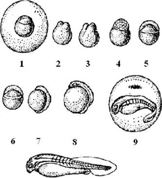
Fig. 3.3. Embryonic development period of the grass carp:
Stage I. Hydration of the peri-yolk cavity. Phases: 1 — unhydrated egg, diameter 1.3 mm; 2 — onset of peri-yolk cavity hydration, age 10 min; 3 — final egg hydration, diameter 6 mm, age 40 min.
Stage II. Blastodisc cleavage. Phases: 4 — two blastomeres, age 1 h; 5 — four blastomeres, age 1 h 20 min; 6 — eight blastomeres, age 1 h 40 min; 7 — sixteen blastomeres, age 2 h; 8 — macrocellular morula, age 2 h 30 min; 9 — microcellular morula, age 4 h 50 min; 10 — blastula, age 6 h 30 min.
Stage III. Gastrulation. Phases: 11 — onset of gastrulation, age 7 h 10 min; 12 — yolk plug, age 10 h; 13 — completion of gastrulation (closure of the yolk plug), age 12 h 10 min.
Stage IV. Organogenesis. Phases: 14 — formation of optic vesicles, onset of mesoderm segmentation, age 15 h; 15 — formation of optic cups, age 18 h.
Stage V. Separation of the embryonic tail region. Phases: 16 — onset of tail separation, age 1 day 5 h; 17 — straightening and initial body flexions of the embryos, age 1 day 5 h; 18 — onset of active embryonic rotation, age 1 day 8 h.
Stage VI. Embryo hatching from the membrane. Phase 19 — newly hatched embryo, body length 5.2 mm, age 1 day 10 h.
Stage VII. Appearance of a developed embryonic vascular system. Phase: 20 — embryo with a developed vascular system, body length 6.5 mm, age 2 days 3 h.
Stage VIII. Emergence of the movable gill-jaw apparatus. Phases: 21 — onset of mobility in the gill-jaw apparatus, body length 7.4 mm, age 3 days; 22 — reduction of embryonic respiratory organs, formation of the swim bladder, body length 7.5 mm, age 4 days.
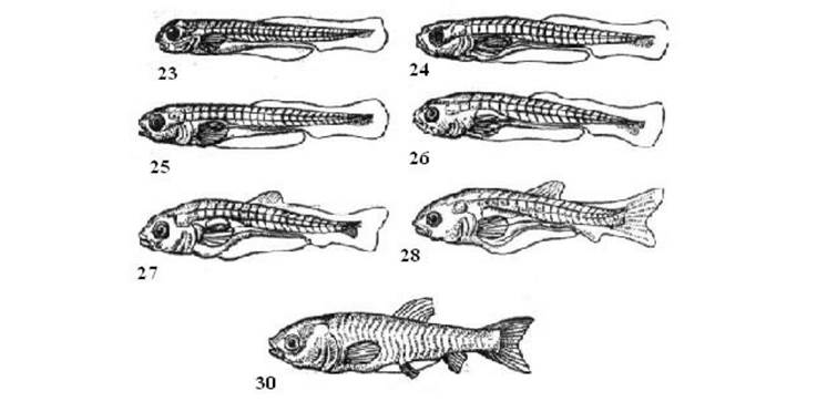
Fig. 3.4. - Larval development period of grass carp:
Stage I. Mixed feeding of the larva. Stages: 23 - onset of mixed feeding of the larva with a predominance of yolk food, body length 7.5 mm, age 4.5 days; 24 - mixed feeding of the larvae with a predominance of external food, body length 7.8 mm, age 6 days.
Stage II. Complete transition to external feeding. Stages: 25 - onset of exclusively external feeding of the larva, body length 7.6 mm, age 7 days.
Stage III. Formation of unpaired fins. Stage 26 - formation of mesenchymal clusters at the sites of Origin of the dorsal and anal fins, body length 8.1 mm, age 9 days; 27 - formation of fin rays in the paired fins, body length 9 mm, age 16 days.
Stage IV. Appearance of the second section of the swim bladder. Stages 28 - appearance of the anterior section of the swim bladder, origin of the pelvic fins, body length 10.2 mm, age 18 days.
Stage V. Formation of fin rays in the paired fins. Stages 29 - appearance of fin rays in the pectoral fins, body length 11.5 mm, age 20 days (not pictured); stages 30 - presence of rays in the pectoral and pelvic fins, body length 14.2 mm, age 22 days.
Table 3.11. - Developmental differences among grass carp, silver carp, and bighead carp
Morphological and morphometric traits |
Grass carp |
Silver carp |
Bighead carp |
Eggs (swollen) |
|||
Average diameter of the yolk sac membrane, mm |
4.38-5.22 1.21-1.36 |
3.80-4.50 1.10-1.20 |
4.82-5.63 1.42-1.50 |
Prolarvae, ind. |
|||
Number of myotomes in the trunk and tail |
29-31 12-14 |
24-26 14-17 |
24-26 14-17 |
Ratio of trunk length to tail length |
2.5 |
2 |
2 |
Presence of black pigment on the yolk sac |
Pigment only anteriorly |
Pigment anteriorly and on the ventral part |
Pigment anteriorly and slightly on the ventral part |
During artificial fish breeding, it is not always possible to provide optimal conditions for the normal course of physiological processes and proper development. This impairs the hatchery quality of the eggs, causes various structural disorders in the embryos, and reduces their viability.
Most often, the cause of unsatisfactory egg quality and larval anomalies is over-ripening of eggs in the body of females after ovulation.
A reliable criterion of egg quality is the fertilization rate and the morphological pattern of development. High-quality eggs have a high fertilization rate (90-95%, and sometimes 100%) and show no developmental disorders. Low-quality eggs often exhibit a high fertilization rate as well, while development may proceed normally. Therefore, the fertilization rate percentage alone cannot serve as a definitive criterion of quality. The process must be monitored at various Selection/3.html">Stages of development to detect characteristic anomalies depending on both egg quality and environmental conditions.
For example, swelling anomalies are observed, which are caused by the size variation of eggs obtained from a single female; such eggs generally fertilize well, but suffer high mortality during incubation and yield a significant number of larvae characterized by various Developmental Anomalies and deviations from normal egg Membrane Structure, leading to egg agglutination. This occurs if the eggs are not thoroughly stirred when water is added after insemination. At the site of adhesion, the outer layer of the membrane ruptures, and the inner layer protrudes through the tear.
Regarding blastodisc cleavage anomalies, the most frequent disorder is the detachment of individual blastomeres and variation in their size (Fig. 3.5).

Fig. 3.5. - Developmental disorders in silver carp:
A - cleavage of blastomeres; B - C - deformed embryo.
Regarding yolk sac anomalies, the yolk in low-quality eggs contains larger and more heterogeneous granules compared to high-quality eggs. Embryos with such yolk alterations usually survive until hatching, but upon transition to the larval developmental stage, they become non-viable and perish en masse.
Pseudodevelopment of unfertilized eggs occurs in a peculiar manner. Upon entering the water, they swell, and a cytoplasmic mound forms at the animal pole, which begins to cleave. However, this cleavage proves to be false, as blastomere division is incomplete, resulting in differently sized, symmetrically or asymmetrically arranged pseudoblastomeres—anucleated cytoplasmic bulges (Fig. 3.6).

Fig. 3.6. - Changes in unfertilized silver carp eggs:
A - B - false cleavage; C - D - false epiboly
During such chaotic pseudo-cleavage, unfertilized eggs are clearly distinguished from fertilized ones, whose blastomeres are uniform in size and have sharp contours. Therefore, the most suitable stages for determining the fertilization rate are the cleavage stages ranging from 4-8 blastomeres to the early morula. Later, the BOUNDARIES OF THE pseudoblastomeres disappear, the plasma acquires a smooth surface, and pseudo-gastrulation begins.
Pseudo-gastrulation culminates in The breakdown of the surface plasma layer and the coagulation of the yolk leaking from the rupture. Mass mortality of unfertilized eggs coincides with the period when the embryo's body begins to form in fertilized eggs, which should be considered as a diagnostic criterion.
A typical developmental anomaly is embryonic hydrops, which usually manifests at the onset of Heart formation. It consists of an excessive enlargement and fluid accumulation in the pericardial cavity, which in most cases leads to significant heart deformation. Hydrops may develop behind or around the pericardial cavity, beneath the anterior or posterior sections of the intestine, in the Urinary Bladder, but its occurrence in other locations is also possible. Only embryos with very minor signs of hydrops, which subsequently disappear, remain viable.
Developmental anomalies and body deformities in embryos are quite common. In such cases, distortions of the trunk and tail regions as well as disproportion of individual body parts are typically observed, which is also generally the case with dropsy.
The cause of these and other anomalies is undoubtedly poor egg quality and a violation of incubation conditions, particularly lowered temperatures and other factors that require further clarification.
One of the criteria for evaluating embryogenesis is the diameter of swollen eggs. Within their natural habitat, the formation process of the faunistic complex, which includes herbivorous fish, takes place in water bodies characterized by low water mineralization. Herbivorous fish are freshwater species known for their considerable plasticity.
During the acclimatization process, the range of these species underwent significant changes, and herbivorous fish, demonstrating high ecological plasticity,
showed the ability to grow and develop across various ecosystems against the backdrop of a fairly wide range of water mineralization. Meanwhile, the vast majority of semi-migratory and migratory fish species, despite being adapted to water with increased mineralization, choose freshwater areas of river systems as spawning grounds during the spawning season. In this regard, herbivorous fish, being resident freshwater species, should exhibit high conservatism toward increased water mineralization during embryogenesis and early post-embryogenesis. This issue is of distinct theoretical interest and exceptionally significant from a practical standpoint. Its urgency stems from the fact that in a number of regions that have successfully adopted and cultivated herbivorous fish, there is a trend not only toward increasing water mineralization in rearing and fattening water bodies, but also in water bodies used as sources of water supply for incubation facilities. Consequently, there is an obvious need to establish the limiting threshold for normal embryogenesis of herbivorous fish based on water mineralization, which is also relevant for other aquaculture ichthyofauna species.
To obtain objective information, special studies were conducted. The incubation of herbivorous fish eggs took place within optimal temperature ranges, the active reaction of the water was slightly alkaline to alkaline, and permanganate oxidizability corresponded to the upper limits of permissible norms, Table 3.12.
Table 3.12. Some abiotic environmental parameters during embryogenesis
Variants |
Average water temperature, °C |
pH |
Permanganate oxidizability, mg O/L |
Mineralization, g/L |
1 (freshwater) |
23.6 |
9.1 |
15.4 |
0.3 |
24.0 |
8.4 |
16.4 |
0.5 |
|
2 (brackish) |
23.2 |
8.8 |
17.9 |
1.1 |
25.5 |
7.7 |
10.1 |
1.7 |
|
27.7 |
8.1 |
10.0 |
2.7 |
The development of herbivorous fish eggs incubated under optimal temperatures followed a typical pattern. With an increase in water temperature, some acceleration in development was noted. At the same time, the swelling of grass carp and silver carp eggs varies depending on water mineralization. This process occurs most intensively in fresh water (0.3–0.5 g/L) and lasts 1 hour 40 minutes to 2 hours. As water mineralization increases to 1.1–1.7 g/L, its duration reaches 2 hours 10 minutes to 2 hours 30 minutes, and at 2.7 g/L, it takes 3 hours 30 minutes (r = 0.96).
The increase in mineralization of water used for supplying incubation facilities not only inhibits the egg swelling process but also exerts a decisive influence on the size of the swollen eggs. It has been established that a close inverse correlation exists between the average diameter of swollen eggs and salt concentration (r = 0.88), Table 3.13.
Table 3.13. Diameter of herbivorous fish eggs incubated at different water mineralizations
Variants |
Mineralization, g/L |
M±m |
Diameter, mm |
|
lim |
CV |
|||
1 (freshwater) |
0.3 |
4.67±0.06 |
4.30-4.90 |
4.97 |
0.5 |
4.32±0.03 |
4.00-4.50 |
2.62 |
|
1.1 |
3.75+0.04 |
3.30-4.10 |
5.46 |
|
2 (brackish) |
1.7 |
3.63±0.03 |
2.90-4.15 |
9.51 |
2.7 |
3.09±0.05 |
2.80-3.50 |
7.28 |
|
The EFFECT OF WATER mineralization ($X$, g/L) on egg size ($y_1$, mm) is indicated by a relative approximation error of 3.4%. The relationship between these indicators is described by the power equation $y_1 = 3.82 X^{-0.17}$. The decrease in egg diameter with increased water mineralization affects their total volume, leading to an actual reduction in the load on the incubation apparatus. The dependence of the calculated stocking rates ($y_2$, g/L) on water mineralization ($X$, g/L) is described by the equation $y_2 = 10.97 X^{0.45}$, which has a small relative approximation error of 7.7%.
In this regard, the stocking rates of incubation apparatuses relative to water mineralization are of definite practical interest, Table 3.14.
Thus, it is evident that embryogenesis proceeds normally under various water mineralizations and varying degrees of egg swelling. Furthermore, the decrease in egg swelling should not be regarded as an anomaly. Moreover, incubating eggs in brackish water opens up opportunities for more efficient utilization of incubation facility capacities, though this requires increased water flow rates for the effective removal of Metabolic waste products.
Table 3.14. Calculation of incubation apparatus stocking rates depending on water mineralization and egg size
Variants |
Water mineralization, g/L |
Egg diameter, mm |
Egg volume, mm3 |
Stocking in a 100 L capacity incubation apparatus |
||
egg volume, L |
egg mass, g |
quantity, thousand pcs |
||||
0.3 |
5.0 |
65.4 |
600 |
450 |
||
1 (freshwater) |
0.5 |
4.5 |
50.9 |
775 |
620 |
|
1.1 |
4.1 |
36.1 |
31.4 |
1090 |
870 |
|
2 (brackish) |
1.7 |
3.9 |
31.0 |
1250 |
1000 |
|
2.7 |
3.3 |
18.8 |
2000 |
1600 |
||
The diameter of swollen eggs can be considered one of the criteria for quality, but it is not a decisive one. In some cases, the egg diameter, having failed to reach maximum size upon completion of swelling, may be larger or smaller than established perceptions. In such instances, it is necessary to analyze abiotic environmental parameters and not rule out individual characteristics of certain females capable of producing eggs of larger or smaller diameters without a decline in their quality traits, which is confirmed not only by normal embryogenesis and high survival rates in early post-embryogenesis, but also by the viability and quality of the offspring. The studies performed using herbivorous fish as viable material are likely applicable to several other fish species that serve as objects of artificial fish breeding. However, a sufficiently high species specificity is evident for each specific fish species.
Periods and Stages of Ontogeny. The issues surrounding various aspects of fish breeding are largely determined by the biology and ecology of individual development. The Diversity of the ichthyofauna species composition, living conditions, and species-specific morphological, physiological, and biotechnological characteristics have proven to be objective prerequisites that determine the lifespan of species, the age at which they reach sexual maturity, and the features of embryogenesis and early post-embryogenesis.
The aforementioned has allowed for the accumulation of significant material characterizing fish ontogeny. However, it was V. V. Vasnetsov who, analyzing existing data and carrying out a large volume of special research, developed the THEORETICAL FOUNDATIONS OF the Stages of fish development, formulating the core tenets of this theory.
The main Concept of the theory he proposed is that, regardless of the species, fish development throughout ontogeny proceeds not only gradually and continuously, but also discontinuously and saltationally, clearly demonstrating the philosophical category of the transition from quantitative changes to qualitative ones.
As already noted, the embryonic, larval, fry, and adult (sexual maturity) periods are distinguished in the ontogeny of fish. Each period is characterized by qualitative specificity. During embryogenesis, typical endogenous nutrition occurs at the expense of nutrients from the yolk sac. Larvae feed on organisms living in the surrounding environment (exogenous nutrition), but in the initial period (from free embryo to larva), feeding has a mixed character (endogenous-exogenous), and the transition to external feeding occurs only after the complete resorption of the yolk sac. The larval period of fish life is characterized by the presence of specific larval organs that later disappear. With the onset of the fry period, this process is completed, the larval organs disappear completely, and new ones appear that make the fry a miniature copy of the adult fish of the given species.
During each period, a series of successive stages takes place, which make up a specific period. In turn, each stage consists of developmental steps corresponding to the developmental period. A developmental period is understood as a group of successive stages united by the same qualitative specificity.
From this, it is evident that the theory of developmental stages in fish implies an alternation of slow, gradual development (quantitative changes) and discontinuous, saltational (qualitative) changes during ontogeny. At the same time, sharp changes are observed in individual organs and systems. These changes are rapid and occur within a few hours or even minutes. It must be taken into account that morphological changes are most closely linked to changes in a number of biological characteristics of fish, including at the biochemical and physiological levels.
Qualitative, or saltational, changes during development occur quite rarely; between them, sequential growth and slow, yet noticeable quantitative changes take place. Slow, sometimes latent changes occur within specific limits of ontogeny without altering the main quality characterizing the given interval of fish development. Meanwhile, the qualitative features of the organism and its relationship with the external environment remain unchanged. The intervals of relative stability in fish ontogeny between two different qualitative changes are termed stages.
These stages are sequential in nature and exhibit specific Structural Features of the organism's body, characterized by specific biological properties and imposing precise environmental requirements. The absence of the necessary conditions for the normal progression of a given stage leads to developmental arrest at a specific point, accompanied by suppressed growth or complete cessation of growth, and ultimately resulting in death.
Examining the theory of developmental stages in fish, we observe that ontogenesis is characterized by both continuous and discontinuous dynamics. This process is divided into a series of successive stages during which mass and linear dimensions increase, accompanied by slow, gradual changes, while fundamental shifts in the morphology, physiology, and biology of the fish are excluded. Ontogenesis, or fish development, is the sequential adaptation of the organism to its habitat across specific developmental stages. Further elaboration of the core principles of fish developmental stages is associated with the research of S.G. Kryzhanovsky, who established a clear distinction in the progression of individual stages, determined their varying duration ranges, and identified specific features dependent on a number of factors. This principle is of vital importance for the development of fish-farming technologies. Therefore, further study of all related aspects concerning species targeted for artificial breeding remains essential.
It is important to note that research conducted in this direction has successfully characterized the biological and Ecological features of fish development across various stages.
Understanding the characteristics of these stages has made it possible to develop guidelines for rearing juvenile anadromous fish up to the downstream migration stage. The transition to this stage is marked by complex and profound changes, The Essence of which is preparing the organism for existence in a new environment. From a practical perspective, information characterizing the IMPACT OF ENVIRONMENTAL factors on fish at different developmental stages is of exceptional importance.
The theory of developmental stages should be regarded as the foundation and core upon which modern biotechnology for rearing juveniles of valuable fish species is based, thereby ensuring the efficiency of artificial fish farming.
Last update: 08/08/2026
Editorial and Educational Adaptation: This material has been compiled based on the primary/original source text. The project team performed an editorial review, corrected technical inaccuracies, structured sections, and adapted the content for an educational format.
What was processed:
- elimination of formatting defects (OCR errors, structural breaks, corrupted characters);
- editorial organization of content;
- standardization of terminology in accordance with academic sources;
- verification of factual statements against the original source text.
All mentions of the author, publication year, and origin of the primary text have been preserved in accordance with the source.