LEHNINGER PRINCIPLES OF BIOCHEMISTRY - VOL. 1. THE FOUNDATIONS OF BIOCHEMISTRY: STRUCTURE AND CATALYSIS - 2011
PART I. STRUCTURE AND CATALYSIS
10. LIPIDS
10.3. Lipids as Signaling Molecules, Cofactors, and Pigments
The two functional classes of Lipids discussed thus far—storage lipids and structural lipids—are major cellular components; Membrane Lipids account for 5% to 10% of the dry mass of most Cells, while stored lipids make up more than 80% of an adipocyte's mass. With few exceptions, these lipids play a passive role within The Cell: lipid fuels are stored until oxidized by Enzymes, and membrane lipids form impermeable barriers around cells and cellular compartments. Another group of lipids, present in much smaller amounts, plays an active role in METABOLISM as metabolites and messengers. Some act as potent signaling molecules, such as Hormones transported through the Blood from one tissue to another, or as intracellular messengers generated in response to an extracellular signal (a hormone or growth factor). Others function as enzyme Cofactors in Electron transfer reactions in Chloroplasts and Mitochondria, or in The transfer of sugar residues in numerous glycosylation reactions. A third group comprises lipids with a system of conjugated double bonds: pigment molecules that absorb visible light. Some of these act as light-absorbing pigments in Vision and Photosynthesis; others provide coloration, such as the orange color of pumpkins and carrots and the yellow color of canary feathers. Finally, a large group of volatile lipids is synthesized by plants as airborne signals that enable plants to communicate with one another, attract friendly animals, and deter enemies. In this section, we describe several such biologically active lipids. Their synthesis and biological roles will be discussed in greater detail in subsequent chapters.
Phosphatidylinositols and Sphingosine Derivatives Function as Intracellular Signals
Phosphatidylinositol and its phosphorylated derivatives are involved in several levels of Regulation of cellular Structure and metabolism. Phosphatidylinositol 4,5-bisphosphate (Fig. 10-9) on the cytoplasmic (inner) surface of The Plasma Membrane serves as a reservoir of messenger molecules released into the cell in response to extracellular signals acting on specific receptors located on the outer surface of the plasma membrane. Extracellular signals, such as the hormone vasopressin, activate phospholipase C in the membrane. It hydrolyzes phosphatidylinositol 4,5-bisphosphate, releasing two products that act as intracellular messengers: Inositol 1,4,5-trisphosphate (IP3), which is Water-soluble, and diacylglycerol, which remains bound to the plasma membrane. The IP3 molecule triggers the release of Ca2+ from The Endoplasmic reticulum, and the combination of diacylglycerol and an elevated cytosolic Ca2+ concentration activates the enzyme protein kinase C. This enzyme phosphorylates specific Proteins,
thereby eliciting a cellular response to the extracellular signal. This signaling mechanism is described in more detail in Chapter 12 (see Fig. 12-10).
Inositol Phospholipids also serve as "anchors" or nucleation sites for the assembly of certain supramolecular complexes involved in signal Transduction or exocytosis. Specialized signaling proteins bind specifically to phosphatidylinositol 3,4,5-trisphosphate in the plasma membrane, initiating The formation of multienzyme complexes on the cytosolic surface of the membrane. The production of phosphatidylinositol 3,4,5-trisphosphate in response to extracellular signals holds proteins together in signaling complexes at the plasma membrane surface (see Fig. 12-16).
Membrane Sphingolipids can also serve as a source of intracellular messengers. Both ceramide and sphingomyelin (Fig. 10-13) are potent regulators of protein Kinases; ceramide and its derivatives are known to be involved in the Regulation of Cell division, differentiation, migration, and programmed cell death (apoptosis; see Chapter 12).
Eicosanoids Transmit Signals to Neighboring Cells
Eicosanoids are paracrine hormones—substances that act only on cells close to the site of hormone synthesis, rather than being transported by the blood to act on cells in other Tissues or Organs. These fatty acid derivatives exert diverse and powerful effects on vertebrate tissues. They are known to be involved in reproductive Functions; in inflammation, fever, and pain associated with injury or disease; in blood clot formation and Blood Pressure Regulation; in gastric acid secretion; and in numerous other processes in both health and disease.
All eicosanoids are derivatives of arachidonic acid (20:4(Δ5,8,11,14)) (Fig. 10-18), a 20-carbon polyunsaturated fatty acid from which they derive their collective name (from the Greek word for twenty). There are three classes of eicosanoids: Prostaglandins, thromboxanes, and Leukotrienes.
Class="center">Figure 10-18. Arachidonic acid and some eicosanoids. Arachidonic acid (arachidonate ion at pH 7) is the precursor of eicosanoids, including prostaglandins, thromboxanes, and leukotrienes. In prostaglandin E1, C-8 and C-12 of the arachidonate ion are joined to form a characteristic five-membered ring. In thromboxane A2, C-8 and C-12 are connected, and an additional oxygen atom is present, forming a six-membered ring. Leukotriene A4 has three conjugated double bonds. Nonsteroidal anti-inflammatory drugs (NSAIDs), such as aspirin and ibuprofen, block the formation of Prostaglandins and thromboxanes from the arachidonate ion by inhibiting the enzyme cyclooxygenase (prostaglandin H2 synthase).

John Vane, Sune Bergström, and Bengt Samuelsson

Prostaglandins (PG) contain a five-carbon ring derived from arachidonic acid. Their name originates from the Prostate Gland, from which they were first isolated by Bengt Samuelsson and Sune Bergström. Initially, two groups of prostaglandins were identified: PGE (ether-soluble) and PGF (phosphate buffer-soluble, from the Swedish *fosfat*). Each group contains numerous subtypes, designated PGF1, PGF2, and so forth. Prostaglandins perform many functions. Some stimulate the contraction of uterine smooth Muscle during menstruation and labor. Others affect the Blood supply to individual organs, the Sleep-wake cycle, and the sensitivity of various tissues to hormones such as epinephrine and Glucagon. A third group of prostaglandins raises body Temperature, leading to fever, and causes inflammation and pain.
Thromboxanes have a six-membered ring containing an ether bond. They are produced by Blood Platelets and are involved in blood clot formation and the reduction of blood flow to the site of clot formation. As demonstrated by John Vane, NSAIDs such as aspirin, ibuprofen, and naproxen inhibit the enzyme prostaglandin H2 synthase (cyclooxygenase, COX), which catalyzes the early stage of The conversion of the arachidonate ion into prostaglandins and thromboxanes (Fig. 10-18; see also Fig. 21-15).
Leukotrienes, first discovered in leukocytes, contain three conjugated double bonds. They are potent biologically active signaling molecules. For example, leukotriene D4, a derivative of leukotriene A4, causes contraction of the smooth muscle lining the Airways to the lung. Overproduction of leukotrienes triggers asthmatic spasms, which is why leukotriene synthesis is a target for anti-asthmatic drugs such as prednisone. The severe contraction of pulmonary smooth Muscles that occurs during anaphylactic Shock in allergic individuals (hypersensitive to bee stings, penicillin, or other substances) can be fatal. ■
Steroid Hormones Transmit Signals Between Tissues
Steroids are oxidized derivatives of sterols; they possess a sterol Nucleus but lack the alkyl chain attached to ring D of Cholesterol, making them more polar than cholesterol. Steroid hormones are transported via the bloodstream (carried by carrier proteins) from their site of synthesis to target tissues, where they enter cells, bind to highly specific receptor proteins in The Nucleus, and initiate changes in Gene Expression and metabolism. Because these hormones have a very high affinity for their receptors, low concentrations of hormones (nanomolar or lower) are sufficient to elicit responses in target tissues. The principal group of steroid hormones comprises the male and Female Sex Hormones, along with the hormones cortisol and aldosterone produced by the adrenal cortex (Fig. 10-19). Prednisone and prednisolone are steroid drugs with potent anti-inflammatory activity, which is partly related to the inhibition of arachidonate ion release by phospholipase A2 (Fig. 10-18) and the resulting inhibition of leukotriene, prostaglandin, and thromboxane synthesis. They are widely used in medicine, including for the Treatment of asthma and rheumatoid Arthritis. ■
Vascular plants contain a steroid-like substance, brassinolide (Fig. 10-19), a potent growth regulator that increases The rate of stem elongation and influences the orientation of Cellulose microfibrils in The Cell wall during growth.
Figure 10-19. Steroids as derivatives of cholesterol. Testosterone, the male sex hormone, is produced in the Testes. Estradiol, one of the female sex hormones, is produced in the Ovaries and Placenta. Cortisol and aldosterone are hormones synthesized in the adrenal cortex; they regulate glucose metabolism and salt excretion. Prednisolone and prednisone are synthetic steroids used as anti-inflammatory drugs. Brassinolide is a growth regulator found in vascular plants.

Vascular plants use thousands of volatile signaling substances
Plants synthesize literally thousands of different lipophilic substances and volatile compounds that serve to attract pollinators, deter herbivores, recruit organisms that protect plants from herbivores, or act as communication signals with other plants. Jasmonate, derived from the 18:3(∆9,12,15) fatty acid in membrane lipids, is chemically similar to eicosanoids found in animal tissues; it acts as a potent signaling molecule that activat-
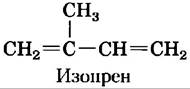
es plant defense in response to insect damage. Methyl jasmonate possesses the characteristic aroma of jasmine oil, which is widely used in perfumery. Many of the volatile compounds synthesized by plants are derivatives of Fatty acids or compounds formed by the Condensation of five-carbon isoprene units. Notable Examples among these compounds include geraniol (the characteristic scent of geranium), β-pinene (pine), limonene (lime), menthol, and carvone (see Fig. 1-23, a).
Vitamins A and D as Hormone Precursors
■ In the first third of the 20th century, the primary goal of research in physiological chemistry was the identification of vitamins—compounds that are essential for the health of humans and other vertebrates, yet cannot be synthesized by these animals and therefore must be obtained from the diet. Early nutritional studies revealed two Major Classes of such compounds: Fat-soluble vitamins, which dissolve in nonpolar organic Solvents, and Water-Soluble Vitamins, which can be extracted from foodstuffs with aqueous solvents. The fat-soluble vitamins include vitamins A, D, E, and K. All of these are isoprenoid compounds synthesized through the repeated condensation of isoprene units. Vitamins D and A function as hormone precursors.
Vitamin D3, or cholecalciferol, is normally formed in the Skin from 7-dehydrocholesterol via a photochemical reaction triggered by the ultraviolet (UV) component of sunlight (Fig. 10-20, a). Vitamin D3 itself is biologically inactive, but it is converted by Liver and Kidney enzymes into 1,25-dihydroxycholecalciferol, a hormone that regulates intestinal calcium absorption as well as calcium levels in the Kidneys and bones. Vitamin D deficiency leads to impaired skeletal development and Rickets, a condition rapidly cured by vitamin D administration (Fig. 10-20, b). Vitamin D2 (ergocalciferol) is produced commercially by UV irradiation of Yeast ergosterol. Structurally, vitamin D2 is analogous to D3, differing only in the side chain of the sterol D-ring. Both compounds produce identical biological effects, and D2 is commonly added to milk and butter as a dietary Supplement. Like steroid hormones, the vitamin D metabolite 1,25-dihydroxycholecalciferol regulates gene expression by interacting with nuclear receptor proteins (see Section 28.3).
Fig. 10-20. Formation and metabolism of vitamin D3. (a) Cholecalciferol (vitamin D3) is produced in the skin upon UV irradiation of 7-dehydrocholesterol, which induces bond Cleavage (highlighted in pink). A hydroxyl group is added to C-25 in the liver (pink); a second hydroxylation at C-1 in the kidney (pink) yields the active hormone 1,25-dihydroxycholecalciferol. This hormone regulates Ca2+ metabolism in the kidneys, intestines, and Skeleton. (b) Dietary vitamin D prevents rickets—a disease once prevalent in cold climates where heavy, thick clothing blocked the UV component of sunlight required for vitamin D3 production in the skin. Shown here is a detail from the mural "The Beneficial Influence of Biochemical Research," painted by John Steuart Curry in 1943 on a wall in the Biochemistry Building at the University of Wisconsin-Madison, USA. The people and animals depicted on the left show the effects of malnutrition (note the bowed legs of the rachitic boy). Those on the right represent individuals and animals rendered healthy through the outcomes of scientific research, including The Use of vitamin D to prevent and treat rickets.


Vitamin A (retinol) in various forms functions as the visual pigment in the vertebrate eye (Fig. 10-21). Acting via receptor proteins in the Cell Nucleus, the vitamin A derivative retinoic acid regulates gene expression during The Development of Epithelial Tissues, including the skin. Retinoic acid is the active ingredient in tretinoin (Retin-A), a medication used to treat acne and photoaged skin. Another vitamin A derivative, retinal, is the pigment that initiates the light response in retinal rod and cone cells, transmitting nerve impulses to the Brain. This role of retinal is discussed in detail in Chapter 12.
Fig. 10-21. Vitamin A1, its precursor, and derivatives. (a) β-Carotene is a precursor of vitamin A. Isoprene structural units are outlined with red dashed lines. Cleavage of β-carotene yields two molecules of vitamin A1 (retinol). (b) Oxidation at C-15 converts retinol to the aldehyde retinal (c), and further oxidation yields retinoic acid (d), a hormone that regulates gene expression. Retinal combines with the protein opsin to form rhodopsin (not shown), the visual pigment. In the dark, the retinal of rhodopsin is in the 11-cis conformation (c). When a rhodopsin molecule is excited by visible light, 11-cis-retinal undergoes a series of photochemical reactions that convert it entirely to all-trans-retinal (e), forcing the entire rhodopsin molecule to change its shape. This transformation in vertebrate retinal rods generates an electrical signal to the brain, which forms The basis of visual transduction. We will explore this topic in greater depth in Chapter 12.

Vitamin A was first isolated from fish liver oil; liver, eggs, and whole milk are excellent dietary sources of vitamin A. In vertebrates, β-carotene—the pigment responsible for the characteristic color of carrots, sweet potatoes, and other yellow vegetables—can be enzymatically converted into vitamin A. Vitamin A deficiency in humans leads to a wide range of symptoms, including dry skin, eyes, and mucous membranes; stunted GROWTH AND DEVELOPMENT; and night blindness, an early diagnostic sign of vitamin A deficiency. ■
Vitamins E and K and Lipid Quinones as Redox Cofactors
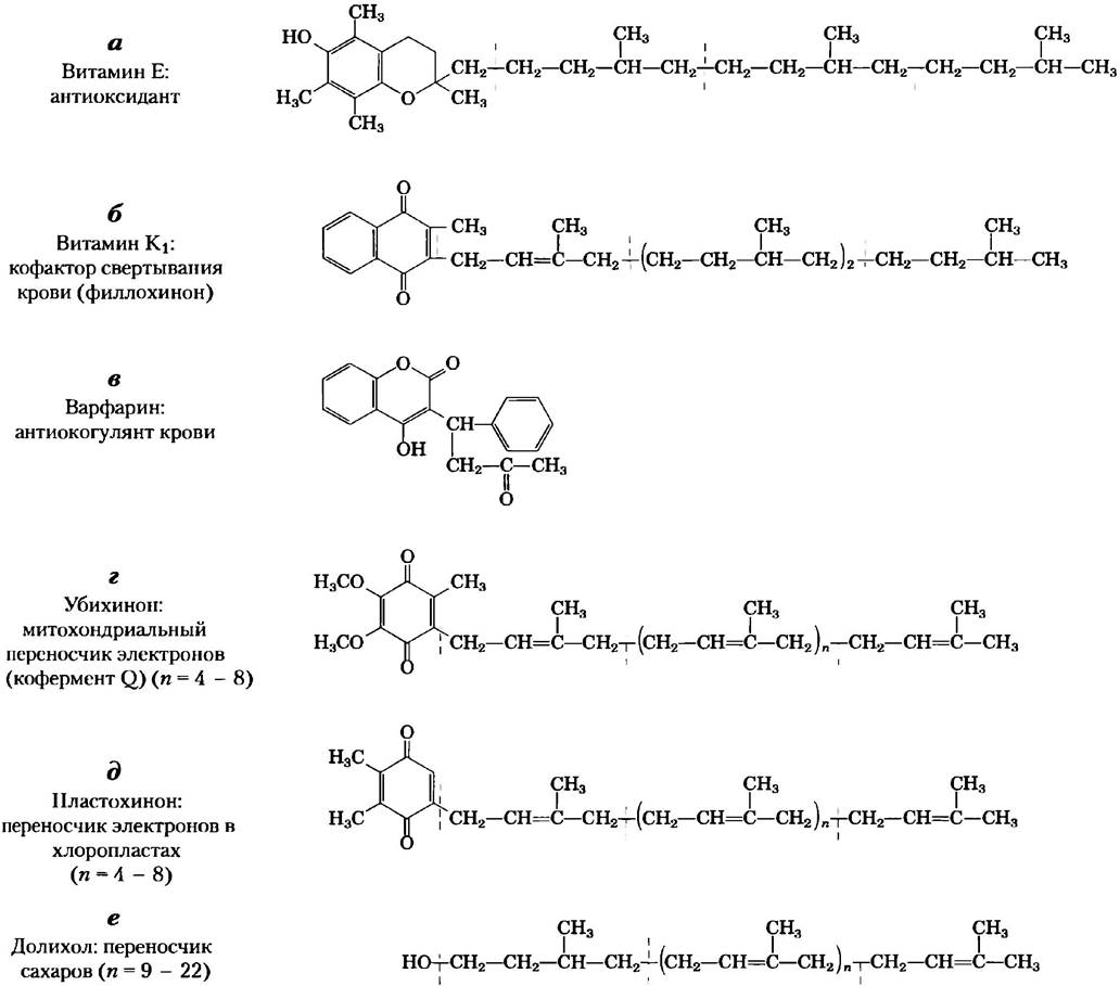
Vitamin E is the generic term for a group of closely related lipids known as tocopherols, which feature a substituted aromatic ring and a long isoprenoid side chain (Fig. 10-22, a). Tocopherols are hydrophobic and associate with cell membranes, lipid deposits, and blood Lipoproteins. They function as biological antioxidants. The aromatic ring reacts with and neutralizes highly reactive oxygen species and other free radicals, thereby protecting Unsaturated fatty acids from oxidation and preventing oxidative damage to membrane lipids that could lead to cell death. Tocopherols are found in eggs and vegetable oils, and are particularly abundant in wheat germ. Laboratory animals fed a vitamin E-deficient diet exhibit skin scaliness, muscular weakness, wasting, and sterility. Vitamin E deficiency in humans is extremely rare; the primary symptom is shortened erythrocyte lifespan.
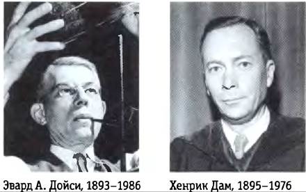
The aromatic ring of vitamin K (Fig. 10-22, b) undergoes a cycle of Oxidation and reduction during the formation of active prothrombin, a Blood Plasma protein essential for blood clot formation. Prothrombin is a proteolytic enzyme that cleaves peptide bonds in the blood protein fibrinogen, converting it into fibrin—an insoluble fibrous protein that holds blood clots together. Henrik Dam and Edward A. Doisy independently discovered that vitamin K deficiency slows blood clotting, which can be fatal to the Organism. Vitamin K deficiency is very rare in humans, occurring only in a small number of newborns suffering from hemorrhagic disease of the newborn, a potentially fatal condition. In the United States, newborns are routinely administered 1 mg of vitamin K. Vitamin K1 (phylloquinone) is found in green plant leaves, whereas a related form, vitamin K2 (menaquinone), is synthesized by Bacteria inhabiting the vertebrate intestine.
Warfarin (Fig. 10-22, c) is a synthetic compound that inhibits the formation of active prothrombin. It is widely used as an effective rat poison, causing death by internal Hemorrhage. Ironically, this potent rodenticide also serves as a crucial anticoagulant—a medication used to treat patients at risk of excessive blood clotting, such as those undergoing surgery, as well as individuals with coronary thrombosis. ■
Ubiquinone (also known as coenzyme Q) and plastoquinone (Fig. 10-22, d, e) are Isoprenoids that function as lipophilic electron carriers in the redox reactions of ATP Synthesis in Mitochondria and chloroplasts, respectively. Both ubiquinone and plastoquinone can accept either one or two electrons and one or two protons (Fig. 19-2).
Fig. 10-22. Several other biologically active isoprenoid compounds and derivatives. Isoprene structural units are highlighted with dashed red lines. In most mammalian tissues, ubiquinone (coenzyme Q) contains 10 isoprene units. Animal dolichols contain 10 to 17 isoprene units (85 to 105 carbon atoms), bacterial dolichols contain 11, and plant and fungal dolichols contain 14 to 24 units.
Dolichols Activate Sugar Precursors for Biosynthesis
During the assembly of complex CARBOHYDRATES in the Introduction/37.html">Bacterial cell wall and the attachment of polysaccharide units to certain proteins (Glycoproteins) and lipids (Glycolipids) in eukaryotes, the incoming sugar residues are chemically activated by linkage to isoprenoid alcohols known as dolichols (Fig. 10-22, f). These compounds engage in strong hydrophobic interactions with the membrane, anchoring the attached sugars within the bilayer where they participate in sugar-transfer reactions.
Many Natural Pigments Are Lipids with Conjugated Double Bonds
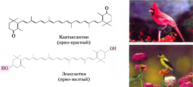
In the carbon chain of conjugated dienones, single bonds alternate with double bonds. Such a structure readily undergoes electron delocalization, allowing these compounds to transition into an excited state upon exposure to low-energy electromagnetic radiation (visible light), which makes them visible to humans and animals. Carotene (Fig. 10-21) has a yellow-orange color. Similar compounds make bird plumage bright orange, red, and yellow (Fig. 10-23). Like sterols, steroids, dolichols, vitamins A, E, D, and K, ubiquinone, and plastoquinone, these pigments are synthesized from five-carbon isoprene derivatives. This biosynthetic pathway is described in detail in Chapter 21.
Fig. 10-23. Lipids as pigments in plants and bird plumage. Compounds with extended systems of conjugated bonds absorb light in the visible region of the spectrum. These substances, which differ slightly in structure, serve as natural pigments responsible for strikingly vibrant and diverse colors. Bird plumage acquires a red or yellow hue when birds consume plants containing carotenoid pigments, such as canthaxanthin and zeaxanthin. The difference in coloration between male and female birds is attributed to variations in the uptake and metabolism of carotenoids.
Summary of Section 10.3 Lipids as Signaling Molecules, Cofactors, and Pigments
■ Certain types of lipids, although present in relatively small amounts, play critical roles as cofactors or signaling molecules.
■ Phosphatidylinositol bisphosphate is hydrolyzed to yield two intracellular messengers, diacylglycerol and inositol 1,4,5-trisphosphate. Phosphatidylinositol 3,4,5-trisphosphate serves as a scaffold for the assembly of supramolecular Protein Complexes involved in biological signal transduction.
■ Prostaglandins, thromboxanes, and leukotrienes (eicosanoids), derived from arachidonic acid, are exceptionally potent hormones.
■ Steroid hormones, derived from sterols, act as powerful biological signaling molecules (for example, sex hormones).
■ Vitamins D, A, E, and K are fat-soluble compounds built from isoprene units. All of them play essential roles in animal metabolism or physiology. Vitamin D is the precursor to a hormone that regulates Calcium Homeostasis. Vitamin A provides the visual pigment in the vertebrate eye and acts as a regulator of gene expression during epithelial cell growth. Vitamin E protects membrane lipids from oxidative damage, and, finally, vitamin K is essential for blood clotting.
■ Ubiquinones and plastoquinones, along with other isoprenoid compounds, function as electron carriers in mitochondria and chloroplasts, respectively.
■ Dolichols activate and anchor sugars to cell membranes for use in the synthesis of certain complex carbohydrates, glycolipids, and glycoproteins.
■ Lipids with conjugated diene structures function as pigments in flowers and fruits, and they are also responsible for the vibrant coloration of bird plumage.
Last update: 06/08/2026
Editorial and Educational Adaptation: This material has been compiled based on the primary/original source text. The project team performed an editorial review, corrected technical inaccuracies, structured sections, and adapted the content for an educational format.
What was processed:
- elimination of formatting defects (OCR errors, structural breaks, corrupted characters);
- editorial organization of content;
- standardization of terminology in accordance with academic sources;
- verification of factual statements against the original source text.
All mentions of the author, publication year, and origin of the primary text have been preserved in accordance with the source.