LEHNINGER PRINCIPLES OF BIOCHEMISTRY - VOLUME 1. THE FOUNDATIONS OF BIOCHEMISTRY, STRUCTURE AND CATALYSIS - 2011
PART I. STRUCTURE AND CATALYSIS
10. LIPIDS
10.2. Structural Lipids in Membranes
Introduction/36.html">Biological Membranes are composed of a lipid bilayer that acts as a barrier to the passage of polar molecules and ions. Membrane Lipids are amphipathic: one end of the molecule is hydrophobic, and the other is hydrophilic. Driven by hydrophobic interactions with Water, these lipid molecules spontaneously form sheet-like aggregates called membrane bilayers. In this section, we examine the five main Types of Membrane lipids: Glycerophospholipids, in which the hydrophobic regions consist of two fatty acid residues attached to glycerol; galactolipids and sulfolipids, which also contain two Fatty acids esterified to glycerol but lack the phosphate group found in Phospholipids; archaeal tetraether lipids, in which two very long alkyl chains are ether-linked to glycerol at both ends; Sphingolipids, in which a single fatty acid is linked to a long-chain amino alcohol, sphingosine; and sterols, compounds characterized by a rigid system of four fused hydrocarbon rings.
The hydrophilic regions of these amphipathic compounds can be quite simple, such as a single -OH group at one end of a sterol ring system, or they can be much more complex. In glycerophospholipids and some sphingolipids, the polar HEAD group is joined to the hydrophobic moiety via a phosphodiester bond; these are phospholipids. Other sphingolipids lack phosphate and instead feature a simple sugar or a complex oligosaccharide at their polar ends; these are Glycolipids (Fig. 10-7). Membrane lipids display remarkable diversity, arising from various combinations of fatty acid "tails" and polar "heads." The arrangement of phospholipids within membranes, along with their Structural and functional roles, will be explored in the next chapter.
Class="center">Figure 10-7. Common types of storage and membrane lipids. All lipids shown here are built upon either a glycerol or a sphingosine backbone (pink bars), linked to one or more long-chain alkyl groups (yellow) and polar head groups (blue). In triacylglycerols, glycerophospholipids, galactolipids, and sulfolipids, the alkyl chains derive from fatty acid residues attached via ester bonds. Sphingolipids contain a single fatty acid linked via an amide bond to the sphingosine backbone. Archaeal membrane lipids contain very long, branched alkyl chains, with each end forming an ether bond with a glycerol moiety. In phospholipids, the polar head group is attached through a phosphodiester bond, whereas in glycolipids, a glycosidic bond directly links the sugar head group to glycerol.

Glycerophospholipids Are Derivatives of Phosphatidic Acid
Glycerophospholipids, also known as phosphoglycerides, are membrane lipids in which two fatty acid residues are attached by ester bonds to the First and Second carbon atoms of glycerol, while a highly polar or charged group is attached to the third carbon via a phosphodiester bond. Glycerol is a prochiral Structure; it has no asymmetric carbon atoms, but the attachment of a phosphate group to one end turns it into a chiral compound, correctly designated as L-glycerol 3-phosphate or D-glycerol 1-phosphate, or more commonly as sn-glycerol 3-phosphate (Fig. 10-8). Glycerofosfolipidy are named as derivatives of the parent compound, phosphatidic acid (Fig. 10-9), depending on the Chemical Nature of the polar alcohol head group.
Figure 10-8. L-Glycerol 3-phosphate, the backbone of phospholipids. Glycerol is not intrinsically chiral because it possesses a plane of Symmetry passing through C-2. However, glycerol can be rendered chiral by introducing a substituent, such as a phosphate group, to either -CH2OH group, making glycerol prochiral. The DL-nomenclature is used for glycerophosphates (see p. 117), in which isomers are named according to their stereochemical relationship to glyceraldehyde stereoisomers. In this nomenclature, the stereoisomer of glycerol phosphate found in most lipids is properly designated as L-glycerol 3-phosphate or D-glycerol 1-phosphate. Alternatively, stereospecific numbering (the sn-system) is used, by which C-1 is defined as the group of the prochiral compound that occupies the pro-S position. In this nomenclature, the glycerol phosphate in phospholipids is termed sn-glycerol 3-phosphate.
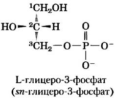
Figure 10-9. Glycerophospholipids. Among glycerophospholipids, diacylglycerols with head-group alcohol moieties attached via phosphodiester bonds are the most common. Phosphatidic acid, a phosphomonoester, serves as the parent compound. Each derivative is named after the polar alcohol head group (X), with the prefix "phosphatidyl-". In cardiolipin, two phosphatidic acid molecules share a single glycerol. R1 and R2 represent fatty acid residues.

For example, phosphatidylcholine and phosphatidylethanolamine contain Choline and ethanolamine in their polar head groups, respectively. In all these compounds, the head group is joined to glycerol through a phosphodiester bond, with the phosphate group carrying a negative charge at neutral pH. The polar alcohol may carry a net negative charge (as in phosphatidylinositol 4,5-bisphosphate), be neutral (phosphatidylserine), or carry a positive charge (phosphatidylcholine, phosphatidylethanolamine). As we will see in Chapter 11, these charges contribute significantly to the Surface Properties of membranes.
Glycerophospholipids can contain any of a variety of naturally occurring fatty acids. For instance, the phospholipid phosphatidylcholine can exist in multiple molecular species, each featuring a unique combination of fatty acids. The distribution of these molecular species is specific to different organisms, different Tissues within the same Organism, and different glycerophospholipids within the same Cell or tissue. Typically, glycerophospholipids contain a C16 or C18 saturated fatty acid at C-1 and a C18 to C20 unsaturated fatty acid at C-2. With rare exceptions, the exact Biological Significance of these variations in fatty acid composition and head groups remains unclear.
Some Phospholipids Contain Ether-Linked Fatty Acids
Certain animal tissues and unicellular organisms are rich in lipids in which one of the two acyl chains is attached to glycerol via an ether bond rather than an ester bond. This ether-linked chain may be saturated, as in alkyl ether lipids, or it may contain a double bond between C-1 and C-2, as in plasmalogens (Fig. 10-10). Vertebrate Heart tissue is uniquely rich in Ether-linked Lipids; approximately half of heart lipids are plasmalogens. The membranes of halophilic Bacteria, Protozoa, and certain invertebrates also have a high content of ether-linked lipids. The functional role of ether lipids in these membranes is not yet fully understood; their resistance to phospholipases—Enzymes that cleave ester-linked fatty acids from membrane lipids—may be crucial for certain specialized Functions.
Figure 10-10. Ether lipids. Plasmalogens contain an ether-linked alkenyl chain at the position where most glycerophospholipids have an ester-linked fatty acid (compare with Fig. 10-9). Platelet-activating factor features a long ether-linked alkyl chain at the C-1 position of glycerol, but carries an ester-linked acetate residue at the C-2 position, making this compound far more water-soluble than most glycerophospholipids and plasmalogens. The head-group alcohol in both plasmalogens and platelet-activating factor is choline.
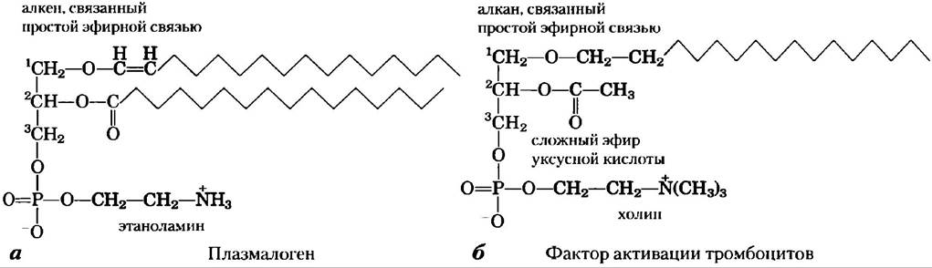
■ At least one ether lipid, platelet-activating factor, functions as a potent signaling molecule. Released from leukocytes known as basophils, it stimulates platelet aggregation and the release of serotonin (a vasoconstrictor). It also exerts diverse physiological effects on the Liver, smooth Muscle, heart, uterine, and lung tissues, and plays a key role in inflammatory and allergic responses. ■
METABOLISM/14.html">Chloroplasts Contain Galactolipids and Sulfolipids
A second major group of membrane lipids is the galactolipids, which predominate in plant Cells. In these lipids, one or two galactose residues are connected by a glycosidic linkage to the C-3 atom of 1,2-diacylglycerol (Fig. 10-11; see also Fig. 10-7). Galactolipids are localized in the thylakoid membranes (internal membranes) of chloroplasts, accounting for 70% to 80% of the total membrane lipids in vascular plants, making them arguably the most abundant membrane lipids in the biosphere. Because soil phosphate availability is often limited, evolutionary pressures to conserve phosphate for more critical metabolic needs likely drove plants to evolve these non-phosphorus-containing lipids. Plant membranes also contain sulfolipids, in which a sulfonated glucose residue is joined to diacylglycerol via a glycosidic bond. In sulfolipids, the sulfonate head group carries a fixed negative charge, much like the phosphate group in phospholipids (Fig. 10-11).
Figure 10-11. Three glycolipids from chloroplast thylakoid membranes. In monogalactosyldiacylglycerols (MGDG) and digalactosyldiacylglycerols (DGDG), nearly all acyl groups derive from linoleic acid (18:2(∆9,12)), and the head groups are uncharged. In the sulfolipid 6-sulfo-6-deoxy-α-D-glucopyranosyldiacylglycerol, the sulfonate group bears a fixed negative charge.

Archaea contain unique membrane lipids
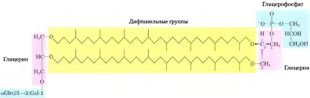
In Archaea, the majority of which inhabit ecological niches with extreme conditions—such as high temperatures (boiling water), low pH, or high Ionic strength—membrane lipids contain long-chain (32 carbon atoms) branched Hydrocarbons connected at each end to glycerol (Fig. 10-12). The simple ether bonds of these compounds are far more resistant to Hydrolysis at low pH and high Temperature than the ester Bonds Found in bacterial and eukaryotic lipids. In their fully extended conformation, archaeal lipids are twice as long as Phospholipids and Sphingolipids, spanning the entire width of the outer membrane. At each end of this elongated molecule is a polar "head group" consisting of glycerol linked to either phosphate or sugar residues. The general term for these compounds, glycerol dialkyl glycerol tetraether (GDGT), reflects their unique structure. The glycerol moiety of archaeal lipids is a different stereoisomer from that in bacterial and eukaryotic lipids; the central carbon is in the R-configuration in Archaea, but in the S-configuration in the other "domains" (Fig. 10-8).
Fig. 10-12. A typical archaeal membrane lipid. In this diphytanyl tetraether lipid, the diphytanyl moieties (yellow) are long hydrocarbons composed of eight five-carbon isoprene units condensed head-to-tail (for the Condensation of isoprene units, see Fig. 21-36; compare also the diphytanyl groups with the 20-carbon phytyl side chain of chlorophyll in Fig. 19-47a). In this extended conformation, the diphytanyl groups are roughly twice as long as the 16-carbon fatty acid typically found in bacterial and eukaryotic membrane lipids. The glycerol moieties in archaeal lipids have the R-configuration, in contrast to bacterial and eukaryotic lipids, where they have the S-configuration. Archaeal lipids also differ in the substituents on the glycerol moiety. In the molecule shown here, one glycerol is linked to the disaccharide α-glucopyranosyl-(1->2)-β-galactofuranose; the other glycerol is connected to a glycerophosphate head group.
Sphingolipids are derivatives of sphingosine
Sphingolipids—the fourth major class of membrane lipids—also consist of a polar head group and two nonpolar tails, but, unlike glycerophospholipids and galactolipids, they contain no glycerol. Sphingolipids comprise one molecule of the long-chain amino alcohol sphingosine (4-sphingenine) or one of its derivatives, one molecule of a long-chain fatty acid, and a polar head group linked by glycosidic or phosphodiester bonds (Fig. 10-13).
Fig. 10-13. Sphingolipids. The first three carbons at the polar end of sphingosine are analogous to the three carbons of glycerol in glycerophospholipids. The amino group at C-2 forms an amide linkage with a fatty acid. This fatty acid is typically saturated or monounsaturated, with 16, 18, 22, or 24 carbon atoms. Ceramide is the simplest compound of this group. Other sphingolipids differ in the polar head group (X) attached to C-1. Gangliosides have very complex oligosaccharide head groups. Standard Abbreviations for sugars are used here (see Table 7-1).
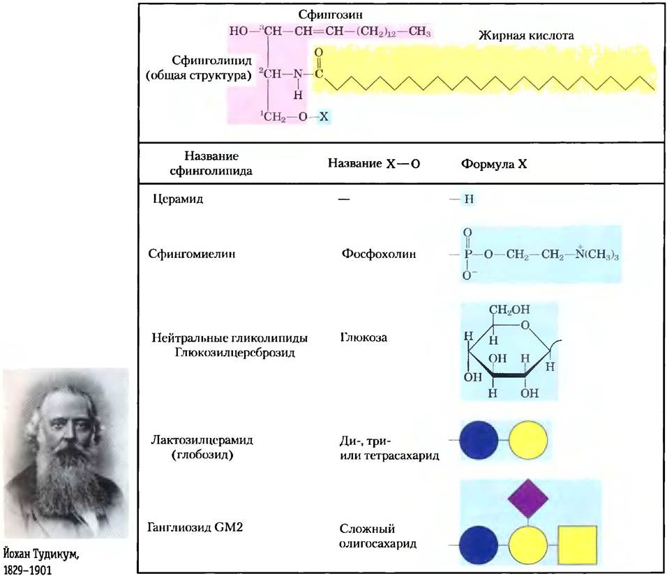
Carbons C-1, C-2, and C-3 of sphingosine are analogous to the three carbons of glycerol in glycerophospholipids. When a fatty acid is attached via an amide bond to the -NH2 group at C-2, the result is a ceramide, which is structurally similar to diacylglycerol. Ceramide is the structural "parent" of sphingolipids.
There are three subclasses of sphingolipids, all derived from ceramide but differing in their polar head groups: sphingomyelins, neutral (uncharged) glycosphingolipids, and gangliosides. Sphingomyelins contain phosphocholine or phosphoethanolamine as their polar head groups and are therefore classified as phospholipids alongside glycerophospholipids (Fig. 10-7). Indeed, in their properties and three-dimensional structure, sphingomyelins closely resemble phosphatidylcholines, and their polar head groups are likewise uncharged (Fig. 10-14). Sphingomyelins are present in the Plasma Membranes of animal cells and are particularly abundant in myelin, the membranous sheath that surrounds and insulates the axons of certain Neurons—hence the name "sphingomyelins."
Fig. 10-14. The similarity in shape and molecular structure between phosphatidylcholine (a glycerophospholipid) and sphingomyelin (a sphingolipid) is clearly evident when comparing their structural formulas and space-filling models.

Glycosphingolipids, found predominantly on the outer surface of plasma membranes, contain head groups composed of one or more sugars linked directly to the —OH group of the C-1 carbon of the ceramide moiety; they lack phosphate. Cerebrosides have a single sugar linked to ceramide; galactocerebrosides are characteristic of plasma membranes of neural cells, whereas glucocerebrosides are found in the plasma membranes of cells of other tissues. Globosides are neutral (uncharged) glycosphingolipids with two or more sugars, usually D-glucose, D-galactose, or N-acetyl-D-galactosamine. Cerebrosides and globosides are sometimes referred to as neutral glycolipids because they lack any charge at pH 7.
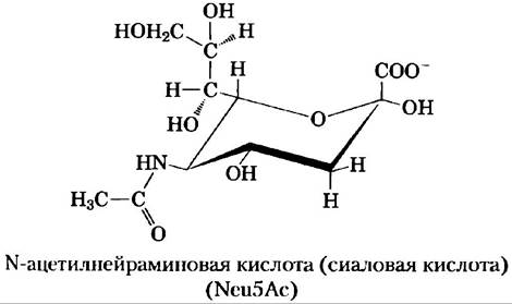
Gangliosides, the most complex sphingolipids, have Oligosaccharides as their polar head groups with one or more residues of N-acetylneuraminic acid (Neu5Ac)—often simply called sialic acid—at the termini. Sialic acid imparts a negative charge to gangliosides at pH 7, distinguishing them from globosides. Gangliosides with one sialic acid residue are designated the GM series (M for mono-), those with two residues are of the GD series (D for di-), and so forth (GT designating three sialic acid residues, GQ four).
Sphingolipids at The Cell Surface: Sites of Biological Recognition
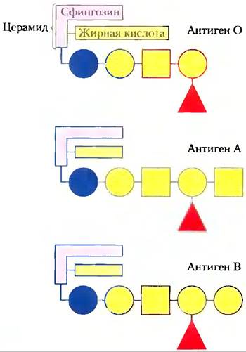
When the physician and chemist Johann Thudichum discovered sphingolipids over 100 years ago, their biological role seemed as enigmatic as the Sphinx (hence their name). At least 60 different sphingolipids have been identified in human cell membranes. Many of these are particularly abundant in the plasma membranes of neurons, and some certainly serve as recognition sites on the cell surface, yet the specific functions of only a few sphingolipids are known to date. The carbohydrate moieties of certain sphingolipids define human Blood Groups and thus determine the type of blood a person can safely receive in transfusions (Fig. 10–15).
Fig. 10–15. Glycosphingolipids as determinants of blood group. Human blood groups (O, A, B) are determined in part by the oligosaccharide head groups (shown in blue) of these glycosphingolipids. In addition, three specific oligosaccharides are found attached to certain blood Proteins in individuals with blood types O, A, and B, respectively. Standard sugar designations are used here (see Table 7–1).
Gangliosides are concentrated on the outer surface of cells, acting as recognition points for extracellular molecules or surfaces of neighboring cells. The types and concentrations of gangliosides in The Plasma Membrane undergo dramatic changes during embryonic development. Tumor formation induces the synthesis of a new Complement of gangliosides, and very low concentrations of a specific ganglioside have been found to induce differentiation in cultured neural tumor cells. Elucidating the BIOLOGICAL FUNCTIONS OF various gangliosides remains a fertile ground for future research.
Phospholipids and Sphingolipids Are Degraded in Lysosomes
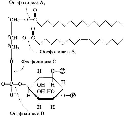
In most cells, membrane lipids are continually degraded and replaced by new ones. For every bond hydrolyzable in a lipid, lysosomes contain a specific hydrolytic enzyme (Fig. 10–16). Phospholipases of the A type remove one of the two fatty acids, yielding a lysophospholipid (these esterases do not cleave the ether bond in plasmalogens). Lysophospholipases remove the remaining fatty acid.
Fig. 10–16. Specificity of phospholipases. Phospholipases A1 and A2 hydrolyze the ester bonds of intact glycerophospholipids at the C-1 and C-2 carbons of glycerol. Once one fatty acid is removed by an A-type phospholipase, the second fatty acid is cleaved from the molecule by a lysophospholipase (not shown). Phospholipases C and D each cleave one of the phosphodiester bonds in the head group. Some phospholipases act only on a single type of glycerophospholipid—such as phosphatidylinositol 4,5-bisphosphate (shown here) or phosphatidylcholine—whereas others are less specific.
Gangliosides are degraded by a set of lysosomal enzymes that catalyze the stepwise removal of sugar units, ultimately leaving a ceramide. A genetic defect in any of these hydrolytic enzymes leads to the accumulation of gangliosides within cells, resulting in severe pathological consequences (Box 10–2).
Sterols have four fused carbon rings
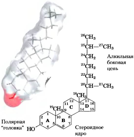
Structural lipids known as sterols are present in the membranes of most Eukaryotic cells. The hallmark of this fifth group of membrane lipids is the steroid Nucleus, consisting of four fused rings: three six-carbon rings and one five-carbon ring (Fig. 10-17). The steroid nucleus is nearly planar and relatively rigid; the fused rings make rotation about the C-C bonds impossible. Cholesterol, the major sterol in animal tissues, is amphipathic, with a polar "head" (the hydroxyl group at C-3) and a nonpolar hydrocarbon body (the steroid nucleus and the branched hydrocarbon side chain at C-17) roughly equal in length to a 16-carbon fatty acid. Similar sterols are found in other eukaryotes,
such as stigmasterol in plants and ergosterol in Fungi. Bacteria cannot synthesize sterols, although a few species can incorporate exogenous sterols into their membranes. The sterols of all eukaryotes, as well as the Fat-soluble Vitamins, Quinones, and dolichols described in Section 10.3, are synthesized from simple five-carbon isoprene units.
Fig. 10-17. Cholesterol. A stick representation of cholesterol is superimposed on a space-filling model of the molecule. In the chemical structure, the rings are labeled A-D to simplify the nomenclature of steroid derivatives, and the carbon atoms are numbered (in blue). The hydroxyl group (pink in both molecular representations) at C-3 is the polar "head". For storage and transport of the sterol, this hydroxyl group is condensed with a fatty acid to form a sterol ester.
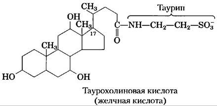
In addition to their role as membrane components, sterols serve as precursors for A wide variety of products with specific biological activities. For example, Steroid Hormones are potent biological signaling molecules that regulate Gene Expression. Bile acids are polar derivatives of cholesterol that act as detergents in the intestine, emulsifying dietary fats to make them more accessible to digestive lipases.
We will return to Cholesterol and other sterols in subsequent chapters to examine the Structural Role of cholesterol in biological membranes (Chap. 11), steroid hormone signaling (Chap. 12), and the remarkable pathway of Cholesterol Biosynthesis and its transport by lipoprotein carriers (Chap. 21).
Box 10-2. MEDICINE. Genetic Defects in Human Membrane Lipid Metabolism
Polar membrane lipids undergo a continuous metabolic turnover, The rate of synthesis generally being balanced by the rate of breakdown. Lipid degradation is carried out by hydrolytic enzymes in lysosomes, each enzyme being capable of hydrolyzing only one specific bond. If sphingolipid degradation is impaired due to a defect in one of these enzymes (Fig. 1), products of incomplete breakdown accumulate in tissues, causing serious diseases.
Fig. 1. Degradation pathways of GM1, globoside, and sphingomyelin leading to ceramide. Red circles with crosses indicate enzymatic defects at specific hydrolysis steps, accompanied by the name of the disease caused by the accumulation of the incomplete breakdown product.
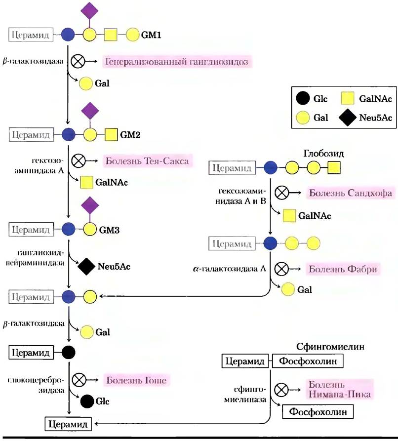
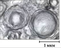
For example, Niemann-Pick disease is caused by a rare genetic defect in the enzyme sphingomyelinase, which removes phosphocholine from sphingomyelin. Sphingomyelin accumulates in the Brain, Spleen, and liver. The disease manifests in early childhood and leads to delayed mental development and early death. Tay-Sachs disease is more common; in this condition, the ganglioside GM2 accumulates in the brain and spleen (Fig. 2) due to a deficiency of the enzyme hexosaminidase A. Symptoms include progressive developmental delay, paralysis, blindness, and death by age 3 to 4.
Fig. 2. Electron micrograph of a brain cell from an infant with Tay-Sachs disease, showing abnormal deposits of gangliosides in lysosomes.
Genetic Counseling can predict and prevent many inherited disorders. Testing prospective parents can reveal abnormal enzymes, and subsequent DNA analysis can determine the precise Nature of the genetic defect, making it possible to assess the risk to offspring. During Pregnancy, samples obtained from the Placenta (chorionic villus sampling) or from the fluid surrounding the fetus (amniocentesis) can be tested in the same way.
Summary of Section 10.2 Membrane Structural Lipids
■ Polar lipids with polar "heads" and nonpolar "tails" are the major components of membranes. The most common are glycerophospholipids, which contain fatty acids esterified to two hydroxyl groups of glycerol, and a second alcohol (the polar "head") attached to the third glycerol hydroxyl via a phosphodiester bond. Other polar lipids include sterols.
■ Glycerophospholipids vary in The structure of their polar "head"; the most prevalent phospholipids are phosphatidylethanolamine and phosphatidylcholine. At pH near 7, the polar "heads" of glycerophospholipids carry an electrical charge.
■ Chloroplast membranes are extremely rich in galactolipids, consisting of diacylglycerol attached to one or two galactose residues, and sulfolipids, which are diacylglycerols with a sulfonated sugar residue that provides a negatively charged "headgroup".
■ Archaea contain unique membrane lipids with long-chain alkyl groups ether-bonded to glycerol at both ends, and sugar and/or phosphate residues attached to the glycerol to form a polar or charged "head". These lipids are stable under the extreme conditions in which archaea thrive.
■ Sphingolipids do not contain glycerol; instead, they contain sphingosine, a long-chain aliphatic amino alcohol. In addition to phosphoric acid and choline, sphingomyelin contains two long hydrocarbon chains—one derived from a Fatty acid and the other from sphingosine. The other three classes of sphingolipids are cerebrosides, globosides, and gangliosides, all of which contain sugar moieties.
■ Sterols possess four fused rings and a hydroxyl group. Cholesterol, the major animal sterol, serves both as a structural component of membranes and as a precursor for a variety of Steroids.
Last update: 06/08/2026
Editorial and Educational Adaptation: This material has been compiled based on the primary/original source text. The project team performed an editorial review, corrected technical inaccuracies, structured sections, and adapted the content for an educational format.
What was processed:
- elimination of formatting defects (OCR errors, structural breaks, corrupted characters);
- editorial organization of content;
- standardization of terminology in accordance with academic sources;
- verification of factual statements against the original source text.
All mentions of the author, publication year, and origin of the primary text have been preserved in accordance with the source.