Fundamentals of Biochemistry - Filippovich, Y. B. 1999
Proteins
Protein Isolation
Although about two dozen Proteins (such as Insulin, Ribonuclease, Lysozyme, cytochrome c, somatotropic hormone, ß-lipotropin, acyl carrier protein, a-bungarotoxin, cobrotoxin, cardiotoxin, gastrin inhibitor, Trypsin inhibitor, apolipoprotein, ferredoxin, and others) have been successfully synthesized—marking the first step on a very difficult path toward their artificial creation—these syntheses remain extremely complex, labor-intensive, time-consuming, and expensive. Therefore, the only viable method for obtaining proteins is their isolation from natural sources. However, this is also a challenging task, as proteins are highly sensitive to most chemical Reagents (acids, alkalis, organic Solvents, etc.) and are degraded by standard Procedures used in substance purification (heating, distillation, sublimation, extraction, etc.). Autolysis (self-Digestion) of the isolated proteins is also a possibility. Proteins easily lose their native, natural properties (such as solubility and biological activity) and transition into a denatured state. To prevent Protein Denaturation during isolation, all procedures are carried out under mild conditions: at low temperatures (not exceeding +5° C) and avoiding harsh chemical reagents.
Proteins (gluten) were first isolated from wheat flour by J. Beccaria in 1728, a date widely considered the birth of Protein Chemistry. Studies on milk protein—casein—began in 1762 (A. Haller), followed by Blood proteins in 1789 (A. Fourcroy). Over the course of two and a half centuries, hundreds of different proteins have been obtained from natural sources and their properties thoroughly studied.
Successful Protein Isolation from biological tissue requires extremely fine tissue grinding to the point of Cell wall disruption. For this purpose (Fig. 6), specialized roller (A) or ball (B) mills are used, in which the starting material is repeatedly pressed between closely spaced rollers or crushed by continuously colliding balls. Homogenizers (C and D) are also widely used; in these devices, the material is either chopped by ultra-high-speed rotating blades (12,000 rpm or more), ground between the precision-ground surfaces of a Glass tube and pestle, or frozen and extruded through the dies of a specialized press (E).
Cell walls can also be effectively disrupted by alternating cycles of freezing and thawing. Here, ice crystals act as the "working tool," tearing The Cell walls and releasing the intracellular contents. Another approach is the "nitrogen bomb" method, which involves saturating suspended Cells with gaseous nitrogen under high pressure, followed by a rapid pressure release. The nitrogen that has penetrated the cells expands into gas and effectively "explodes" them.
A Classification of Methods for disrupting (disintegrating) biological material, illustrating the diverse approaches used to isolate proteins and Other Compounds from natural sources, is given in Table 2.
Class="center">
Fig. 6. Design of devices for grinding biological material during protein isolation;
A — roller mill (a1 and a2 — rollers rotating towards each other; the straight arrow indicates the entry direction of the material being ground); B — ball mill (b1 — housing; b2 — balls; the arrow indicates the direction of housing rotation); C — manual homogenizer (c1 — pestle; c2 — housing); D — mechanical homogenizer (d1 — blade; d2 — chamber for grinding material; d3 — housing with electric motor and starter switch; d4 — lid); E — working chamber of the press-grinding device (e1 — space filled with biological material; e2 — plungers for extruding the material; e3 — chamber walls; e4 — partition with microscopic pores)
Once fine grinding of the material is achieved, the next stage—Protein Extraction—begins. Proteins are most commonly extracted using 8–10% salt solutions, as the vast majority of proteins are readily soluble in such concentrations. Different salts exhibit varying solvating effects on proteins. Below is a series of cations and anions arranged in order of decreasing solubilizing efficiency:

Table 2 Classification of disintegrating treatments by their nature (according to B. A. Fichte and G. A. Gurevich, 1988)
|
Physical |
Chemical |
Enzymatic |
Biological |
|
Mechanical |
Non-mechanical |
|||
Ballistic; extrusion; ultrasonic; gas decompression; hydraulic Shock; electro-hydraulic shock; combined |
Osmotic, thermal, or cold shock; freeze — thaw; freeze — drying; dehydration — rehydration; slow gas decompression; phase transitions at high pressures |
Action of alkalis, acids, salts, detergents, Antibiotics, chelating agents, organic solvents |
Action of bacteriological, Yeast-lytic, mycolytic Enzymes, and immobilized lytic enzymes |
Action of phages, bacteriocins, and other killer factors, intracellular parasites, plasmid-like factors. Inhibition of cell wall synthesis. Autolysis |
Thus, lithium pyrophosphate should exhibit the maximum solubilizing effect, while sodium chloride shows the minimum.
Since Protein solubility is strongly influenced by environmental pH, most salts are applied as buffer mixtures (phosphate, acetate, borate, citrate, etc.). Recently, buffer systems formulated with Organic compounds have gained widespread use, including tris-(hydroxymethyl)-aminomethane (HOCH2)3CNH2 and its salt with HCl (Tris buffer); diethylbarbituric acid and its sodium salt (veronal-medinal buffer); N,N-bis-(2-hydroxyethyl)-Glycine (HOCH2CH2)2NCH2COOH (bicine buffer); N-[tris-(hydroxymethyl)-methyl]-glycine (HOCH2)3CNHCH2COOH (tricine buffer), and others.
Good results are also obtained by extracting proteins with alcohol-salt mixtures. The resulting Metalloproteins exhibit varying solubility in alcohol, which allows individual proteins to be extracted at different alcohol concentrations. Glycerol is very widely used for protein extraction as it protects them against denaturation.
Protein extraction from biological material is facilitated by Treatment with detergents, such as sodium dodecyl sulfate, sodium deoxycholate, Triton X-100, alkyl Glycosides, and trialkylammoniopropanesulfonates:

These detergents weaken hydrophobic protein-lipid and Protein-Structure/156.html">Protein Interactions, resulting in the disruption of Introduction/36.html">Biological Membranes and the release of Structural and functional protein components.
The principles of protein isolation discussed above primarily apply to animal Tissues. Isolating proteins from plant material is immeasurably more difficult: Plant Cell Walls are harder to break, and protein denaturation is more likely due to interactions with Tannins and other compounds. Therefore, isolating proteins from Vegetative plant Organs requires specific techniques: treating tissues with a Water-ether mixture that sharply increases plant cell wall permeability (Chibnall's method), extracting proteins with a mixture of phenol, acetic acid, and water (Singch's method), and others.
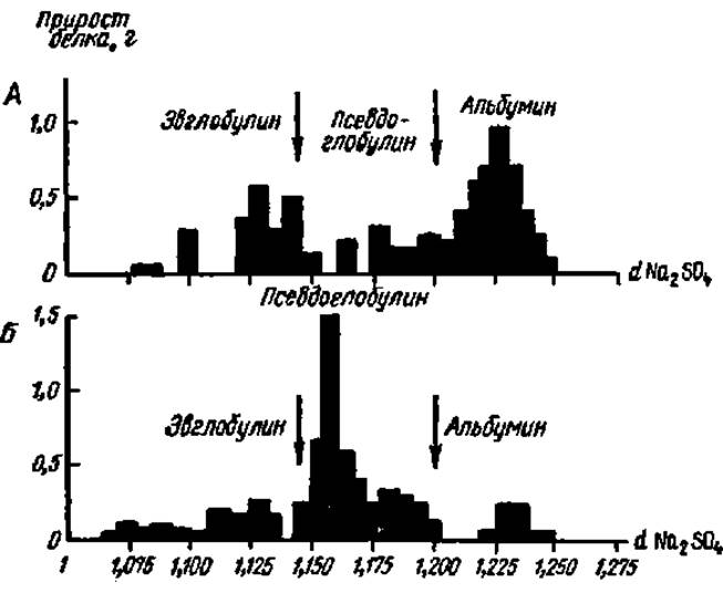
Fig. 7. Difference diagrams of protein salting-out:
A — human blood Serum proteins; B — silkworm hemolymph proteins. Each bar on the diagram corresponds to The amount of protein precipitated by changing the Na2SO4 concentration by 0.005 (in solution density units). Arrows indicate the Na2SO4 concentrations that serve as boundaries for separating the fractions from one another
Following the extraction of a protein mixture from biological material, the resulting mixture is fractionated into individual proteins, since PLANT AND ANIMAL tissues contain a multitude of different proteins extracted in varying proportions during the isolation process. Protein fractionation is carried out using various methods: salts, organic solvents, Electrophoresis, chromatography, molecular sieving, etc.
The method of fractionating proteins using salt solutions is based on the principle that each individual protein in a mixture precipitates out at a specific concentration of a given salt, while other proteins remain in solution at that concentration. The process of precipitating a protein from solution via salt action is called salting-out. Upon further salt saturation, the next individual protein precipitates; thus, by step-by-step increases in the salt concentration of the reaction medium, relatively pure individual Proteins can be isolated one after another. To determine the salt concentration boundaries at which a specific protein precipitates, difference diagrams are constructed (Fig. 7).
Among organic solvents, methyl and ethanol alcohols are widely used for protein fractionation (the process is conducted at around 5°C to prevent protein denaturation). Difference diagrams are also preliminarily constructed in these cases. Fig. 8 presents data on ethyl alcohol concentrations required to precipitate various protein fractions from human blood serum. This method has found widespread application in The production of blood substitutes, as it allows essential ingredients to be extracted and preserved for long periods. Protein fractionation sometimes combines salting-out and alcohol precipitation through The Use of alcohol-salt solutions.
The heavy metal ion precipitation method has limited application in Protein Fractionation and is mostly used in combination with other fractionation techniques (such as salting-out or organic solvent precipitation). This method is based on the interaction of Hg, Zn, Ca, Ba, Pb, Fe, Cu, U, and other heavy Metal Ions with reactive amino acid residues of the protein molecule (e.g., Hg2+ interacts with sulfhydryl groups, Zn2+ with imidazole groups, and Ca2+ and Ba2+ with phosphoserine residues).
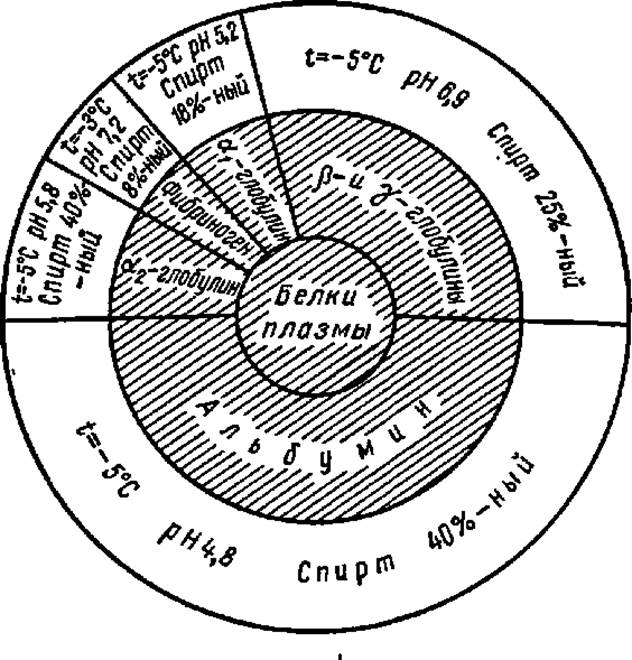
Fig. 8. Fractionation diagram of human Blood Plasma Proteins using ethanol
The electrophoresis method is based on the ability of various proteins to move under METABOLISM/18.html">The Influence of an electric field at different speeds (and sometimes in opposite directions) within a solution, on moist filter paper, or in another solid supporting medium. The voltage is typically set from several hundred to several thousand volts, with a current of several tens of milliamperes. The migration rate of a specific type of protein molecule toward the anode or cathode is a function of its electrical charge, molecular weight and shape, Ionic strength, pH, and the COMPOSITION OF THE buffer solution, as well as the applied potentials. The combination of these factors is always specific to each individual protein, meaning that different proteins possess distinct electrophoretic mobilities.
Electrophoresis in solid supporting media—such as Cellulose, cellulose acetate, Agar gel, starch gel, and polyacrylamide gel—is the most widely used approach. Proteins on electrophoretograms are visualized using stains such as Amido Black 10B, bromophenol blue, Coomassie Brilliant Blue, and others:

However, detecting proteins by staining them in gels with ammoniacal silver complexes is 100–200 times more sensitive.
Electrophoresis in solid media prepared from a three-dimensional copolymer of acrylamide and methylenebisacrylamide holds particular promise:

Depending on The ratio of acrylamide to N,N-methylenebisacrylamide in the reaction medium, the three-dimensional structure formed during copolymerization features fine, medium, or large pores. During electrophoresis, protein molecules pass through these pores with varying ease, which facilitates protein fractionation. For instance, the fractionation of human serum proteins reveals 6 fractions via paper electrophoresis, 10 fractions in starch gel, and 16 fractions in polyacrylamide gel (Fig. 9).
An even higher degree of protein mixture fractionation, down to the isolation of individual proteins, is achieved by performing electrophoresis in supporting media with a pH gradient. Because a protein's position at a certain level within the Column or thin-layer carrier is determined by its isoelectric point, this method is known as isoelectric focusing (also referred to as electrofocusing or isotachophoresis). The pH gradient is established using polyaminopolycarboxylic acids—ampholines—specifically synthesized for this purpose by O. Vesterberg, whose composition is expressed by the following formula:

where x = 2–3, and R is H or —(CH2)x—COOH. The Molecular Weight of ampholines ranges from 300 to 1000, and depending on the ratio of tertiary nitrogen atoms to carboxyl groups in their molecules, an aqueous ampholine solution can provide a pH range from 3 to 10.

Fig. 9. Resolving power of solid supporting media in the fractionation of human blood serum proteins by electrophoresis:
(A — cellulose; B, C — starch and polyacrylamide gels). On electrophoretograms A, B, and C, the large protein fraction on the left is albumin; the one on the right is the γ-globulin complex; and the fractions between them correspond to other globulins. In all cases, the same preparation was applied to the origin zone at amounts of 100, 80, and 5 µL, respectively

Fig. 10. Mechanism of Ion-exchange chromatography of proteins
The Mechanism of ion-exchange chromatography involves the displacement of counterions bound to the anionic and cationic sites of the ion exchanger (I) by ionized groups of protein molecules (II), followed by the binding of the latter to the ion exchanger via Electrostatic Interactions. Depending on the type of protein and, in particular, the ratio of dicarboxylic and diamino acid residues within it, the net effect of these bonds varies. Consequently, different proteins are displaced from the ion exchanger by developer ions (III and IV) at varying rates and elute from the column separately. The ion exchanger is then regenerated. Cellulose and Sephadex derivatives (see p. 325) bearing ion-exchange groups (aminoethyl, sulfoethyl, carboxymethyl, etc.), calcium phosphate gel, silica gel, and other carriers are used as adsorbents. When carriers selectively bind strictly specific proteins, the Fractionation of Protein mixtures is highly efficient. This type of chromatography is referred to as Affinity Chromatography. Over the past 5 years, immobilized metal ion affinity chromatography (IMAC) of proteins has become widespread, which is based on their ability to reversibly adsorb onto transition metal ions by utilizing the latter's free coordination bonds.
One of its modifications is Hydrophobic Interaction Chromatography, which relies on the interaction between a carrier (phenyl- or octyl-Sepharose) and the corresponding nonpolar amino acid residues of protein molecules. The use of efficient sorbents (various silica gel derivatives) and high pressure during protein elution has led to The Development of several variants of High-Performance Liquid Chromatography.
The mechanism of protein ion-exchange chromatography is illustrated in Fig. 10.
Salt solutions with varying concentrations and pH values are used to elute adsorbed proteins from the column. If the concentration of the salt solution is changed while eluting proteins from the same column, such elution is termed gradient elution. The eluate is collected in small portions (a few milliliters each) into individual test tubes using a specialized device—a fraction collector. Typically, several hundred such fractions are obtained during the chromatographic fractionation of proteins. A colorimetric protein assay or the measurement of the ultraviolet absorbance of each solution determines the protein content in every fraction. Based on these data, a curve is plotted that reveals how many protein fractions are present in the eluate and which series of test tubes contains each fraction. Identical fractions are pooled, and the pure protein is isolated from the resulting solution.
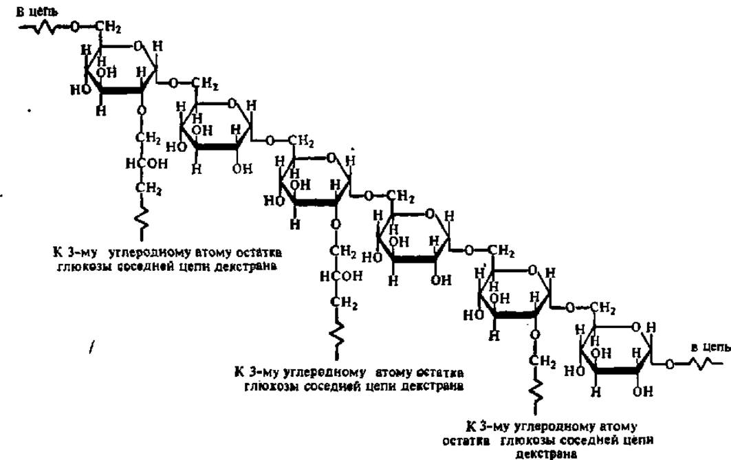
Protein fractionation by molecular sieve chromatography (Gel filtration) is based on the differential rate at which protein molecules (depending on their molecular weight) pass through a column packed with a specialized material—Sephadex1. Sephadex is produced by treating the polysaccharide dextran with epichlorohydrin, which introduces a varying number of cross-links between the dextran molecules:
1 Its name is derived from the initial syllables of three words: Separation, Pharmacia (the name of the Swedish company manufacturing the reagents), and dextran. It is also applied using simple equipment. The distribution of proteins between phases is determined by differences in the PHYSICOCHEMICAL PROPERTIES OF the fractionated proteins and the polymers comprising the upper and lower phases: interactions based on a combination of their properties (such as The formation of ionic and Hydrogen Bonds, and hydrophobic interactions) lead to the accumulation of a specific protein or group of proteins in one of the polymer phases.
Similarly, the treatment of another polysaccharide, agarose, with 2,3-dibromopropanol under strongly alkaline conditions yields a cross-linked polymer, Sepharose, which forms gels suitable for the fractionation of proteins with molecular weights in the hundreds of thousands of Daltons.
Depending on the degree of cross-linking in Sephadex and the size of the protein molecules, the latter penetrate the Sephadex beads packed in the column to a varying extent. Large protein molecules, unable to pass through the mesh into the bead, are rapidly eluted from the column by the eluent flow; smaller protein molecules that have penetrated inside the Sephadex beads are retained for some time and emerge from the column with subsequent portions of the eluate. Thus, the Sephadex column acts as a molecular sieve, sorting protein molecules by size and—paradoxically—eluting the largest molecules first (Fig. 11). Since Sephadex beads swell considerably in solvents to form a gel, this technique is also referred to as gel filtration.
Of particular interest is the method of protein fractionation proposed by P. A. Albertsson, which utilizes two-phase systems composed of aqueous polymer solutions. This fractionation technique operates under mild conditions (physiological ionic strength and pH), and the aqueous polymer solutions exert a stabilizing effect on the separated proteins. The method can be scaled for any amount of substance. Materials similar to Sephadex include Molselect (produced by Reanal, Hungary), Acrylex and Bio-Gel P (based on polyacrylamide), Sephacryl (obtained from allyldextran and N,N'-methylenebisacrylamide), Sepharose (derived from agarose), and others. These materials are widely used not only for protein fractionation but also for the separation of Nucleic Acids, higher CARBOHYDRATES, and other Biopolymers (see also p. 325).

Fig. 11. Mechanism of molecular weight-based substance separation on a Sephadex column:
A — column at THE START OF the Procedure: light circles represent Sephadex beads; dark dots represent protein molecules of varying molecular weight newly applied to the top of the column; B — as the eluent moves down the column, protein molecules are transported, but only the smaller protein molecules penetrate inside the Sephadex beads; C — small protein molecules lag significantly behind the large ones due to retention within the Sephadex beads. The large molecules are the first to emerge from the column
Last update: 06/08/2026
Editorial and Educational Adaptation: This material has been compiled based on the primary/original source text. The project team performed an editorial review, corrected technical inaccuracies, structured sections, and adapted the content for an educational format.
What was processed:
- elimination of formatting defects (OCR errors, structural breaks, corrupted characters);
- editorial organization of content;
- standardization of terminology in accordance with academic sources;
- verification of factual statements against the original source text.
All mentions of the author, publication year, and origin of the primary text have been preserved in accordance with the source.