Biochemistry and Molecular Biology - Belyasova N.A. 2002
Metabolism. Energy-Yielding Processes
Capture of Light Energy by Biomolecules
Photosynthesis
Photosynthesis is a highly efficient process that captures visible light energy and transforms it into the chemical bond energy of biological molecules. This process occurs in the Cells of plants, Algae, and phototrophic Bacteria. In most of these organisms, photosynthesis is accompanied by The Use of Water as an electron donor and the release of molecular oxygen (Equation 13.1). Only in anoxygenic bacteria belonging to the Class Anoxyphotobacteria can certain other reduced compounds, such as hydrogen sulfide, serve as electron Donors instead of water. In this case, no O2 is produced (Equation 13.2).
CO2 + H2O → (CH2O) + O2 (13.1)
CO2 + 2H2S → (CH2O) + H2O + 2S (13.2)
This observation was made by Cornelis van Niel, who, while still an undergraduate thesis student between 1931 and 1933, investigated the characteristics of photosynthesis in various bacteria and put forward a bold hypothesis that revolutionized researchers' views on the mechanisms of photosynthesis. Based on the photosynthesis equation he derived for purple sulfur bacteria (13.2), C. van Niel hypothesized that it is water, rather than carbon dioxide, that splits during plant photosynthesis to yield molecular oxygen, and he proposed a general equation for photosynthesis (13.3):
CO2 + 2H2A → (CH2O) + H2O + 2A (13.3)
In the latter equation, H2A represents an electron donor, which is water in oxygenic organisms and other substances (such as hydrogen sulfide, molecular hydrogen, or isopropanol) in anoxygenic bacteria. This process releases a dehydrogenated electron donor (A) and reduces carbon dioxide to an organic compound (CH2O). These transformations constitute the core of the photosynthetic process.
It is worth noting that water is a very poor electron donor, and none of the oxidizing agents available to Living organisms are powerful enough to strip hydrogen atoms from an H2O molecule. This becomes possible only through the photochemical transformations of specific chlorophyll molecules, which thereby become powerful oxidizing agents and acquire The ability to oxidize water.
Photosynthesis strictly requires the involvement of membranes. In prokaryotes, this role is performed by invaginations of Cell/30.html">The Plasma Membrane, whereas in eukaryotes, it is carried out by thylakoid membranes located within METABOLISM/14.html">Chloroplasts. Thylakoids reside in the chloroplast stroma, forming stacks known as grana. The internal space of the thylakoids is called the lumen.
The thylakoid membranes house electron transport components grouped into Two Photosystems, along with ATP synthase. The photosystems are used for electron transport coupled with the pumping of protons into the lumen, which establishes a proton gradient across the thylakoid membrane. The energy of this proton gradient drives ATP synthesis, much like what occurs during Oxidative Phosphorylation. However, unlike the Respiratory Chain, electrons in photosystems move from a poor donor (water molecules) to a poor acceptor (NADP+), i.e., in the opposite direction compared to Respiration. Consequently, energy must be expended to drive this "uphill" electron flow. Indeed, to reduce NADP+, electrons must be excited by light twice.
Characteristics of photosystem components. The components of photosystems are Structure/178.html">Protein Complexes containing photosensitive molecules (pigments) and Carriers of Reducing equivalents.
Pigments are represented mainly by chlorophylls, carotenoids, and phycobilins. The Central Role in photosynthesis is played by chlorophyll a, a green, magnesium-containing tetrapyrrole pigment (Fig. 13.1). This chromophore is a magnesium porphyrin and is structurally similar to heme (an iron porphyrin). However, chlorophyll a has several key differences: a fifth cyclopentane ring is fused to one of the pyrrole rings; one of the pyrrole rings is partially reduced; and one of the acidic side chains is esterified with phytol, a twenty-carbon, highly hydrophobic alcohol. The phytol tail imparts amphiphilic properties to the chlorophyll molecule and acts as an "anchor" that keeps the chromophore embedded in The Lipid Bilayer of the thylakoid membrane. Meanwhile, the hydrophilic HEAD (magnesium porphyrin) lies on the membrane surface facing the aqueous phase of the stroma and is oriented parallel to the membrane plane. This spatial orientation optimizes light energy capture in chloroplasts.

Fig. 13.1. STRUCTURE OF THE chlorophyll a molecule
Other chlorophylls of green plants (b, c, pheophytin), as well as those of certain algae and bacteria (Chlorobium chlorophyll, bacteriochlorophylls), differ from chlorophyll a in The structure of the substituents on their pyrrole rings, the absence of a magnesium ion (pheophytin), and their absorption spectra. The coexistence of chlorophylls with distinct absorption spectra within a cell broadens the range of harvested radiation.
Carotenoids and phycobilins serve as accessory pigments. They further expand THE SPECTRUM OF energy that an Organism can absorb and, in addition, protect chlorophylls from excess light and oxidation by the oxygen generated during photosynthesis. Carotenoids are yellow, orange, red, or brown pigments that strongly absorb in the violet region of the spectrum. Chloroplasts contain two groups of carotenoids: carotenes (Hydrocarbons, mostly tetraterpenes, discussed in detail in Chapter 17) and xanthophylls (chemically similar to carotenes but containing oxygen). Phycobilins are characteristic of cyanobacteria and the chloroplasts of red algae.
To capture light energy more efficiently, pigments in the photosystems are organized by Proteins into antenna complexes. Each antenna complex contains several hundred pigment molecules, and their primary function is to transfer the absorbed energy to chlorophyll a. Thus, an antenna complex can be compared to a funnel that "channels" energy toward the reaction center chlorophyll of the photosystem. Excitation energy (an exciton) is transferred from molecule to molecule via the electric field of an excited electron (essentially, an excited electron in a molecule that has absorbed a light quantum transfers its energy to a neighboring molecule in such a way that an electron in the neighbor is also excited).
The electron-transporting Components of the photosystems include the cytochrome b/f complex (an aggregate of integral Membrane Proteins containing two Cytochromes: b563 and f), pheophytin, membrane-bound plastoquinones (QA and QB), and mobile carriers such as plastoquinone QP, plastocyanin, and ferredoxin. Plastoquinone closely resembles ubiquinone in Structure and function (Fig. 12.1). Plastocyanin is a protein containing a single copper atom coordinated with amino acid side chains: during electron transfer, Cu alternates between the +1 and +2 oxidation states. Ferredoxin is an iron-sulfur protein of the 4Fe, 4S2- type that carries electrons. The chain of carriers terminates with an enzyme that transfers electrons to NADP+.
It has been established that photosynthesis requires two photosystems: II and I. Photosystem II contains chlorophyll a with an absorption optimum at 680 nm (P680) in its reaction center, whereas the reaction center of Photosystem I (P700) contains chlorophyll a with an absorption optimum at 700 nm.
Light reactions of photosynthesis. The photosynthetic process can be broadly divided into two stages: light reactions and dark reactions. The light reactions require visible light and take place in the thylakoid membranes, resulting in the reduction of NADP+ and the synthesis of ATP. The dark reactions can occur in the absence of visible light within the stroma and generally refer to the reduction of CO2 utilizing ATP and NADPH.
In plants, photosynthetic electron transport begins with Photosystem II. The energy of absorbed light is funneled from the antenna complexes to the reaction center chlorophyll a (P680), exciting one of its electrons. This excited electron is immediately transferred to a nearby pheophytin molecule, leaving behind a positively charged "hole" in the chlorophyll molecule with a very high electron affinity—effectively creating a positively charged radical, P680•+. This "hole" is rapidly filled by an electron extracted from water by the water-splitting enzyme (H2O → 2H+ + SO2 + 2e-) with the participation of manganese ions. The excited electron migrates along the chain of carriers (Fig. 13.2) and reaches a second "hole" in P700 of Photosystem I, which in turn was formed when a "hot" electron was transferred to an electron acceptor (likely plastoquinone). Here, the electron is excited a second time and is subsequently passed via Photosystem I carriers to NADP+ (Fig. 13.2). This directional electron movement along the carrier chain is called noncyclic flow, or the Z-scheme. A key distinguishing feature of this process is that the electron is excited twice in the reaction centers of the two photosystems, providing sufficient energy to reduce NADP+. The formation of NADPH is catalyzed by ferredoxin-NADP+ reductase, which contains FAD as a prosthetic group. Notably, the reduction of nicotinamide Cofactors requires hydride ions: two electrons from two molecules of reduced ferredoxin converge, and a proton is taken up from the stroma.

Fig. 13.2. Schematic representation of electron pathways in the photosystems of thylakoid membranes. Jagged arrows symbolize the photochemical excitation of electrons and their transition to a higher energy level; solid arrows indicate the path of electrons in noncyclic flow; the dashed arrow indicates the path of electrons entering cyclic flow
In addition to producing NADPH, noncyclic electron flow drives proton pumping into the lumen, establishing an electrochemical gradient across the thylakoid membrane. This occurs during The transfer of hydrogen from reduced plastoquinone QP to the cytochrome b/f complex, which Functions as an electron carrier (Chapter 12). When protons flow down their electrochemical gradient from the lumen back into the stroma via Facilitated Diffusion, they pass through ATP synthase channels, driving ATP synthesis. This energy-storage mechanism is known as Photophosphorylation and is fundamentally similar to oxidative phosphorylation.
Besides the noncyclic flow described above, cyclic electron flows can also occur in thylakoid membranes. In these pathways, an electron is excited only once—in the reaction center of Photosystem I—and no NADPH is generated. Electron flow becomes cyclic when electrons from ferredoxin are redirected back to plastoquinone QP rather than being transferred to NADP+ (Fig. 13.2, 13.3, dashed arrows). This movement of electrons provides The Cell with ATP but not NADPH. Cyclic electron flow predominates under conditions of low NADP+ availability, which results from the intracellular accumulation of NADPH.
Figure 13.3 illustrates the arrangement of photosystem components, their substrates, and products. As shown, the primary products of the photosynthetic light reactions—NADPH and ATP—accumulate in the stroma, exactly where they are consumed in the dark reactions.
Dark reactions of photosynthesis. The dark reactions of photosynthesis involve the fixation—meaning the reduction and incorporation into Organic compounds—of CO2. In most photosynthetic organisms, this process takes place via The Calvin Cycle (named after Melvin Calvin, who was awarded the Nobel Prize for its discovery).
The Calvin cycle can be conventionally divided into three stages: 1) carboxylation of ribulose bisphosphate; 2) reduction of 3-phosphoglycerate to an aldehyde; 3) regeneration of ribulose bisphosphate, the CO2 acceptor.
Carbon dioxide reduced in the Calvin cycle enters the stroma of chloroplasts in green plants through Stomata in leaves and green stems, while in algal and cyanobacterial cells it enters in dissolved form. The key reaction of CO2 reduction is the carboxylation of ribulose bisphosphate. This reaction is catalyzed by an unusual enzyme, ribulose bisphosphate carboxylase/oxygenase, which exhibits two catalytic activities: carboxylase and oxygenase (Fig. 13.4).
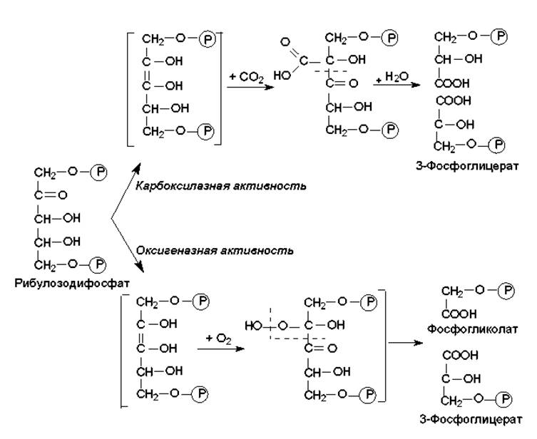
Fig. 13.4. Reactions of ribulose bisphosphate conversion catalyzed by ribulose bisphosphate carboxylase/oxygenase
The oxygenase activity is manifested in the absence of CO2 and in the presence of O2, and the products of this reaction are involved in Photorespiration. Ribulose bisphosphate carboxylase/oxygenase operates very slowly: its catalytic rate is hundreds of times lower than that of most Other Enzymes. Therefore, this enzyme can account for up to half of the total protein in chloroplasts. It is considered to be the most abundant protein on Earth, quantitatively exceeding any other protein. The rate of the Calvin cycle is regulated at the level of this enzyme.
The reduction of the products of the carboxylase reaction (two molecules of 3-phosphoglycerate) proceeds with the participation of phosphoglycerate kinase and glyceraldehyde-3-phosphate dehydrogenase. The reverse Reactions Catalyzed by these enzymes take place in Glycolysis (Fig. 9.3). This process consumes ATP energy and reducing equivalents supplied by NADPH.
The regeneration of ribulose bisphosphate is accomplished through intermolecular rearrangements involving transaldolases and transketolases (which also take part in the Pentose Phosphate Pathways, described in Chapter 9). Three compounds enter these transformations: glyceraldehyde-3-phosphate, dihydroxyacetone phosphate formed from it via isomerization, and fructose-1,6-bisphosphate, which is formed in an aldol Condensation reaction from two triose phosphates (see Glycolysis, Chapter 9).
As a result of the described transformations, 1 hexose molecule is synthesized from 6 molecules of CO2 over 6 turns of the Calvin cycle. This process requires 18 ATP molecules and 12 NADPH molecules (Fig. 13.5). Thus, carbon dioxide fixation is energetically very costly for the cell: the incorporation of just a single CO2 molecule into an organic compound consumes 3 ATP molecules and 2 NADPH molecules.
The Calvin cycle is depicted as closed; however, much like the TCA cycle, many of its intermediates serve as precursors for The Biosynthesis of cellular compounds. For instance, 3-phosphoglycerate can be converted into Pyruvate (glycolysis reactions); erythrose-4-phosphate into aromatic Amino Acids; ribose-5-phosphate into NUCLEOTIDES; and hexose phosphates into Polysaccharides (the latter three intermediates are formed during the stage of intermolecular rearrangements). Just as in other cycles, there are anaplerotic reactions that prevent the cycle from being interrupted.
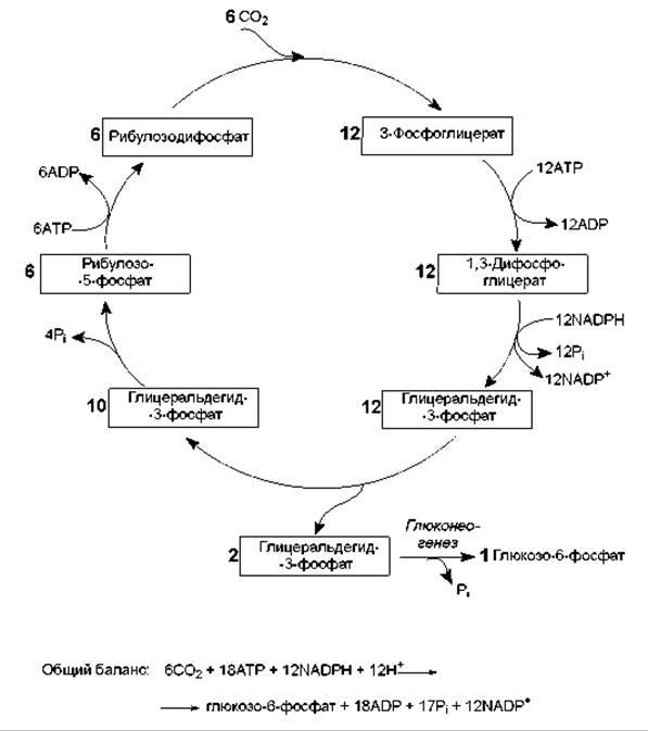
Fig. 13.5. The Calvin cycle
The rate of the Calvin cycle is strictly regulated, since excessive cellular energy should not be wasted on CO2 fixation. The core principle of this regulation is to match the rate of CO2 fixation with the rate of the light reactions of photosynthesis, which supply the ATP and NADPH required for fixation. Therefore, despite the fact that the Calvin cycle does not require direct visible light, it proceeds most intensively under illumination. The primary rate-limiting step of carbon dioxide fixation is the carboxylation of ribulose bisphosphate. Carboxylase activity increases significantly upon illumination due to the following factors:
1) carboxylase is allosterically activated by fructose-6-phosphate and inhibited by fructose-1,6-bisphosphate. In turn, the levels of these products are controlled by the enzyme fructose-1,6-bisphosphatase, which is activated by light and catalyzes The breakdown of fructose bisphosphate;
2) another allosteric activator of carboxylase is NADPH, The amount of which increases in the light under a high non-cyclic electron flow;
3) the rate of this enzymatic reaction increases as the pH rises from 7 to 9. The enzyme functions in the stroma, and its alkalinization is a consequence of lumen acidification during the ESTABLISHMENT OF THE proton gradient across the thylakoid membrane;
4) carboxylase is activated by manganese ions, which are released into the stroma when protons are transferred into the lumen during electron transport.
In addition, light activates another Calvin cycle enzyme—glyceraldehyde-3-phosphate dehydrogenase—which apparently changes its coenzyme Specificity (from NADH to NADPH) under the action of light.
The C4 pathway of CO2 fixation. The process described above—incorporation of CO2 into 3-phosphoglycerate (the result of the carboxylase reaction of ribulose bisphosphate carboxylase/oxygenase)—is called the C3 pathway, and plants that utilize it are termed C3 plants. At the same time, there is an alternative C4 pathway (the Hatch–Slack cycle) characterized by an additional initial stage: the fixation of CO2 into a four-carbon compound (oxaloacetate). This reaction is catalyzed by phosphoenolpyruvate carboxylase, an enzyme that operates much faster than ribulose bisphosphate carboxylase/oxygenase. As a result, C4 plants incorporate carbon dioxide into oxaloacetate much more efficiently than C3 plants incorporate it into 3-phosphoglycerate. However, the C4 pathway requires extra stages and energy costs: fixing one molecule of CO2 consumes 5 ATP molecules instead of 3 in the C3 pathway (Fig. 13.6). Nevertheless, C4 plants (maize, sugarcane, sorghum, many temperate weeds, etc.) grow much faster than C3 plants (wheat, rye, oats, rice, etc.). The reason for this disparity is photorespiration—a wasteful process characteristic of all C3 plants and virtually absent in C4 plants.
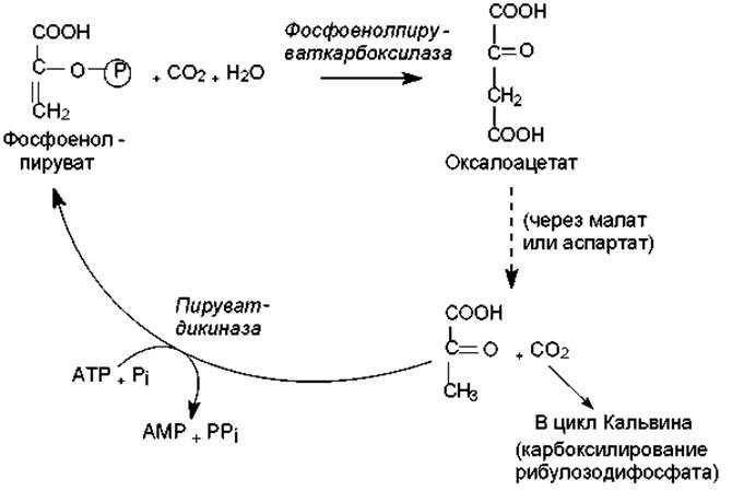
Fig. 13.6. The Hatch–Slack cycle (C4 pathway of CO2 fixation)
Photorespiration. This process stems from the oxygenase activity of ribulose bisphosphate carboxylase/oxygenase. Molecular oxygen competes with CO2 for the Active Site of this enzyme, and a portion of ribulose bisphosphate is converted into phosphoglycolate (Fig. 13.4). This does not occur in C4 plants, because most of them originate from tropical regions, where high moisture evaporation forced plants to evolve a mechanism for closing their stomata—through which gaseous tissue ventilation occurs—during the hottest hours of the day (under maximum sunlight). To prevent this mechanism from reducing the rate of CO2 fixation, plants store carbon dioxide as malate or aspartate (formed from oxaloacetate in the Hatch–Slack cycle) and subsequently consume it as needed via decarboxylation reactions. In this case, the concentration of CO2 in the green PARTS OF THE plants remains consistently high, and there is no competition from O2 for the enzyme's active site, since molecular oxygen enters the cells in limited amounts when the stomata are closed.
The substrate for photorespiration is glycolate, which is formed by the dephosphorylation of phosphoglycolate. Unlike mitochondrial respiration (which also occurs in plants in the dark), photorespiration involves O2 consumption and CO2 release that are not coupled with energy storage; instead, reducing equivalents and ATP are consumed, and the carbon fixed in the Calvin cycle is futilely lost as CO2. It has been shown that photorespiration can lead to the re-oxidation and release of up to 50% of the carbon fixed in the Calvin cycle. The biological rationale behind such a wasteful process that limits plant growth efficiency remains unsolved to this day.
The sequence of events in photorespiration is illustrated in Fig. 13.7. As can be seen, individual stages of this process take place in different Organelles. The net result of photorespiration is the formation of one molecule of phosphoglycerate (3 carbon atoms) from two molecules of glycolate (4 carbon atoms), while the involvement of oxygen leads to The oxidation of the fourth carbon atom into carbon dioxide. Apparently, the purpose of this complex sequence of transformations in photorespiration is to recover at least a fraction of the carbon from glycolate, which accumulates in excess.
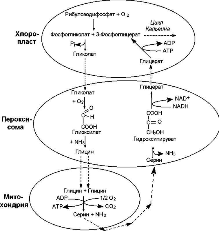
Fig. 13.7. Photorespiration
During photorespiration, one carbon atom is lost for every four, and it should be borne in mind that energy has already been expended on the fixation of this carbon atom in the Calvin cycle. In addition, the oxidation of Glycine and deamination of Serine release ammonia, the incorporation of which into amino acids will again require Energy Expenditure. As a result, photorespiration reduces the potential yield of C3 plants by 30–40%.
Last update: 06/08/2026
Editorial and Educational Adaptation: This material has been compiled based on the primary/original source text. The project team performed an editorial review, corrected technical inaccuracies, structured sections, and adapted the content for an educational format.
What was processed:
- elimination of formatting defects (OCR errors, structural breaks, corrupted characters);
- editorial organization of content;
- standardization of terminology in accordance with academic sources;
- verification of factual statements against the original source text.
All mentions of the author, publication year, and origin of the primary text have been preserved in accordance with the source.