Fundamentals of Biochemistry - A. A. Anisimov 1986
Biomembranes
Membrane Structure
Membranes are an essential component of all Cells. They enclose The Cell, separate one cellular compartment from another, and form the structures of various cell Organelles. The network of intracellular membrane systems is particularly well-developed in highly differentiated cells. Membranes are complex structures, 6–10 nm thick, composed primarily of Proteins and Lipids. In addition, they contain CARBOHYDRATES, inorganic salts, Water, and various Other Compounds; some membranes, for instance, have been found to contain traces of RNA (up to 0.1%).
Currently, the universally accepted model of Membrane Structure is the fluid-mosaic model, proposed in 1972 by S. Singer and G. Nicolson. According to this model, the membrane is a dynamic mosaic formed by a viscous lipid phase and proteins embedded within it (Fig. 9.1). Some proteins are deeply embedded in the lipid layer or even span it entirely—these are integral Membrane Proteins—while others only partially contact the lipids and are loosely associated with the membrane—peripheral proteins.
Class="center">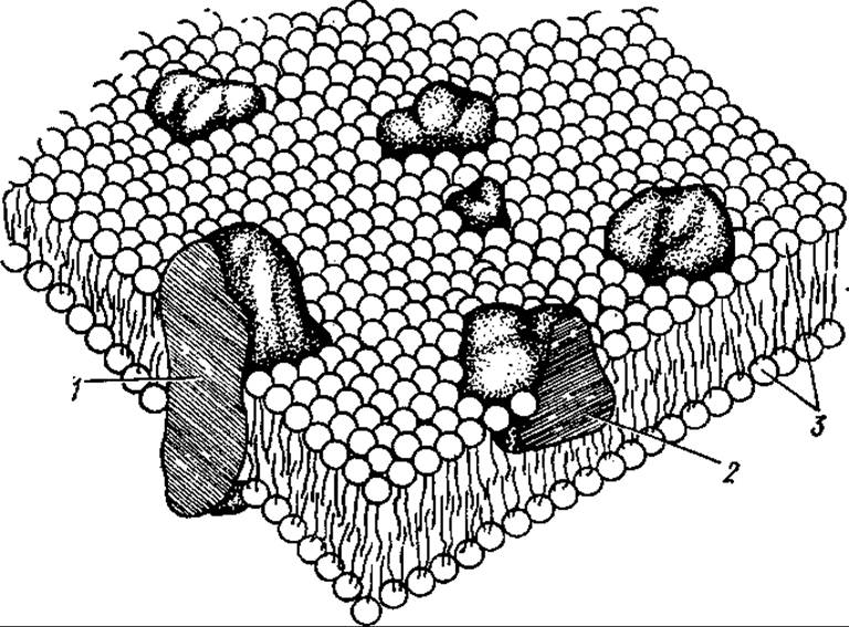
Fig. 9.1. The fluid-mosaic model of membrane structure:
1 — integral protein, 2 — peripheral protein, 3 — lipid bilayer
Among membrane proteins, only a very small fraction (mainly peripheral proteins) is water-soluble and easily extracted from membranes. Integral Proteins can be separated from lipids only by using detergents or organic Solvents. Proteins can account for 20 to 80% of the total mass of proteins and lipids. Their content depends on the origin, structure, and function of specific membranes. Phospholipids make up the major part of the membrane lipid fraction (up to 80–90% of the total lipids). In addition, neutral lipids such as Glycolipids and Cholesterol are frequently found in membranes. In some cases, mono-, di-, and triacylglycerols may constitute up to 16% of the lipids.
The Membrane Lipids of many prokaryotes contain species-specific Fatty acids, such as branched-chain and cyclopropane fatty acids. The Lipid Composition of membranes—particularly the degree of unsaturation of fatty acid residues—changes as organisms adapt to new environmental temperatures, which represents one of the core Functions of lipids. The lipid matrix of Introduction/36.html">Biological Membranes is a bilayer in which the long hydrocarbon tails of fatty acids face inward to form the Hydrophobic core of the membrane, while the polar HEAD groups (such as phosphates and alcohols like Choline or ethanolamine) are oriented outward. Integral membrane proteins likewise feature both hydrophobic and hydrophilic domains: the former interact directly with the hydrophobic regions of lipid molecules, whereas the latter reside in the surface layer of the membrane, interacting with the polar lipid head groups.
Recent X-Ray Diffraction data indicate that membrane protein chains fold in such a way that their α-helical and β-sheet regions are embedded within the hydrophobic core of the membrane, whereas the portions extending outside the membrane consist predominantly of unstructured regions.
Biological membranes exhibit distinct outer and inner surfaces that, in most cases, differ in composition, meaning Membranes are asymmetric. The lipids and proteins located on the outer leaflet of The Plasma Membrane typically feature covalently attached carbohydrates. Conversely, the inner surface of the plasma membrane and intracellular membranes generally lack carbohydrates. The carbohydrate moiety is represented by Polysaccharides comprising usually no more than 15 monosaccharide residues, which frequently form branched structures. The plasma membrane of Eukaryotic cells commonly contains galactose, mannose, fucose, N-acetylglucosamine, N-acetylgalactosamine, arabinose, xylose, and neuraminic acid. Glycolipids include glycosyldiacylglycerols (found predominantly in bacterial membranes) and glycosphingolipids such as cerebrosides and gangliosides (found mainly in eukaryotic cells).
The membrane is a dynamic structure, with lipids serving as its most mobile component. Lipids diffuse quite freely within the plane of the lipid layer (lateral diffusion), exchanging places with neighboring molecules on average 106 times per second. Protein molecules are also capable of lateral mobility within the plane of the membrane. Furthermore, protein molecules may undergo rotational motion around axes perpendicular and parallel to the plane of the bilayer, which can be crucial for the proper functioning of macromolecules and the membrane as a whole.
However, membrane proteins are not distributed randomly; rather, they form specialized domains with distinct functions. In other words, protein molecules cannot diffuse completely freely within the membrane plane, as interactions may occur between individual protein molecules or between membrane proteins and the cellular Cytoskeleton, including structural proteins, microfilaments, and microtubules underlying the inner surface of the membrane. In turn, the spatial arrangement of protein molecules within the membrane influences the distribution and orientation of lipid molecules, depending on the specific affinity between particular proteins and lipids.
The mobility of membrane molecules depends heavily on fatty acid composition. Membrane structures enriched in saturated fatty acids within their phospholipids are more ordered and stable, whereas those containing substantial amounts of Unsaturated fatty acids are less ordered. At optimal physiological temperatures for living organisms, the membrane typically exists in a liquid-crystalline state (intermediate between liquid and solid). This state is primarily maintained by the lipid-protein-water system within the membrane, which forms various types of ordered structures that simultaneously retain considerable mobility. Such a membrane state profoundly influences overall function and accounts for the high sensitivity of membranes to various external factors.
Adjacent Cells of the same tissue must communicate with one another to coordinate their vital activity and function as a unified whole in accordance with tissue Specificity. Such communication is achieved through specialized short tubes assembled into disc-shaped structures at sites known as Gap Junctions. Each tubule consists of two cylindrical protein molecules called connexons. A connexon molecule is partially embedded in The cell membrane, and its protruding part is capable of binding in the intercellular space with a connexon from the adjacent cell, thus forming a continuous channel connecting the internal spaces of the two cells.
The membranes of various cells and intracellular organelles exhibit a certain specificity determined by their structure, chemical composition, and functions. The following main groups of membranes are distinguished in eukaryotic organisms: the plasma membrane (outer cell membrane, Plasmalemma), nuclear membrane, Endoplasmic reticulum, membranes of the Golgi apparatus, Mitochondria, METABOLISM/14.html">Chloroplasts, myelin sheaths, and excitable membranes. In prokaryotic organisms, besides the plasma membrane, there are intracytoplasmic membrane formations; in heterotrophic prokaryotes, these are called mesosomes. The latter are formed by the invagination of the outer cell membrane inward and in some cases retain their connection with it.
A good object for studying the Structure and properties of the plasmalemma is The erythrocyte membrane; it is relatively easy to obtain in pure form because erythrocytes lack intracellular membranes. The erythrocyte membrane consists of proteins (50%), lipids (40%), and carbohydrates (10%). The bulk of the carbohydrates (93%) is bound to proteins, and the rest to lipids. The membrane is asymmetrical, as the carbohydrate components are located exclusively on the outer surface. The membrane is also asymmetrical with respect to enzymatic activity and lipid composition. The inner layer of the erythrocyte membrane contains mainly sphingomyelin, phosphatidylethanolamine, and phosphatidylserine, while the outer layer contains phosphatidylcholine.
A well-studied integral protein of Erythrocyte membranes is Glycophorin, an amphipathic protein. Its N- and C-termini are hydrophilic and located, respectively, on the outer and inner sides of the membrane. The middle region is hydrophobic and spans the membrane (Fig. 9.2). Attached to the N-terminal part are 20–30 polysaccharide molecules consisting predominantly of N-acetylgalactosamine, galactose, and sialic acid located at the polysaccharide terminus. The carbohydrate components of glycophorin perform a receptor function for Influenza Viruses, phytohemagglutinins, and A number of Hormones.
Another integral protein containing few carbohydrates and spanning the membrane has been discovered in the erythrocyte membrane. It is referred to as the tunnel protein (component a) because it is hypothesized to form an anion channel. A peripheral protein associated with the inner side of the erythrocyte membrane is spectrin, a protein that performs a structural role. It is tightly associated with Actin-like Proteins of the erythrocyte membrane, forming an ATP-dependent system similar to Actomyosin.

Fig. 9.2. Arrangement of the glycophorin molecule in the erythrocyte membrane
Myelin membranes surrounding neuron axons are multi-layered and contain a high proportion of lipids (about 80%, half of which are phospholipids). The proteins of these membranes are essential for anchoring the superimposed membrane layers together.
Chloroplasts are enclosed by a double-layer membrane. The outer membrane bears some similarity to that of mitochondria (see Section 7.2). In addition to this surface membrane, chloroplasts contain an internal membrane system—the lamellae. The lamellae form either flattened vesicles, known as thylakoids, which stack up on top of one another to form grana or construct the stroma membrane system (stroma lamellae). The grana and stroma lamellae are formed by globular lipoprotein subunits. Hydrophilic groups of proteins, galactolipids, and sulfolipids are concentrated on the outer side of the thylakoid membrane. The phytol moiety of the chlorophyll molecule is embedded in the globule and contacts the hydrophobic groups of proteins and lipids. The porphyrin rings of chlorophyll are predominantly localized between the touching membranes of grana thylakoids.
The inner (cytoplasmic) membrane of Bacteria is structurally similar to the inner membranes of chloroplasts and mitochondria. It localizes the Enzymes of the Respiratory Chain, Oxidative Phosphorylation, and Active Transport, as well as enzymes involved in The formation of membrane components, such as Glycosyltransferases, which catalyze the synthesis of outer membrane lipopolysaccharides. The predominant component of bacterial membranes is protein: the protein-to-lipid ratio (by weight) is 3:1. The composition of membrane proteins is regulated According to the needs of the cell and the external environment. Compared to the cytoplasmic membrane, the outer membrane of Gram-negative bacteria contains fewer distinct phospholipids and proteins. Both membranes differ in lipid composition. The outer membrane contains proteins that form pores for the penetration of many low-molecular-weight substances. A characteristic component of the outer membrane is also a specific lipopolysaccharide. A number of outer membrane proteins serve as phage receptors.
Among viruses, membrane structures are characteristic of those containing a nucleocapsid composed of PROTEIN AND NUCLEIC acid. This viral "core" is surrounded by a membrane (envelope). It also consists of a lipid bilayer with embedded Glycoproteins located mainly on the membrane surface. In a number of viruses (toga- and microviruses), 70–80% of all proteins are integrated into the membranes, while the remaining proteins are contained within the nucleocapsid.
Viral membrane proteins are specific proteins, such as hemagglutinin and neuraminidase. Carbohydrates account for a significant portion of the mass of the viral membrane. The specificity of Oligosaccharides on the viral surface is determined to some extent by the host cell, as host cell sugar transferases participate in attaching carbohydrates to envelope proteins. The lipids of viral membranes are likewise derived from the membranes of infected cells.
Mitochondrial membranes are described in Section 7.2.
Last update: 06/08/2026
Editorial and Educational Adaptation: This material has been compiled based on the primary/original source text. The project team performed an editorial review, corrected technical inaccuracies, structured sections, and adapted the content for an educational format.
What was processed:
- elimination of formatting defects (OCR errors, structural breaks, corrupted characters);
- editorial organization of content;
- standardization of terminology in accordance with academic sources;
- verification of factual statements against the original source text.
All mentions of the author, publication year, and origin of the primary text have been preserved in accordance with the source.