Protein Chemistry - Part 1 - General Protein Chemistry - Ashmarin I. P. 1968
Spatial Organization of the Protein Molecule
Main Types of Bonds in a Protein Molecule and Levels of Its Organization
As noted above, Proteins are linear copolymers built from Amino Acids connected by peptide bonds. Constructing the peptide backbone from repeating structural units gives protein molecules a certain resemblance to synthetic linear polymers. However, comparing various PHYSICOCHEMICAL PROPERTIES OF Proteins and synthetic polymers shows that this similarity is purely superficial.
First and foremost, the repeating structural element in proteins is a single peptide group with one carbon atom (the alpha-carbon atom) between adjacent bonds. The amino acid side chains do not participate in forming the polypeptide chain, acting merely as its radicals. Recall that in nylon, there are six carbon atoms between adjacent peptide bonds. At the same time, protein molecules feature a compact Structure and contain roughly 3 to 4 times less bound Water (20–30%) than the loose coils of linear polymers. As a result, protein macromolecules are smaller in linear dimensions, and proteins do not form the viscous solutions characteristic of conventional polymers. This indicates that proteins possess an incomparably higher degree of Organization than linear polymers. Currently, four levels of Cell/13.html">Protein Structure, or organization, are known; all of them are distinct and defined primarily by Different types of molecular interactions, or types of bonds, within the molecule.
The first type includes covalent bonds formed through the interaction of alpha-carboxyl and alpha-amino groups of amino acids, known as peptide bonds, which are located along the polypeptide chain. Thus, the primary level of organization is the polypeptide chain itself. This level of structure is referred to as the Primary Structure.
In their studies of the crystal structures of various Amino Acids and Peptides, Pauling and Corey demonstrated that the dimensions of peptide groups are roughly identical and do not depend on which specific amino acids form a given group. The distance between the Carbon and Oxygen atoms was found to be 1.24 Å, whereas the sum of covalent double Bond Lengths should equal only 1.21 Å. Similarly, the carbon-nitrogen bond length in the amide group is 1.32 Å, which is also less than the sum of single bond lengths (1.47 Å). This proves that the C—N bond has 40% double-bond character, and the carbonyl group bond has 60% double-bond character, resulting from Resonance between the bonds (electron migration from nitrogen to oxygen via the carbon atom).
Class="center">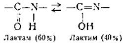
Since the C—N bond partially possesses double-bond character, one can assume that the amide group adopts a planar configuration. This assumption has been confirmed by a range of experimental data. In all peptide structures studied, the peptide group proved to be planar (within a few degrees), and upon deviation from coplanarity by 10–20°, the displacement energy equaled 1–3.5 kcal/mol, respectively. These deviations can affect the energy of the peptide bond, which is typically 2.5–3.5 kcal/mol, but may range from 1.1 to 4.1 kcal/mol.
A third feature of the amide group is that it adopts a trans configuration. This means that the alpha-carbon atoms are attached to the planar amide group on opposite sides of the C—N bond.
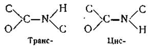
This crystal structure analysis is supported by dipole moment measurements of simple peptides. It turned out that the trans configuration of the amide group is more stable than its cis configuration. Infrared absorption spectra of amides and N-substituted amides have similarly demonstrated the greater Stability of the peptide trans configuration (the energy difference between these configurations exceeds 2 kcal/mol). All these Structural Features of the amide group are illustrated in Fig. 16.
Finally, the last characteristic feature of the amide group stems from the presence of a hydrogen atom bonded to a nitrogen atom, which enables The formation of a Hydrogen bond. Each of these peptide groups can form two Hydrogen Bonds with other groups. The formation of this bond type can be explained by electrostatic and covalent interactions.
It is known that a hydrogen atom attached to an electronegative atom in a molecule can drive an interaction with an electronegative atom in either another or the same molecule. This interaction is called a hydrogen bond. Such electronegative atoms include F, O, and N. The hydrogen attached to them is stripped of its electron shell, acting essentially as a proton. Thus, if a second molecule containing an electronegative atom is nearby, the proton will be attracted to it. Consequently, these two groups will bind, with attractive forces balanced by repulsive forces arising from the interpenetration of their electron clouds. Obviously, the bond strength will be greater the higher the positive charge of the hydrogen atom and the negative charge of the acceptor. That is precisely why the strongest Hydrogen bonds are formed by the most electronegative atom—fluorine (F>O>N).
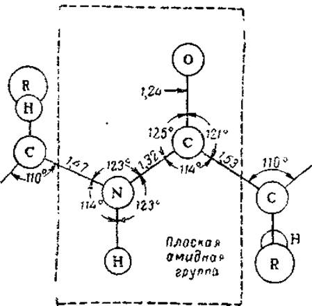
Fig. 16. Section of a polypeptide chain and its key parameters calculated from X-Ray Diffraction data of amino acids and simple peptides (after Pauling, 1955).
The covalent factor also plays a significant role in hydrogen bond formation. In covalent compounds, F, O, and N atoms have 3, 2, and 1 lone electron pairs, respectively. Therefore, when a proton is shared between adjacent electronegative atoms, its electronic configuration is supplemented by one of these pairs.
Consequently, a hydrogen bond arises because The Nucleus of a hydrogen atom located within a polar group is temporarily shared between two electronegative atoms, while its electronic configuration is completed by a lone electron pair from one of them. Calculations of hydrogen bond formation energy have shown that in proteins, H-bonds are driven primarily by electrostatic forces, whereas the covalent factor accounts for no more than 10%.
Hydrogen bonds in proteins possess several distinct features. First of all, the hydrogen atom lies closer to nitrogen than to oxygen. The bond energy is relatively low at 1.4 kcal/mol, and its length is on the order of 2.8 Å. Due to its polarity, all atoms involved in this bond must lie on a straight line (i.e., be collinear), although some deviation is permissible. Evaluating the loss in hydrogen bond energy when the CO and NH groups are at an angle to each other, Pauling concluded that the deviation from collinearity cannot exceed 20°.
Hydrogen bonds can form both between separate polypeptide chains and between units of a single chain. Since the H-bond energy is 1.4 kcal/mol, the more such bonds form within a molecule, the lower its energy and the higher its stability. As a result, polypeptide chains tend to form ordered, rigid helices with the maximum possible number of hydrogen bonds. This ordered helical STRUCTURE OF THE polypeptide chain, stabilized by intramolecular hydrogen bonds, is called the Introduction/11.html">Secondary structure of proteins. It is also frequently referred to as intramolecular crystallization, as such a molecule truly resembles a crystal (exhibiting a melting point for the secondary structure, high rigidity, and high order). However, the ordered helix with intramolecular hydrogen bonds is not the sole configuration of protein polypeptide chains. Alongside it, a structure is known in which extended polypeptide chains are linked to one another by intermolecular (interchain) hydrogen bonds.*
Evidence for the existence of hydrogen bonds in Proteins can be obtained by studying The rate of isotopic exchange of imide hydrogen with deuterium- or tritium-labeled water. This technique was employed in the laboratories of Linderstrøm-Lang and Bresler. It is known that in low-molecular-weight peptides, this hydrogen atom exchanges with water extremely rapidly. In high-molecular-weight Polypeptides with A large number of hydrogen bonds, imide hydrogen exchange is drastically slowed down. For example, in polyalanine with 28 peptide bonds, only 5 to 6 imide hydrogens exchange rapidly. This indicates that nearly all peptide groups are connected by hydrogen bonds, and the chain itself is folded into an ordered helix. In Insulin, out of 49 imide hydrogens, 30 exchange slowly, meaning the degree of helicity is roughly 60%. Another confirmation of hydrogen bonds in proteins is their breakdown and Denaturation under METABOLISM/18.html">The Influence of agents with a high capacity to form hydrogen bonds with the amide group (concentrated solutions of urea, guanidine, trifluoroacetic and formic acids, etc.).
* See details in § 2
Alongside hydrogen bonds, other bonds play a crucial role in the ORGANIZATION OF THE protein molecule. Chief among these are Disulfide Bonds, which are formed via The oxidation of two Cysteine residues:
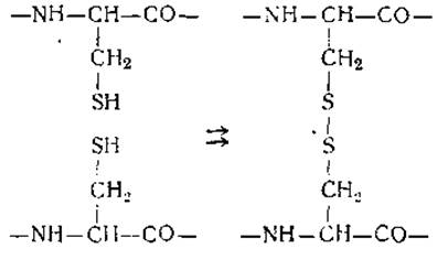
Disulfide —S—S— bonds can link multiple separate polypeptide chains together as well as anchor individual points of a single chain, leading to the formation of loops within it. Both types of bonds are present in the insulin molecule, where two disulfide bridges connect chains A and B, while a third causes the formation of a loop containing 6 amino acid residues in chain A (Fig. 17).
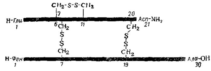
Fig. 17. Schematic diagram of the insulin structure featuring interpeptide and intrapeptide disulfide bonds (after Oncley, 1961).
The numbers indicate the positions of the amino acid residues.
Stresses arise at the localization sites of disulfide bridges, weakening Hydrogen bonds and disrupting the helical structure. This is supported by experiments on protein deuteration rates. For instance, in insulin itself, out of 30 slowly exchanging hydrogen atoms at 0°, 7 begin to exchange rapidly at 20°. Obviously, these include the hydrogen atoms of those peptide groups that are located close to disulfide bridges and are therefore weakly bound by hydrogen bonds. Thus, the presence of disulfide bonds results in The polypeptide chains containing amorphous regions alongside helical segments, imparting a certain degree of flexibility. At the same time, these very bridges link individual polypeptide chains into a single protein macromolecule.
Another type of secondary bonds—those forming loops inside peptide chains or connecting separate chains—involves interactions mediated by large nonpolar groups. The latter are represented by hydrocarbon radicals of amino acids such as leucine, isoleucine, phenylalanine, and Tryptophan, whose maximum length and volume can reach 8.1 Å and 175.5 Å3, respectively. Just as hydrocarbon molecules in water tend to coalesce into spheres to minimize surface energy, these nonpolar protein radicals tend to merge into a symmetrical droplet.
The reason for this behavior is easy to understand. Water possesses a high degree of internal hydrogen bonding, which leads to the formation of a partially ordered lattice. Since Hydrocarbons are not effective hydrogen bond Donors or acceptors, the existence of their chains in direct contact with water would reduce the total number of hydrogen bonds, an energetically unfavorable outcome. Conversely, ionic groups can readily interact with water, and their interaction energy with polar H2O molecules is quite sufficient to compensate for any suppression of H-bond formation. Droplet formation leads to the partial shielding of hydrocarbon radicals from contact with the solvent, replacing it with hydrocarbon-hydrocarbon contacts.
The interaction of several nonpolar groups, accompanied by the release of water and the formation of new hydrogen bridges within it, is sufficient to create a stable cross-link. This is due to the fact that the association of hydrocarbon chains in an aqueous medium releases an energy of approximately 1.2 kcal per 1 mole of CH2 groups. Consequently, under certain conditions, the interaction of nonpolar radicals can be equivalent to the formation of a hydrogen bond both within a protein molecule and between specific molecules. In this context, specific spatial arrangements of large nonpolar residues in peptide chains can ensure the stability of structures incompatible with a helical configuration. Admittedly, the attraction of such residues is hindered by A number of factors: the close approach of groups bearing like charges, the detachment of water dipoles from charged groups, the rupture of hydrogen bonds, and the disruption of the helical structure—in other words, all processes that require an energy input.
Thus, the interaction of hydrocarbon radicals, much like the formation of disulfide bridges, counteracts the tendency of the polypeptide chain to form rigid helices with the maximum possible number of hydrogen bonds, resulting in the presence of amorphous regions within the macromolecule. Within these regions, polypeptide chains exhibit considerable flexibility and are capable of bending or kinking. Consequently, side-chain interactions and the formation of S—S bonds cause the chains to fold, packing their helical and amorphous segments into a compact body—a globule. It is precisely this spatial packaging of alternating helical and amorphous sections of the primary chain into a symmetrical globule that constitutes the Tertiary Structure of a protein molecule. Therefore, secondary bonds take a direct part in the formation and stabilization of the protein's tertiary structure.
In addition to the association of nonpolar side radicals and disulfide bridges, other covalent bonds—such as phosphoester bonds—can also participate in forming the tertiary structure. Orthophosphate bonds, presumably linking Serine and Threonine residues, have been shown to occur in Pepsin and α-casein; the presence of a phosphoamide cross-link (O—PO2—NH—) in α-casein has also been demonstrated, which appears to be attached to Lysine and Arginine residues. It is possible that similar bonds also exist in Phosphoproteins. However, they are of no significant importance for other proteins.
The attraction or repulsion of polypeptide chains can also be driven by electrostatic forces acting between positively and negatively charged groups of the protein molecule (Coulombic interactions). The magnitude of such forces depends on the Ionic strength of the solution. However, solutions of Globular proteins do not exhibit a significant dependence of electrostatic forces on ionic strength, provided the effective charge of the protein is not excessively large. This suggests that electrostatic forces probably do not play a major role in forming the globular structure. For certain elongated protein molecules, these forces may indeed influence molecular shape, as they are capable of mediating interactions over relatively long distances.
Finally, dipole-dipole interactions and so-called dispersion forces may play a specific role in the spatial configuration of the macromolecule. However, their significance remains poorly understood and, therefore, they will not be discussed in detail here.
A comparison of the secondary bonds involved in the formation and Maintenance of the tertiary structure of a protein molecule shows that the primary role is played not by the peptide backbone, but by the amino acid side chains. Since different proteins contain different side chains in various regions of their polypeptide chains, their interaction results in a unique spatial packing of the chains themselves, strictly specific to each protein. Any uniformity here is impossible, which is precisely why proteins differ so drastically from one another In their tertiary structure.
At present, proteins are conventionally classified into fibrous and globular based on the features of their tertiary structure.
The former comprise insoluble proteins that perform protective Functions and are characterized by a fibrous, oriented structure. The molecules of these Proteins are built from long polypeptide chains and possess an elongated, rod-like tertiary structure (Silk Fibroin, Hair, nail, and feather Keratins). The second group includes soluble proteins of animal Cells, Blood, Lymph, and CEREBROSPINAL FLUID (albumins and globulins). The macromolecules of these proteins feature a compact tertiary structure approaching a sphere or an ellipsoid of revolution. However, this distinction is rather relative, and intermediate between these two classes exist many soluble proteins whose tertiary structure is of a mixed nature. These include, for example, procollagen, fibrinogen, and Myosin. Although the molecules of these proteins feature an elongated, rod-like shape, their polypeptide chains are not stretched out, but instead folded into a topologically complex tertiary structure.
Thus, the organization of a protein molecule arises from various molecular interactions that determine its structural levels. The Primary structure of a protein macromolecule is entirely determined by covalent bonds along the polypeptide chain; the secondary structure, by hydrogen bonds between peptide groups located in adjacent turns of the helix or neighboring regions of different chains; and, finally, the tertiary structure, by chemical "cross-links" between individual segments of a single chain or multiple chains (disulfide bridges, phosphoester bonds) and interaction forces among nonpolar side radicals of Certain amino acids.
In Conclusion, it is necessary to dwell upon The quaternary structure of proteins. This level of organization arises through the association of several (two or more) macromolecules (so-called subunits) into a single complex globule. An example of such an association is the Hemoglobin molecule, which consists of four peptide chains and readily dissociates into two subunits, each containing two polypeptide chains.
The Formation of the quaternary structure involves interaction forces between individual groups located On the surface of protein globules. Such forces may include hydrogen bonds, Electrostatic Interactions between oppositely charged groups, and Van der Waals interactions of amino acid side chains. Often, the association of subunits into complex complexes serves as the basis for their biological activity. For instance, the manifestation of alkaline phosphatase activity requires the prior assembly of its two subunits. Sometimes we observe the reverse picture, where enzymatic activity appears only after the dissociation of the protein molecule's components (phosphoglyceraldehyde dehydrogenase). Nevertheless, it must be emphasized immediately that the functional activity of a given protein is determined not by the quaternary structure alone, but by all four levels of its organization. All these structural levels mutually influence one another, with lower orders of organization dictating higher ones. Thus, the order of succession of individual amino acids in a peptide chain determines its secondary structure and, consequently, the tertiary structure of the protein molecule. Investigating these interactions between higher and lower orders of organization is more conveniently conducted not using natural polypeptides composed of diverse amino acid residues, but rather on their synthetic models. It was precisely through The Study of polypeptides synthesized from a single defined amino acid that the secondary structure of proteins was elucidated.
Last update: 06/08/2026
Editorial and Educational Adaptation: This material has been compiled based on the primary/original source text. The project team performed an editorial review, corrected technical inaccuracies, structured sections, and adapted the content for an educational format.
What was processed:
- elimination of formatting defects (OCR errors, structural breaks, corrupted characters);
- editorial organization of content;
- standardization of terminology in accordance with academic sources;
- verification of factual statements against the original source text.
All mentions of the author, publication year, and origin of the primary text have been preserved in accordance with the source.