Protein Chemistry - Part 1 - General Protein Chemistry - Ashmarin I. P. 1968
Primary Structure of the Protein Molecule
Determination of the Number and Location of Disulfide Bonds
Data on the Amino Acid Sequence of each polypeptide chain of a protein are not yet sufficient to establish its Primary Structure. It is necessary to determine the number and Location of disulfide bridges that link these chains into a single entity. Solving this problem requires very mild Hydrolysis conditions, since Treatment with Reagents such as concentrated Hydrochloric acid leads to The oxidation of cystine to cysteic acid along with several other products. Therefore, the protein is subjected to Enzymatic hydrolysis under the mildest possible conditions and in the presence of thiol inhibitors (e.g., N-ethylmaleimide). Pepsin and Chymotrypsin are frequently used for this purpose, with Digestion carried out at pH 1.9 and 8.0, respectively. The resulting peptide mixture is separated using one or more of the aforementioned techniques, and the fragments containing the disulfide bond are isolated in pure form and oxidized with performic acid. Subsequently, high-voltage paper Electrophoresis is used to obtain Peptides containing cysteic acid. These peptides are subjected to complete acid hydrolysis and identified by their Amino Acid Composition.
In this manner, the following Disulfide Bonds and the peptides linked by them were identified in the Insulin molecule:
Class="center">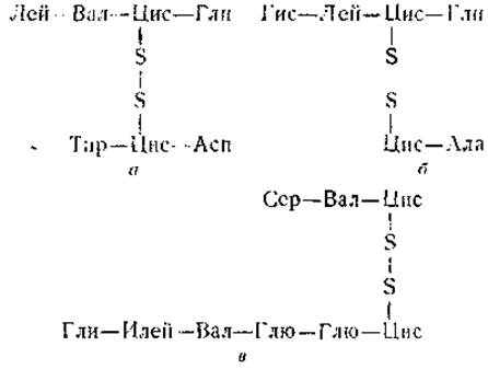
Comparing the structures of these peptides with the known amino acid sequence of the A and B chains of insulin (see Figs. 13 and 14) reveals that bridge a connects amino acid residue No. 19 of the B chain with amino acid residue No. 20 of the A chain. Cystine bridge b links amino acid residue No. 7 in both chains, whereas bridge c forms a disulfide loop between the 6th and 11th Amino Acids of the A chain.
Knowledge of The amino acid residue sequence in the peptide chains, as well as the number and location of disulfide bridges, makes it possible to reconstruct the Introduction/19.html">Primary structure of the protein molecule as a whole. This type of elucidation was first accomplished for the insulin molecule by Sanger around 1955 (Fig. 15).
The research workflow outlined above is not mandatory; various modifications exist depending on The Nature of the protein under investigation. It is evident from the foregoing that despite the application of modern Analytical Methods in all their complexity and diversity, studying the Chemical Structure of Proteins remains a highly challenging and labor-intensive task that demands years of intense effort. For instance, determining The structure of chymotrypsin, which consists of 226 amino acids arranged in a single chain, required approximately 10 years of work by several major laboratories.
Currently, the primary structure is known for only a limited number of proteins, specifically insulin, adrenocorticotropin, Ribonuclease, TMV, chymotrypsin, Trypsin, cytochrome c, Hemoglobin, Myoglobin, and Lysozyme.
Last update: 06/08/2026
Editorial and Educational Adaptation: This material has been compiled based on the primary/original source text. The project team performed an editorial review, corrected technical inaccuracies, structured sections, and adapted the content for an educational format.
What was processed:
- elimination of formatting defects (OCR errors, structural breaks, corrupted characters);
- editorial organization of content;
- standardization of terminology in accordance with academic sources;
- verification of factual statements against the original source text.
All mentions of the author, publication year, and origin of the primary text have been preserved in accordance with the source.