Molecular Biology of the Cell - Volume 3 - Alberts B., Bray D., Lewis J., Raff M., Roberts K., Watson J. 1994
From Cells to Multicellular Organisms
Maintenance of Normal Tissue Organization
Renewal by Simple Duplication
In vertebrates, differentiated Cells are for the most part not permanent—they are constantly dying and being replaced by new ones. Throughout an adult's life, new differentiated cells are generated in one of two ways: (1) by the simple duplication of existing differentiated cells, yielding two daughter Cells of the same type; or (2) from undifferentiated stem cells, a process that, as we will see later, involves A change in cellular phenotype.
The rate of Cell turnover varies greatly from tissue to tissue. Cell Cycle times can range from just a few days, as in the epithelial lining of the Small Intestine (where renewal is driven by stem Cell Division), to a year or more, as in the Pancreas (where cells multiply simply by dividing). Many Tissues that normally renew very slowly can be stimulated to produce new cells more rapidly when the need arises. In this section, we will examine tissue renewal via the simple duplication of differentiated cells, focusing on The Liver and endothelial cells.
17.3.1. The Liver: A Critical Link Between the Digestive Tract and the Bloodstream [9]
Digestion is a complex process. Specialized cells lining the digestive tract secrete various substances, such as Hydrochloric acid and Enzymes, which break down food components into simpler molecules. Other cells absorb these digestion products from the intestinal lumen and transfer them into the bloodstream for use by the rest of the body. All these processes are finely tuned to the composition of ingested food and the concentration of metabolites in circulating Blood. This intricate division of labor is carried out by distinct cell groups: some secrete HCl or enzymes, others absorb nutrients, and still others produce Peptide Hormones (such as gastrin) that regulate digestive and metabolic activity, and so on (Fig. 17-9). Some of these cells are interspersed along the intestinal wall, whereas others are grouped into large glands connected to the intestine, developing embryonically as outgrowths of the intestinal epithelium.
Class="center">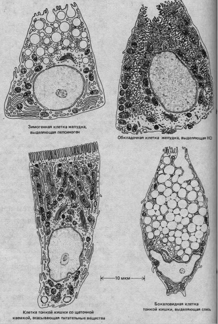
Fig. 17-9. Various types of specialized cells found in the epithelial lining of the gastrointestinal tract. Sections of the epithelium frequently reveal different cell types lying adjacent to one another (see Fig. 17-17B). (From T. L. Lentz, Cell Fine Structure. Philadelphia: Saunders, 1971.)
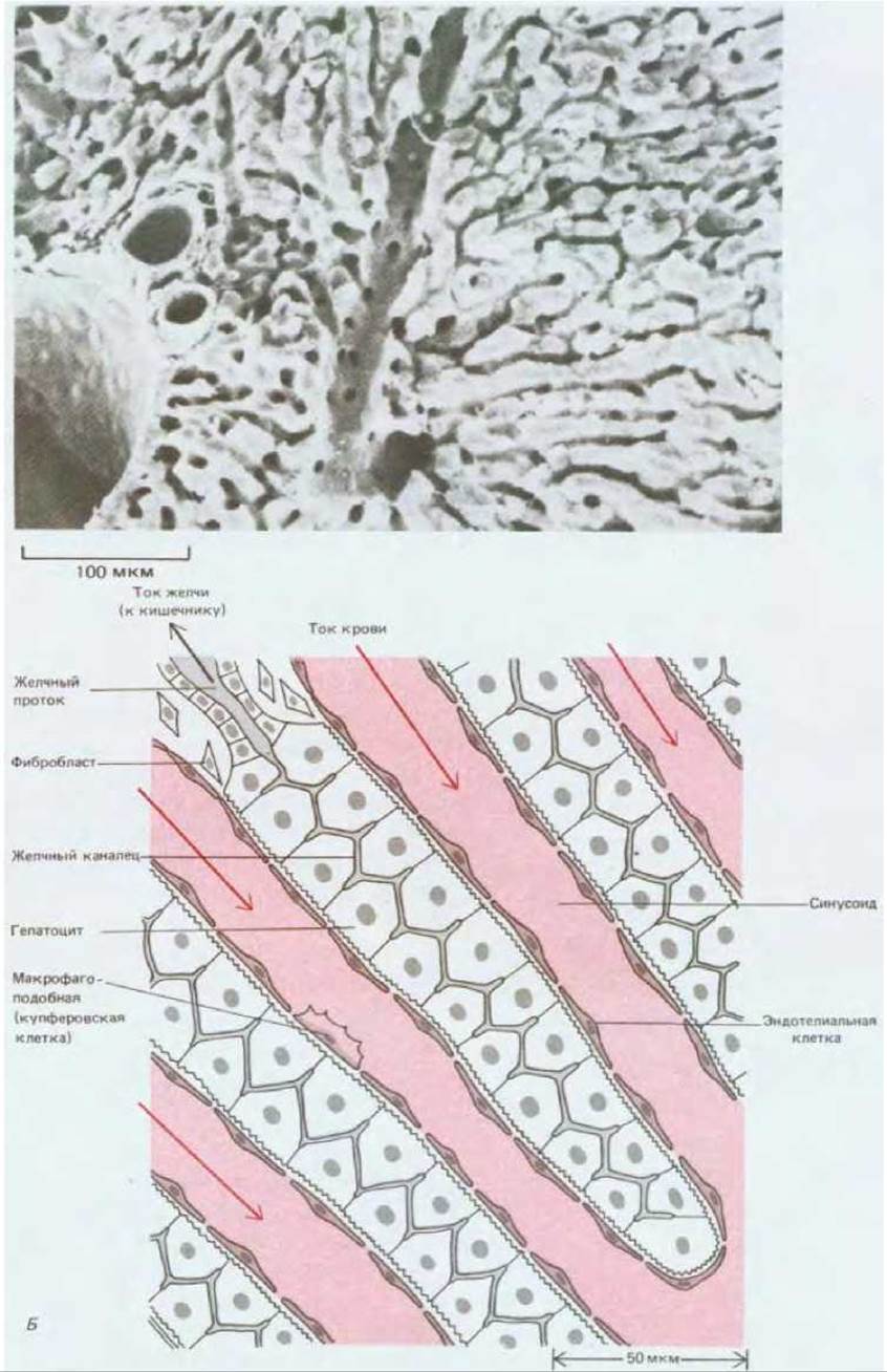
Fig. 17-10. STRUCTURE OF THE liver. A. Scanning electron micrograph of a liver section, showing folded layers of hepatocytes (hepatic cords) and numerous narrow channels (sinusoids) through which blood flows. The wider channels are Blood Vessels that distribute and collect blood flowing through the sinusoids. B. Schematic diagram of the Fine Structure of the liver. Hepatocytes are separated from the bloodstream by only a thin layer of endothelial cells, interspersed with macrophage-like Kupffer cells. Small fenestrations in this layer allow molecules and small particles to pass between hepatocytes and the blood, while shielding the hepatocytes from direct contact with circulating Blood Cells. In addition to exchanging substances with the blood, hepatocytes form a network of extremely narrow Bile canaliculi, into which they secrete bile that flows via bile ducts into the intestine. In reality, the architecture of the liver is less regular than this diagram suggests. (From R. G. Kessel, R. H. Kardon, Tissues and Organs: A Text-Atlas of Scanning Electron Microscopy. San Francisco: Freeman, 1979.)
The largest of these glands is the liver. During embryonic development, it originates at the site where one of the main Veins runs alongside the wall of the primitive gut. Even in the adult Organism, this organ retains an unusually close functional relationship with the blood. Hepatic cells (hepatocytes), derived from the primitive gut epithelium, form folded layers (hepatic cords or trabeculae) adjacent to blood-filled spaces called sinusoids (Fig. 17-10A). The blood is separated from The surface of the hepatocytes by a layer of flattened endothelial cells covering each trabecula (Fig. 17-10B). This architecture facilitates the liver's primary Functions, which rely on the exchange of metabolites between liver cells and the bloodstream.
The liver is a vital organ where nutrients absorbed from the intestine are processed and transformed for use by other body tissues. It receives the majority of its blood supply directly from the digestive tract via the portal vein. Hepatocytes are responsible for the synthesis, breakdown, and storage of a vast array of substances. They play a central role in whole-body carbohydrate and Lipid METABOLISM and synthesize most of the Proteins found in Blood Plasma. At the same time, hepatocytes maintain a connection with the intestinal lumen through a network of microscopic canaliculi and larger ducts (Fig. 17-10B). Through these ducts, hepatocytes excrete Metabolic waste products into the gut, along with bile—an emulsifying agent that facilitates fat DIGESTION AND ABSORPTION. Unlike other Regions of the digestive tract, the hepatocyte population shows no distinct "division of labor": virtually all hepatocytes are capable of performing the same broad spectrum of metabolic and secretory functions.
In terms of their "lifestyle," hepatocytes also differ markedly from the cells lining the intestinal lumen proper. The latter endure a harsh environment; constantly exposed to mechanically and chemically aggressive gut contents, they are short-lived and must be rapidly and continuously replaced by new cells (see Fig. 17-17). Hepatocytes, by contrast, are spared direct contact with the intestinal contents, live considerably longer, and turn over at a slow but tightly regulated rate.
17.3.2. Loss of Liver Cells Stimulates Their Proliferation [10]
Even in a slowly renewing tissue, a small yet persistent imbalance between cell production and cell loss would lead to catastrophic consequences. If 2% of a person's liver cells divided every week while only 1% were lost, the liver would continuously expand, eventually exceeding the weight of the entire body within 8 years. Therefore, a homeostatic mechanism must exist to match the rate of cell division to overall tissue mass. The necessity for such precise control is especially acute in an organ like the liver, whose cells are periodically destroyed by toxins such as alcohol.
The existence of homeostatic control over cell proliferation in the liver has been clearly demonstrated in experiments where a large fraction of hepatocytes is surgically removed or destroyed by carbon tetrachloride administration. Approximately 24 hours after such injury, a wave of cell division sweeps through the remaining hepatocyte population, rapidly replacing the lost tissue. For instance, if two-thirds of a rat's liver is removed, the remaining portion regenerates to its original size in about two weeks. In such cases, a regeneration-stimulating signal can be detected in the blood: if the circulations of two rats are surgically joined (cross-Circulation) and two-thirds of the liver is removed from one of them, mitotic activity is also triggered in the undamaged liver of the second rat. Despite extensive research, the exact circulating factor and its MECHANISM OF ACTION remain elusive. It is possible that the signal is a complex combination of chemical cues rather than a single growth factor. Similar regenerative phenomena are observed in the Kidneys, which appear to possess an analogous growth-control system.
17.3.3. Regeneration Requires Coordinated Growth of Tissue Components [11]
Like most organs, the liver is a composite of several cell types. In addition to hepatocytes and the endothelial cells lining the sinusoids, it contains specialized macrophages (Kupffer cells) that engulf particulate matter from the bloodstream and clear out worn-out red blood cells, as well as a small population of fibroblasts that form a loose Connective Tissue framework (see Fig. 17-10B). All these cell types are capable of division, and for complete and functional regeneration to occur, their proliferation must be properly coordinated.
The Importance of balanced multi-cellular regeneration can be illustrated by the consequences of its failure. For example, if the liver is repeatedly injured by carbon tetrachloride or alcohol at intervals so short that hepatocytes cannot fully recover, fibroblasts may gain a growth advantage; the liver then becomes progressively and irreversibly choked with excess connective tissue, leaving very little room for hepatocytes to expand even after the toxic agent is removed. This condition, known as cirrhosis, is frequently seen in chronic alcoholics. Similarly, the regeneration of severely damaged Skeletal Muscle is often seriously impaired by the overgrowth of connective tissue, resulting in scar tissue replacing muscle fibers. Such imbalances, however, typically arise only from major tissue trauma; under normal conditions of tissue maintenance, as-yet-poorly understood regulatory mechanisms ensure that the appropriate mix of different cell types is preserved.
17.3.4. All Blood Vessels Are Lined with Endothelial Cells [12]
Unlike the aforementioned Examples of uncoordinated fibroblast growth, the endothelial cells that line blood vessels exhibit an extraordinary capacity to alter their number and arrangement in response to local physiological demands. Nearly all tissues require a blood supply, which in turn depends entirely on endothelial cells. These cells construct a highly adaptable life-support network branching into every corner of the body. Without this remarkable ability of endothelial cells to expand and repair the vascular network, tissue growth and wound healing would be impossible.
The largest blood vessels are Arteries and veins, which feature thick, sturdy walls composed of connective tissue and smooth muscle (Fig. 17-11A). The inner surface of this wall is lined by an extremely thin, single layer of endothelial cells resting on a basal lamina. While the thickness of the connective tissue and muscle layers varies depending on the vessel's diameter and function, the endothelial lining is invariably present (Fig. 17-11B). The walls of the tiniest Branches of the vascular tree—capillaries and sinusoids—consist solely of endothelial cells and a basal lamina (Fig. 17-12). Thus, endothelial cells line the entire Cardiovascular system, from The Heart down to the smallest capillaries, regulating the exchange of substances (and white blood cells) between the tissues and the blood. Furthermore, embryological studies have demonstrated that arteries and veins themselves develop from primitive microvessels built exclusively of endothelial cells and a basal lamina; connective tissue and smooth muscle are subsequently added, where appropriate, in response to signals from the endothelium.
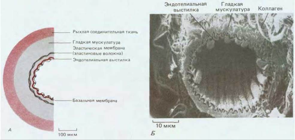
Fig. 17-11. A. Wall of a small artery (schematic cross section). Despite their inconspicuous appearance, endothelial cells are a vital structural component of the vessel wall. Compare with The structure of a capillary in Fig. 17-12. B. Transversely cut arteriole (scanning electron micrograph). The inner endothelial lining is clearly visible, surrounded by layers of smooth muscle and Fibrous connective tissue. The vascular endothelium is folded due to mild contraction of the underlying muscle layer. During fixation, the endothelium shrank and slightly pulled away from the other layers, creating a narrow artifactual gap. (From R. G. Kessel, R. H. Kardon, Tissues and Organs: A Text-Atlas of Scanning Electron Microscopy. San Francisco: Freeman, 1979.)
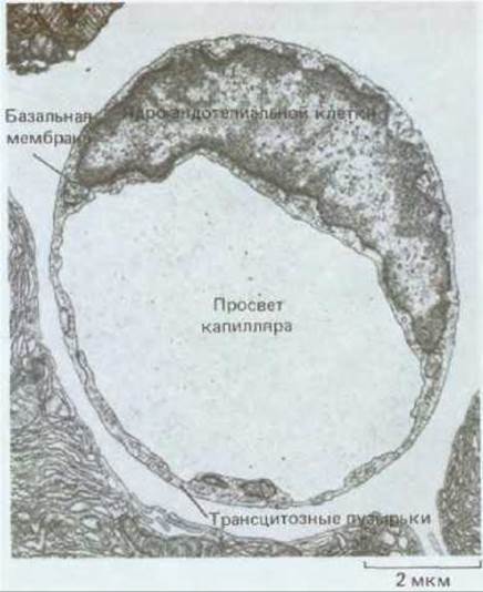
Fig. 17-12. Cytology/practical/72.html">Cross section of a narrow pancreatic capillary (transmission electron micrograph). The wall is formed by a single endothelial cell surrounded by a basal lamina. Note the small (80 nm) "transcytotic" vesicles. These are thought to mediate the Transport of Macromolecules across the Capillary Wall by taking up molecules at the luminal surface and discharging them via exocytosis at the abluminal surface (or vice versa). (From R. P. Bolender, J. Cell Biol., 61, 269–287, 1974. Reprinted by permission of Rockefeller Univ. Press.)
17.3.5. New Endothelial Cells Are Formed by the Simple Division of Existing Endothelial Cells [13]
Throughout the adult Vascular System, endothelial cells retain the capacity for division and migration. If, for instance, a section of the aortic wall is damaged and loses its endothelial lining, new cells are generated in the surrounding endothelium and migrate to cover the affected area. These new cells are even capable of lining the inner surface of plastic tubing used by surgeons to replace damaged segments of blood vessels.
Endothelial cell proliferation can be demonstrated by labeling the cells in S phase with 3H-thymidine. In normal blood vessels, the proportion of labeled endothelial cells is particularly high at arterial branch points, where blood flow turbulence accelerates endothelial wear and tear, thereby apparently stimulating their renewal. Overall, however, these cells renew very slowly: a cell's lifespan is measured in months or even years.
Endothelial cells not only repair the lining of existing blood vessels but also form new ones. This process is essential in the embryo to ensure that the vascular network keeps pace with body growth, as well as in adult tissues undergoing cyclic remodeling and during wound healing.
17.3.6. New capillaries form as sprouts from existing vessels [14, 15]
New vessels initially arise as capillaries that sprout from pre-existing small vessels. This process of angiogenesis represents a response to specific signals and can be readily demonstrated in rabbits. A small hole is pierced in a rabbit's ear, and cover slips are secured on both sides to form a narrow, transparent-walled chamber in which the cells surrounding the wound can grow. Angiogenesis is also conveniently observed in transparent structures such as the cornea. Irritation of the cornea induces the growth of new blood vessels from its vascularized rim toward the center, which is normally virtually devoid of vessels. Thus, corneal vascularization occurs through the ingrowth of endothelial cells into its dense, Collagen-rich tissue.
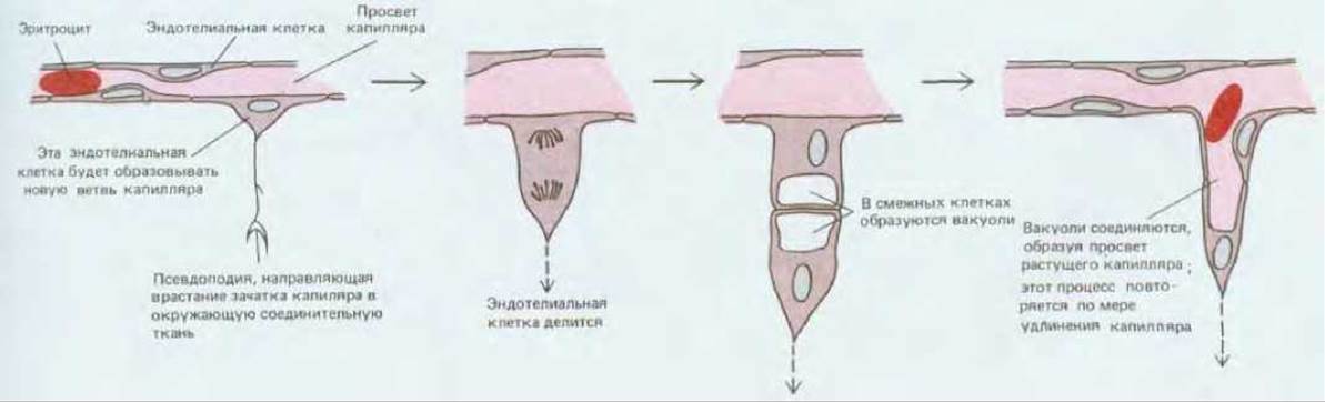
Fig. 17-13. A new blood capillary forms by the "sprouting" of an endothelial cell from the wall of an existing small vessel. This diagram is based on observations of cells in the transparent tail of a living tadpole. (After S. S. Speidel, Am. J. Anat., 52, 1-79, 1933.)

Fig. 17-14. Vacuoles arise spontaneously in cultured endothelial cells and fuse to form a network of capillary-like tubes. Photographs A and B show successive stages of this process. The arrow in (A) indicates a vacuole that has initially appeared within a single endothelial cell. The cultures originated from groups of two to four endothelial cells taken from short capillary segments. These cells attach to a collagen-coated culture dish surface and form a small, flattened colony that gradually enlarges as the cells proliferate. The colony spreads across the dish, and eventually (after approximately 20 days) capillary tubes begin to form in the central regions; branches soon appear, and 5-10 days later an extensive network of tubes is visible (B). (From J. Folkman, C. Haudenschild, Nature, 288, 551-556, 1980. Copyright Macmillan Journals Ltd.)
Such observations indicate that endothelial cells destined to form a new capillary sprout from the wall of an existing capillary or small venule by first extending thin, long pseudopodia (Fig. 17-13); a bulky cellular process then forms, which subsequently becomes hollow and turns into a tube. This process continues to elongate until it encounters another capillary, with which it connects, thereby establishing a pathway for Blood Circulation. As tissue culture experiments have demonstrated, in a medium containing appropriate growth factors, endothelial cells spontaneously form capillary tubes even when isolated from any other cell types. The first sign of such tube formation in culture is the appearance of an elongated vacuole within The Cell, which is initially entirely surrounded by Cytoplasm (Fig. 17-14, A). Similar vacuoles arise in neighboring cells and eventually align end-to-end so that they fuse into a single capillary channel (Fig. 17-14, B). Capillaries formed in pure endothelial cell cultures contain no blood, and no fluid flows through them. Evidently, blood flow and pressure are not required for The formation of the capillary network.
17.3.7. Capillary network growth is regulated by factors secreted by surrounding tissues [15]
In the living organism, endothelial cells form new capillaries wherever they are needed. Apparently, when cells in tissues experience oxygen deprivation, they secrete angiogenic factors that induce new capillary growth. This is likely the reason why almost all vertebrate cells lie no further than 50 µm from a capillary. Similarly, during wound healing, a transient "burst" of capillary growth occurs in the area adjacent to the damaged tissue (Fig. 17-15). Local irritation and local infection also provoke the proliferation of new capillaries, and once the inflammation subsides, many of the newly formed capillaries undergo regression and gradually disappear.
Angiogenesis is also crucial for tumor growth. A tumor growing as a solid mass remains very small until it is supplied by capillaries. Without an internal blood supply, it would rely solely on the diffusion of nutrients from the periphery and thus could not grow larger than a few millimeters in diameter. For further growth, the tumor must induce the formation of a capillary network that sprouts into the tumor mass. A small piece of such a tumor transplanted into the cornea causes rapid capillary growth directed from the vascular margin toward the implant (Fig. 17-16), and the tumor growth rate increases sharply as soon as the vessels reach it.
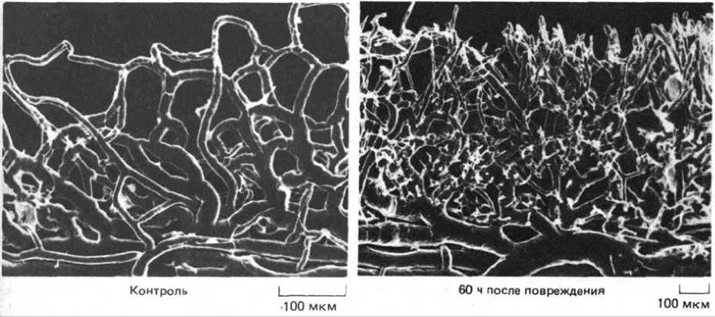
Fig. 17-15. Blood vessel casts of the corneal margin, showing the response to injury (scanning electron micrographs). The casts were prepared by injecting a special resin into the vessels, which subsequently hardens, revealing the shape of the lumen rather than the external cellular contours. Sixty hours after wounding, numerous new capillaries begin to sprout toward the damaged area (just above the upper edge of the photograph). Their directed growth reflects the chemotactic response of endothelial cells to the angiogenic factor released in the wound region. (Courtesy of Peter C. Burger.)
In all these cases, invading endothelial cells must respond to a signal emitted by tissue requiring a blood supply. The endothelial cell response involves at least three components. First, the cells must breach the basement membrane surrounding the existing blood vessel. It has been shown that during angiogenesis, endothelial cells secrete proteases, such as plasminogen activator, which allow them to "eat" their way through the basement membrane of the parent capillary or venule. Second, endothelial cells must migrate toward the signal source. Third, they must divide. Under certain circumstances, one or two of these three response components may be triggered in the absence of the others. For example, new capillaries sometimes form even when endothelial cell proliferation is blocked by irradiation. It has also been shown that a factor present in wound fluid attracts endothelial cells and prompts them to secrete proteases without, however, stimulating proliferation.
Other factors are capable of eliciting all three Components of the endothelial cell response. Examples include acidic fibroblast growth factor (aFGF) and basic fibroblast growth factor (bFGF). These two proteins, which were independently isolated and purified from several different sources and are therefore known by various other names, share similar Amino acid sequences (55% Homology). In addition to their pronounced effects on endothelial cells, they stimulate the proliferation of fibroblasts and several other cell types, and also serve as important regulators of early embryonic development (Section 16.2.3). Which cells secrete them is not entirely clear. Many cell types, including macrophages, mast cells, and adipocytes, can release other substances that act as angiogenic factors during tissue repair, growth, or inflammation. Angiogenesis, much like other cellular proliferation processes, is regulated not by a single signal, but by a complex (and likely redundant) network of signals.
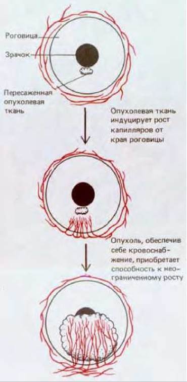
Fig. 17-16. Tumor tissue transplanted into the cornea secretes a factor that induces capillary growth. The capillaries supply the tumor with nutrients from the general circulation, thereby enabling it to grow.
Summary
In vertebrates, most differentiated cell populations undergo renewal. In some cases, fully differentiated cells simply divide to produce daughter cells of the same type. Hepatocytes (liver cells) serve as an example, their division rate being regulated to maintain the required total number. If a significant portion of the liver is destroyed, the division rate of the remaining hepatocytes increases to make up for the loss. However, repair is sometimes unbalanced; for instance, when fibroblasts begin to proliferate too rapidly relative to hepatocytes in a repeatedly injured liver, the hepatic tissue is replaced by fibrous tissue.
Endothelial cells form a single layer lining all blood vessels and regulating the exchange of substances between the blood and surrounding tissues. New blood vessels develop from existing small vessels as endothelial cell outgrowths; these cells are capable of forming hollow capillary tubes even when grown in culture. In the living organism, oxygen-deprived and damaged tissues stimulate angiogenesis by releasing angiogenic factors that attract nearby endothelial cells and stimulate them to proliferate and secrete proteases.
Last update: 12/08/2026
Editorial and Educational Adaptation: This material has been compiled based on the primary/original source text. The project team performed an editorial review, corrected technical inaccuracies, structured sections, and adapted the content for an educational format.
What was processed:
- elimination of formatting defects (OCR errors, structural breaks, corrupted characters);
- editorial organization of content;
- standardization of terminology in accordance with academic sources;
- verification of factual statements against the original source text.
All mentions of the author, publication year, and origin of the primary text have been preserved in accordance with the source.