Molecular Biology of the Cell - Volume 3 - Alberts B., Bray D., Lewis J., Raff M., Roberts K., Watson J. 1994
From Cells to Multicellular Organisms
Maintenance of Normal Tissue Organization
Tissues with Permanent Cells
Not all populations of differentiated Cells in an Organism are subject to renewal. Cells of certain types, formed in the required numbers in the embryo, persist throughout adult life; they never divide and, if lost, cannot be replaced. In this sense, almost all varieties of Nerve Cells are permanent. This category also includes certain other cells, such as mammalian Heart Muscle cells and lens cells.
All of these cells have an extremely long lifespan and are naturally located in sites where they are normally protected from damaging influences, yet in other respects they vary greatly from one another. It is difficult to pinpoint a single reason why these Cells must be permanent while numerous other Cell populations undergo renewal. In the case of cardiac muscle, it is generally hard to grasp the Functional Significance of cellular permanence. As for Neurons (which will be discussed in detail in Chapter 19), it seems understandable why intensive renewal of these cells in the adult organism is impractical: it would be extremely difficult to precisely restore the complex network of neural connections established during development under entirely different conditions. Furthermore, memory traces encoded as subtle alterations in the Structure or connectivity of specific neurons would likely be erased if older cells were replaced by new ones. On the other hand, the permanence of lens cells is apparently a simple and inevitable consequence of the growth pattern of its tissue.
17.2.1. Cells located in the center of the adult lens were formed during the Embryonic period [6]
Very little in the adult organism consists of the exact same molecules that were synthesized during the Embryonic Stage. Rare structures in which neither cellular renewal nor even internal molecular turnover takes place include the crystalline lens of the eye.
The lens develops from the ectoderm at the site of its contact with the developing optic vesicle. Here, the ectoderm thickens and forms an invagination that eventually pinches off, becoming the lens primordium (see Fig. 17-2). Thus, the lens originates as a spherical vesicle composed of a single layer of epithelial cells enclosing a central cavity. Soon, the portion of this epithelium located posteriorly—that is, facing the retina—undergoes a drastic transformation. Its cells begin to synthesize specific lens Proteins, known as crystallins, and become packed with them. Concurrently, the cells elongate remarkably, differentiating into fibers (see Fig. 17-4). Eventually, their nuclei degenerate and Protein Synthesis ceases. In this manner, the portion of the lens vesicle epithelium facing the retina develops into a dense refractive body composed of numerous high prismatic cells devoid of life signs and stacked in an orderly fashion (Fig. 17-5). The central cavity of the vesicle disappears, while the anterior region of the epithelium, facing the external environment, persists as a thin layer of low cuboidal cells. Lens growth depends on the proliferation of these cells in the anterior region, from where they are partially pushed toward the margins of the lens and its posterior surface (see Figs. 17-4 and 17-5, A). During this migration, they cease division, begin synthesizing crystallins, and differentiate into lens fibers. Through this mechanism, additional fibers are added to the lens throughout life, although their rate of formation gradually declines.
Class="center">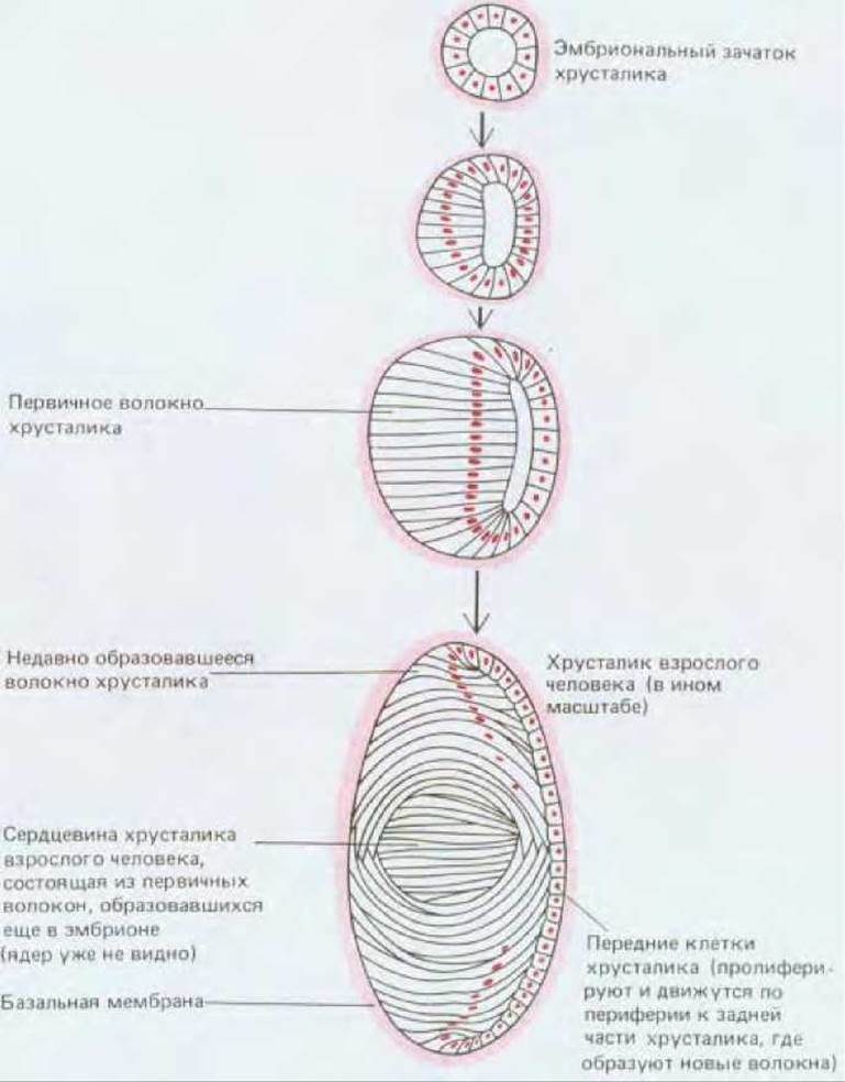
Fig. 17-4. Human lens development (schematic).
Crystallins in lens fibers formed during the early stages differ from those in later fibers, much as fetal Hemoglobins in red Blood Cells differ from adult hemoglobins. However, erythrocytes are continuously replaced by new ones, whereas lens fibers are not. Consequently, the core of the adult lens contains fibers laid down as early as the embryonic stage, which contain specific types of crystallins synthesized during that early period. Differences in refractive index between early embryonic crystallin types and later variants help rid the lens of optical aberrations inherent in simple lenses made of a homogeneous material, such as Glass.

Fig. 17-5. STRUCTURE OF THE adult human lens. A. Light micrograph of a section through a mature lens, showing the junction between the thin epithelium covering the anterior surface of the lens and the differentiated fibers. B. Scanning electron micrograph showing densely packed fibers resembling stacked lumber in a timber yard. Each fiber is a single dead, elongated cell. Individual fibers can reach up to 12 mm in length. (A, courtesy of Peter Gould; B, from R.G. Kessel, R.H. Kardon, Tissues and Organs: A Text-Atlas of Scanning Electron Microscopy, San Francisco: Freeman, 1979.)
17.2.2. The majority of permanent cells renew their constituent parts. Example: retinal photoreceptor cells [7]
Unchanging cells like lens fibers are rare. As a rule, even cells that do not divide throughout the organism's life continuously renew their components. Although cardiac muscle cells and neurons do not divide, they are metabolically active and possess the capacity not only to synthesize new RNAs and proteins but also to alter their Size and Structure over time. For instance, heart muscle cells turn over the bulk of their protein molecules within 1 to 2 weeks, and they can establish a balance between protein Synthesis and degradation such that the cells themselves enlarge if the workload on The Heart increases—for example, during sustained high blood pressure. Nerve cells also continuously replace their protein molecules; moreover, many neurons are capable of regenerating severed axons and dendrites (see Chapter 19).
The process of cellular component renewal is particularly striking in the highly specialized nerve cells that form the photoreceptors of the retina. The neural retina (see Fig. 17-2) consists of several cell layers arranged in what might seem a counterintuitive manner: the neurons that transmit visual signals to the Brain (retinal ganglion cells) lie closest to the external world, meaning that light focused by the lens must pass through them on its way to the photoreceptor cells. The latter are oriented such that their light-receiving tips—the outer segments—are partially embedded in the retinal pigment epithelium (Fig. 17-6). Based on their Morphology, photoreceptors are subdivided into rods and cones. They contain different light-sensitive protein-visual pigment complexes. Rods are exceptionally sensitive under low-light conditions, whereas cones, represented by three distinct types (each tuned to a specific spectral region), serve for Color Vision and perception of fine detail. The outer segment of each photoreceptor type appears to be a modified cilium: it exhibits the characteristic ciliary arrangement of microtubules in the region connecting the outer segment to the rest of The Cell (Fig. 17-7). The main body of the outer segment, however, is almost entirely filled with tightly packed membranes in which the photosensitive visual pigment proteins are embedded. The opposite ends of the photoreceptor cells form synaptic contacts with interneurons of the retina.

Fig. 17-6. Schematic diagram of retinal structure. Upon photoreceptor stimulation, neural signals are transmitted via intermediate neurons to ganglion cells, which in turn Relay them to the brain. The space between neurons and photoreceptors within the neural layer of the retina (light gray in the diagram) is filled with specialized supporting cells, which are omitted from this schematic. (After J.E. Dowling, B.B. Boycott, Proc. R. Soc. Lond. (Biol.), 166, 80-111, 1966.)

Fig. 17-7. Structure of a rod cell. A. In reality, the number of photoreceptor discs in the outer segment reaches approximately one thousand. B. Electron micrograph of a rod region, showing the Base of the outer segment and the modified cilium connecting the outer segment to the inner segment. (A, T.L. Lentz, Cell Fine Structure. Philadelphia: Saunders, 1971; B, M.J. Hogan, J.A. Alvarado, J.E. Weddell, Histology of the Human Eye: An Atlas and Textbook. Philadelphia: Saunders, 1971.)
Photoreceptors are permanent cells incapable of division. Yet the molecules of the photosensitive protein are not permanent. They are constantly being renewed, a process detectable by the continuous incorporation of radioactive Amino Acids. In rods (intriguingly, this does not occur in cones), such renewal proceeds like an assembly line. Pulse-labeling experiments with amino acids allow researchers to track an echelon of labeled protein molecules as they migrate across the entire cell (Fig. 17-8). Following the standard stages of amino acid incorporation into proteins and packaging within the Golgi apparatus in the cell's inner segment, the radioactive material first appears at the base of the membrane stack in the outer segment. From there, it gradually shifts toward the tip of the segment while new material enters the base of the stack. Finally, once the labeled proteins, along with the membrane layers in which they are embedded, reach the apex of the stack (in rats, taking roughly 10 days), they are phagocytosed and digested by the Cells of the pigment epithelium.
Further details regarding photoreceptors and their Functions within The Nervous system can be found in Chapter 19.

Fig. 17-8. Renewal of membrane protein in a retinal rod. Following brief administration of 3H-leucine, autoradiography can be used to monitor its intracellular movement. Red dots indicate sites of radioactivity. The method detects only leucine incorporated into proteins; unincorporated label is washed out during specimen preparation. The incorporated leucine first concentrates near the Golgi apparatus (1); from there, it transitions to the base of the outer segment and enters the newly synthesized photoreceptor membrane disc (2). Here, roughly 3 to 4 new discs are formed per hour (in mammals), pushing older discs toward the pigment epithelium (3-5).
Neurons, cardiac muscle cells, and lens fibers do not divide or undergo replacement throughout the organism's lifespan. In mature lens fibers, cell nuclei have already degenerated and protein synthesis has ceased, meaning that the inner central region of the lens contains proteins synthesized as early as early Embryogenesis. However, in most other permanent cells, metabolic activity persists and cellular components undergo continuous renewal. This is clearly demonstrated in retinal rods, where new layers of photosensitive membrane are synthesized near The Nucleus, steadily migrate toward the cell apex, and are subsequently engulfed and digested by the pigment epithelial cells.
Last update: 12/08/2026
Editorial and Educational Adaptation: This material has been compiled based on the primary/original source text. The project team performed an editorial review, corrected technical inaccuracies, structured sections, and adapted the content for an educational format.
What was processed:
- elimination of formatting defects (OCR errors, structural breaks, corrupted characters);
- editorial organization of content;
- standardization of terminology in accordance with academic sources;
- verification of factual statements against the original source text.
All mentions of the author, publication year, and origin of the primary text have been preserved in accordance with the source.