Molecular Biology of the Cell - Volume 3 - Alberts B., Bray D., Lewis J., Raff M., Roberts K., Watson J. 1994
From Cells to Multicellular Organisms
Maintenance of Normal Tissue Organization
Renewal by Stem Cells. Example: Epidermis
Let us now turn from Cell populations that renew themselves simply by duplicating their Cells to those that are replenished by stem cells. These populations vary widely not only in The properties of their constituent cells and their rates of turnover, but also in the Spatial Organization of this process. For example, in the lining of the Small Intestine, cells form a single-layered epithelium. This epithelium covers The surface of the villi protruding into the intestinal lumen, and it also lines the deep crypts extending into the underlying Connective Tissue (Fig. 17-17). Stem cells reside in a protected niche at the bottom of the crypts. Differentiated cells originating from the stem cells are displaced upward by sliding within the plane of the epithelial layer until they reach the exposed surface of the villi, from the tips of which they are eventually shed. A completely different process can be seen in the Skin: the epidermis is a Stratified Epithelium, and differentiating cells move from their site of origin in a direction perpendicular to the plane of The Cell layers. In hematopoietic Tissues, the spatial pattern of cell production is complex and appears chaotic. But before delving into further details, let us examine what a stem cell actually is.
17.4.1. Stem cells have the capacity for unlimited division and give rise to differentiated progeny [17]
The defining characteristics of a stem cell are as follows:
1) it is not itself terminally differentiated (i.e., it has not completed The pathway of differentiation);
2) it is capable of unlimited division (at least for the lifetime of the Organism);
3) upon division, each daughter cell faces a choice — either to remain a stem cell like the parent cell, or to embark on a pathway that irreversibly leads to terminal differentiation (Fig. 17-18).
Class="center">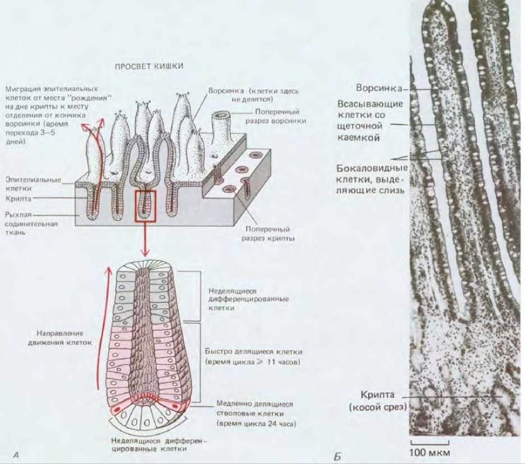
Fig. 17-17. A. Diagram of cell population renewal in the small intestinal lining through stem cell proliferation. B. A section showing villi and crypts. Note the pale goblet cells (which secrete mucus) scattered among the brush-border absorptive cells in the villus epithelium. See also Fig. 17-9, which shows The Structure of these cells. (Courtesy of Peter Gould.)
Stem cells are required wherever There is a continuous demand for new differentiated cells that are themselves incapable of division. In A number of tissues, the state of terminal differentiation is clearly incompatible with Cell Division. For example, cell nuclei may degenerate, as occurs in the outer layers of the epidermis, or be expelled from the cells, as during mammalian erythrocyte maturation. Sometimes, mitosis and cytokinesis are prevented because the Cytoplasm is densely packed with Materials such as the myofibrils of Muscle cells. In other terminally differentiated cells, the inability to divide may be due to more subtle biochemical reasons. In any such case, tissue renewal relies entirely on stem cells.
Stem cells are not meant to perform a specific specialized function themselves, but rather to produce cells that do. Consequently, stem cells often lack any characteristic appearance, making them difficult to identify. However, this does not mean they are all identical. Although not morphologically differentiated, they are nonetheless committed (Section 16.2.8): a satellite cell in Skeletal Muscle serves as a source of muscle fibers, a basal cell of the epidermis as a source of keratinizing epithelial cells, a spermatogonium as a source of sperm, and a basal cell of the olfactory epithelium as a source of olfactory Neurons (Fig. 17-19), and so on. Stem cells that give rise to only a single type of differentiated cell are called unipotent, whereas those that generate multiple types are termed pluripotent.
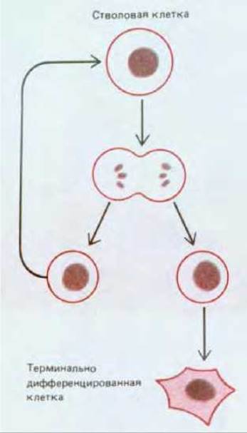
Fig. 17-18. The Fate of stem cell progeny. Each daughter cell produced by stem cell division may either remain a stem cell or embark on a pathway leading to terminal differentiation.
The Study of tissues derived from stem cells raises many profound questions. What determines whether a given stem cell will divide or remain quiescent? What dictates whether a daughter cell will also become a stem cell or begin to differentiate? And once it enters a pathway of differentiation, what regulates this process? We will begin our Discussion with the epidermis, as its simple spatial organization facilitates the study of its stem cell biology and the fate of their progeny.
17.4.2. The epidermis is divided into proliferative units [18, 19]
The epidermal layer of the skin and the epithelial lining of the digestive tract are the two tissues most exposed to direct damaging influences from the external environment. In both tissues, mature differentiated cells wear out rapidly in the most vulnerable areas and are replenished just as quickly through the proliferation of less differentiated cells located in more protected sites.
The epidermis is a stratified epithelium composed primarily of keratinocytes (so named because the synthesis of keratin is the hallmark of their differentiated state) (Fig. 17-20). These cells change their appearance progressively from layer to layer. The deepest of the internal layers is formed by basal cells. It is primarily these cells that divide by mitosis. Above the basal cells lie several layers of larger prickle cells (spinous cells) (Fig. 17-21). They owe this name to their appearance in light Microscopy preparations: their numerous desmosomes, with thick bundles of keratin filaments radiating from them, are barely distinguishable and appear as tiny spines on the cell surface. Above the spinous cells lies a thin layer of granular cells (Fig. 17-20); it forms the boundary between the inner metabolically active zone and the outermost layer composed of dead cells, in which all intracellular Organelles have disappeared. These outer cells are reduced to flat squames filled with densely packed keratin. On the cytoplasmic face, the Plasma Membranes of the squames and outer granular cells are reinforced by a thin (12 nm), rigid, cross-linked layer containing the intracellular protein involucrin. The squames themselves are usually so flattened that their boundaries are barely discernible under a Light Microscope; however, if the preparation is treated with an NaOH solution, these cells swell somewhat, revealing—after proper staining (provided the epidermis is thin in that region)—a remarkably regular geometric cellular pattern: the squames are stacked in hexagonal columns tightly interlocked at their cell edges (Fig. 17-22). The width of these columns is such that about a dozen basal cells lie at the base of each Column. These cells can be subdivided into central and peripheral according to their position at the column's base. Peripheral (but not central!) cells can occasionally be observed transitioning upward from the basal layer into the spinous cell layer. Each such column is termed an epidermal proliferative unit. Although this orderly columnar organization is revealed only in certain Regions of the skin, it serves as an excellent illustration of the General Principles of epidermal cell renewal.
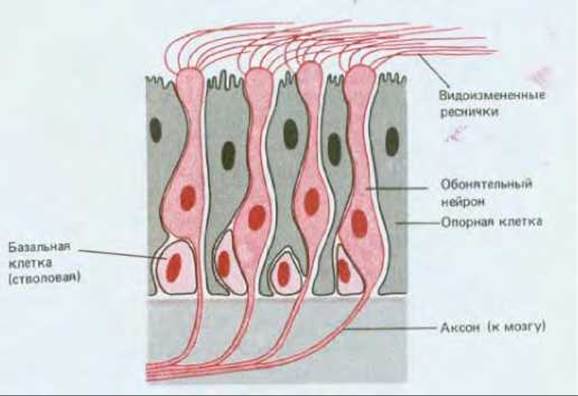
Fig. 17-19. Diagram of the STRUCTURE OF THE olfactory epithelium, specialized for odor reception. Three cell types can be distinguished here: supporting cells, basal cells, and olfactory neurons. Autoradiography experiments demonstrate that basal cells act as stem cells, generating olfactory neurons. This is a rare exception to the rule that neurons are permanent cells. Each olfactory neuron Functions for about a month (in mammals) before being replaced by a new one. From the rounded "HEAD" of the olfactory neuron extend 6–8 modified cilia, which are believed to house the odorant receptors. The axon extending from the other end of the neuron transmits information to the Brain. Every time a basal cell differentiates into an olfactory neuron, a new axon grows out from it to establish the appropriate connections in the brain.
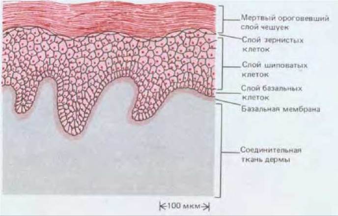
Fig. 17-20. Structure of medium-thickness mammalian epidermis (diagram) (see also Fig. 17-1). Granular cells are located between the spinous cells and the flattened squames. They undergo the penultimate stage of keratinization and contain heavily staining granules of a poorly understood material, keratohyalin, which is involved in the compaction and cross-linking of intracellular keratin. Keratohyalin consists largely of the protein filaggrin. In addition to cells destined for keratinization, the deep layers of the epidermis harbor a small number of entirely different cells (not shown in the diagram): Bone Marrow-derived macrophage-like Langerhans cells; melanocytes derived from the neural crest; and Merkel cells associated with nerve endings in the epidermis.
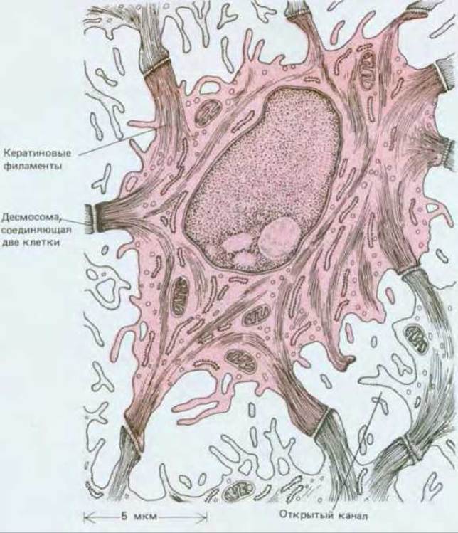
Fig. 17-21. Drawing based on an electron micrograph of a section through an epidermal spinous cell (highlighted). Bundles of keratin filaments are visible, traversing the cytoplasm and running toward the desmosomes that connect the cell to its neighbors. Note the open channels between adjacent cells, allowing nutrients to diffuse freely through the metabolically active layers of the epidermis. Closer to the surface, at the level of the granular cells, there is a waterproof barrier presumably formed by an insulating substance secreted by these cells from specialized vesicles. (R. V. Krstic, ULTRASTRUCTURE OF THE Mammalian Cell: An Atlas. Berlin: Springer, 1979.)
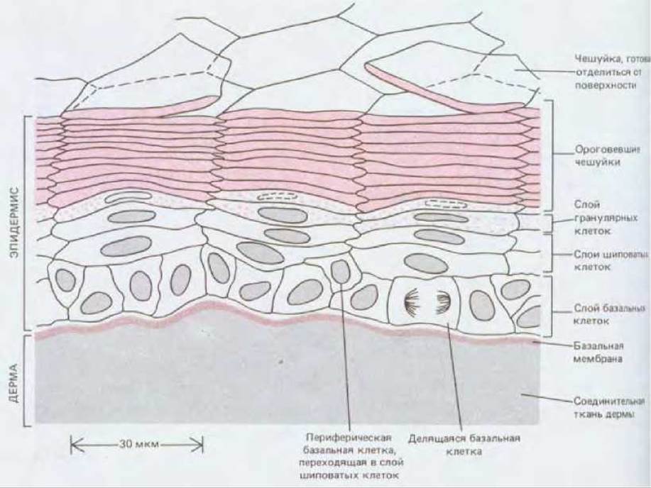
Fig. 17-22. Proliferative units, or columns, in thin-skin epidermis. This structure is revealed upon Swelling the cornified squames in a solution containing NaOH. Such columnar organization is characteristic only of thin regions of the epidermis.
17.4.3. Differentiating epidermal cells sequentially synthesize different Keratins as they mature [19]
Let us move from the static picture described above to dynamics. The central basal cell of a column divides, and some of the daughter cells, having divided in turn, shift toward the periphery of the base. Peripheral basal cells transition from the basal layer into the spinous cell layer—taking the first step on the upward-moving "escalator." Upon reaching the granular layer, spinous cells begin to lose their nuclei and cytoplasmic organelles, gradually transforming into the keratinized squames of the outer layer. Eventually, these scales desquamate and are dispersed by air currents, forming a major component of house dust. In humans, the time span from a cell's birth in the basal layer of the epidermis to its shedding from the skin surface ranges from two to four weeks, depending on the body region.
Concomitant chemical changes can be studied by analyzing thin layers of the epidermis shaved parallel to the surface, or successive layers of cells stripped off by repeatedly applying and removing pieces of adhesive tape. This approach allows the extraction and characterization of keratin molecules, which are abundant in all epidermal layers. There are numerous distinct types of keratin (Section 11.5.1), encoded by a large family of homologous genes; The Diversity of keratins is further amplified by alternative Processing of their transcripts. As a stem cell located at the base of a column differentiates into a squame at the top (Fig. 17-22), it sequentially expresses different subsets from the entire repertoire of homologous keratin genes. Over the course of this process, other characteristic Proteins, such as involucrin, begin to be synthesized as part of the coordinated program of terminal Cell Differentiation.
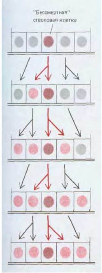
Fig. 17-23. Each proliferative unit must always contain at least one "immortal" stem cell, whose descendants will reside within that unit far into the future. Arrows indicate the Lineage relationships between cells. The stem cell in each cell generation is shown here in the central position. Other basal cells may initially possess distinct chemical properties that predetermine their departure from the basal layer and subsequent differentiation; alternatively, basal cells might be equivalent to immortal stem cells in their properties, yet their progeny could be displaced from the basal layer and shed from the skin, rendering such basal cells "mortal" in this sense.
17.4.4. It is possible that stem cell "immortality" is maintained through contact with the basement membrane [20]
If each epidermal proliferative unit is maintained indefinitely through the proliferation of its basal cells, then among them there must be at least one cell whose progeny does not completely die out before the end of the animal's life. We shall refer to such a cell as an immortal stem cell (Fig. 17-23). In principle, the division of an immortal stem cell could yield two initially identical daughter cells whose subsequent fate would depend entirely on their later living conditions. At the opposite extreme, stem cell division could always be asymmetric, such that one and only one daughter cell inherits the properties necessary for immortality, whereas the other undergoes some alteration at the moment of its birth that forces it to differentiate and ultimately dooms it to death. Under this scenario, the number of immortal stem cells could never increase, which contradicts the facts. When an area of the epidermis is destroyed, tissue continuity is restored by surrounding healthy epidermal cells that migrate and proliferate to close the gap. In doing so, new proliferative units are formed, and their central basal cells must inevitably arise from divisions in which a single immortal cell gives rise to two.
Thus, upon stem cell division, the fate of the daughter cells must depend, at least in part, on extracellular factors. One such factor could be contact with the basement membrane, the disruption of which would trigger terminal differentiation. Tissue culture experiments provide some support for this hypothesis: epidermal cells continue to divide if grown in contact with a suitable substrate (e.g., a fibroblast feeder layer), but immediately begin to differentiate when grown in suspension. However, this type of regulation by external factors fails to account for everything. Other evidence points rather to the reverse direction of causality—namely, that the changes leading to terminal differentiation result in the detachment of cells from the basement membrane, rather than vice versa. According to this hypothesis, only a few basal cells are capable of functioning as stem cells. The surface of these stem cells possesses specialized properties that enable them to attach to the basement membrane. These cells are programmed to produce a specific fraction of progeny "fated" to differentiate, a process that includes the loss of attachment ability. Under special conditions of tissue repair, The ratio of differentiating descendant cells to proliferating cells can be modulated by local growth factors to generate extra cells for wound coverage. Evidence supporting this hypothesis comes from experiments in which keratinocytes were cultured in vitro under calcium-depleted conditions; this maintained them as a monolayer, keeping all cells in a basal state. Nevertheless, some cells under these conditions entered the pathway of terminal differentiation, as indicated by the synthesis of involucrin; these differentiating cells left the basal layer as soon as the $ ext{Ca}^{2+}$ concentration in the medium was raised (Section 14.3.4).
17.4.5. Basal cell proliferation is regulated in accordance with epidermal thickness [21]
Regardless of what determines the choice between maintaining stem cell status and embarking on the fated pathway of terminal differentiation, other factors must operate to regulate The rate of new epidermal cell production. For instance, if the outer layers of the epidermis are scraped away, the basal cells begin to divide more rapidly. After some time, this leads to the restoration of normal epidermal thickness, and proliferation in the basal layer subsides back to its normal level. Everything proceeds as if the removal of the outer differentiated layers relieves the basal cells from METABOLISM/18.html">The Influence of an inhibitory inhibitor factor, which resumes its action as soon as the epidermis is fully regenerated.
Although cultured keratinocytes are known to respond to numerous Hormones and growth factors—including epidermal growth factor, which is of paramount clinical importance—the molecular mechanisms regulating their proliferation in vivo remain unclear. The consequences of disrupted regulation in this process can be observed in psoriasis. In this common skin disorder, basal cell proliferation is drastically accelerated, the epidermis becomes thickened, and cells desquamate from the skin surface within just a week of their formation in the basal layer, before they have undergone complete keratinization.
17.4.6. Cutaneous secretory cells are segregated into glands, and their populations exhibit distinct dynamics [22]
The skin serves not only as a protective barrier; it also performs other functions. In certain specialized regions, alongside the keratinized cells described above, other cell types develop from the epidermis. Specifically, secretory cells are sequestered within deep-lying glands, and their renewal occurs through an entirely different mechanism than in keratinizing areas.
The simplest example of such a structure is a sweat gland. It consists of a long, blind-ended tube formed as an invagination of the epidermis. Sweat is secreted by cells in the lower portion of this tube and reaches the skin surface through an excretory duct (Fig. 17-24). The secretory cells form a single-layered epithelium surrounded by a small number of contractile myoepithelial cells (see Figs. 17-25, B and 17-38, D). The excretory duct is lined by a double-layered epithelium lacking myoepithelial elements. Glands that produce tears, earwax, saliva, and milk are structured in a similarly analogous fashion. At least in salivary and Mammary Glands, the ducts contain stem cells dedicated to replenishing the secretory cell population.
The mammary gland is of particular interest due to the Hormonal Regulation of its cell division and differentiation. Milk production must commence when a baby is born and cease when the child is weaned. When the mammary gland is inactive, its glandular tissue consists of branching duct systems embedded in connective tissue, lined within the secretory regions by a single layer of relatively quiescent epithelial cells. As a preliminary step toward intensive milk synthesis, hormones circulating in the Blood during Pregnancy stimulate cell proliferation here; the terminal ends of the ducts grow and branch, forming small expansions called alveoli that contain secretory cells (Fig. 17-25). Milk secretion begins only when these cells are stimulated by the altered profile of hormones in the mother's blood following childbirth. When breastfeeding ceases, the secretory cells degenerate, macrophages clear away their cellular debris, most of the alveoli regress, and the gland enters a resting state.
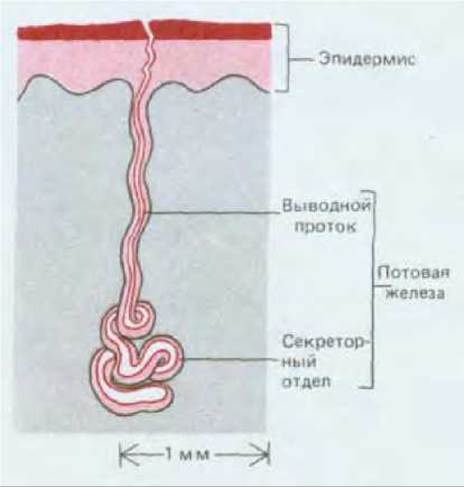
Fig. 17-24. Diagram of the structure of a sweat gland.
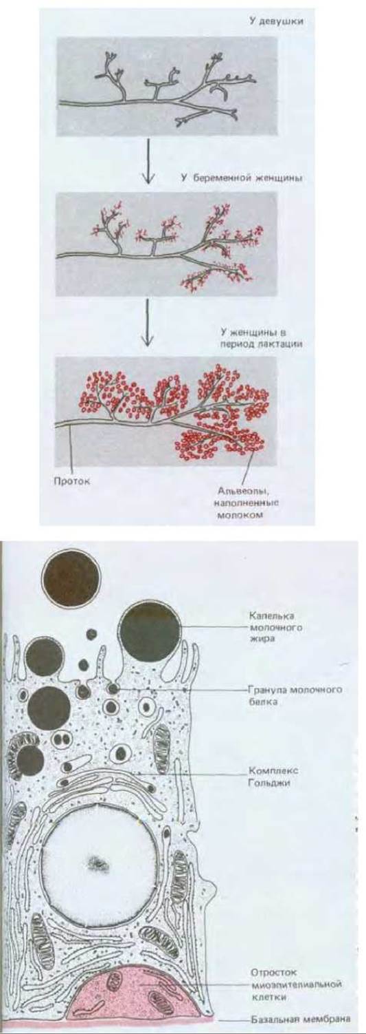
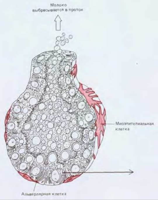
Fig. 17-25. The mammary gland. Top left: Schematic diagram showing The formation of alveoli from mammary ducts during pregnancy and Lactation. Only a small region of the gland is depicted. In the resting state, the gland contains a small number of inactive glandular structures embedded in a mass of adipose connective tissue (shown in grey in the figure). During pregnancy, extensive proliferation of the glandular tissue occurs at the expense of adipose tissue, with predominant Development of the secretory regions and formation of alveoli. Top right: A milk-secreting alveolus of the mammary gland surrounded by a "basket" of myoepithelial cells. Myoepithelial cells contract to expel milk from the alveoli in response to the hormone oxytocin, which is reflexively released in women during nursing. Bottom: Cells of the same type produce both milk proteins and milk fat. Proteins are discharged from the cells via conventional exocytosis, whereas fat is released in the form of lipid droplets enveloped by a Plasma Membrane pinched off from the cell. (Top left after R. Krstic, Die Gewebe des Menschen und der Säugetiere. Berlin: Springer-Verlag, 1978; Bottom after D. W. Fawcett, A Textbook of Histology, 11th ed. Philadelphia: Saunders, 1986.)
Many tissues, particularly those characterized by rapid cellular turnover (such as the intestinal lining, the epidermal layer of the skin, and hematopoietic tissues), are continually renewed with the aid of stem cells. By definition, stem cells are incompletely differentiated cells capable of unlimited division, yielding progeny that partly differentiate while the remainder stay stem cells. Epidermal stem cells reside in the basal layer in direct contact with the basement membrane. The descendants of stem cells differentiate as they depart this layer, sequentially synthesizing various types of keratins as they move further away; subsequently, the cell nuclei degenerate to form an outer layer of dead, cornified cells that are ultimately sloughed off from the surface. In regions where the epidermis is thin, it is clearly subdivided into proliferative units, or columns, containing at least one "immortal" stem cell at the base of each. The fate of stem cell progeny depends partly on extracellular factors that are not yet fully understood. The proliferation rate of stem cells is homeostatically regulated in accordance with epidermal thickness. Associated Glands of the epidermis, such as sweat and mammary glands, also house stem cells, though the organization of their cell population renewal is distinctly different.
Last update: 12/08/2026
Editorial and Educational Adaptation: This material has been compiled based on the primary/original source text. The project team performed an editorial review, corrected technical inaccuracies, structured sections, and adapted the content for an educational format.
What was processed:
- elimination of formatting defects (OCR errors, structural breaks, corrupted characters);
- editorial organization of content;
- standardization of terminology in accordance with academic sources;
- verification of factual statements against the original source text.
All mentions of the author, publication year, and origin of the primary text have been preserved in accordance with the source.