Molecular Biology of the Cell - Vol. 3 - Alberts B., Bray D., Lewis J., Raff M., Roberts K., Watson J. 1994
From Cells to Multicellular Organisms
Maintenance of Normal Tissue Organization
Renewal via Pluripotent Stem Cells. Example: Blood Cell Formation
Blood contains many Cell types performing entirely different functions—ranging from Oxygen transport to antibody production. Some of these Cells function exclusively within the Circulatory system, whereas others use it merely for transport and carry out their functions elsewhere. Nevertheless, The life cycle of all Blood Cells is similar to some extent. Their lifespan is limited, and they are continuously produced throughout the animal's life. Finally, and quite remarkably, all of them descend from the same type of Bone Marrow stem cells. Thus, this hematopoietic, or blood-forming, stem cell is pluripotent, meaning it gives rise to all types of terminally differentiated blood cells.
Blood cells (Fig. 17-26) can be divided into red and white cells: erythrocytes and leukocytes. Erythrocytes remain within Blood Vessels and transport O2 and CO2 bound to Hemoglobin. Leukocytes combat infection, as well as engulf and digest the debris of destroyed cells, by exiting through the walls of small blood vessels into Tissues. In addition, blood contains A large number of platelets, which are not ordinary whole cells, but rather small cellular fragments, or "mini-cells," pinched off from the cortical Cytoplasm of large cells called megakaryocytes. Platelets specifically adhere to the endothelial lining of damaged blood vessels, where they help repair the vessel wall and participate in blood clotting.
Class="center">
Fig. 17-26. Mammalian blood cells within a small blood vessel (scanning electron micrograph). The larger, nearly spherical cells with rough surfaces are leukocytes, while the smaller, smoother, and flatter cells are erythrocytes. (R. G. Kessel, R. H. Kardon, Tissues and Organs: A Text-Atlas of Scanning Electron Microscopy. San Francisco: Freeman, 1979.)
17.5.1. There Are Three Categories of Leukocytes: Granulocytes, Monocytes, and Lymphocytes [23, 24]
While every erythrocyte resembles every other erythrocyte, and every platelet resembles another platelet, leukocytes are divided into A number of distinct classes. Based on morphological features visible under a Light Microscope, they are traditionally subdivided into three main groups: granulocytes, monocytes, and lymphocytes.
All granulocytes contain numerous Lysosomes and secretory vesicles. Phagocytic cells contain specialized Organelles that fuse with newly formed phagocytic vesicles (phagosomes) and attack ingested microorganisms using highly reactive molecules of superoxide (O-2) and hypochlorite (HOCl; the active component of bleach), as well as a concentrated mixture of lysosomal Hydrolases. These vesicles, or granules, gave granulocytes their name due to their distinct staining patterns (Fig. 17-27). Differences in staining reflect important chemical and functional characteristics. Neutrophils (also called polymorphonuclear leukocytes because of their multilobed nuclei) are the most abundant granulocytes; they capture, kill, and digest microscopic organisms, particularly Bacteria. Basophils secrete histamine (and serotonin in some animals), which is involved in inflammatory responses. Eosinophils assist in the destruction of parasites and modulate allergic reactions.
Upon leaving the bloodstream (Fig. 17-27, D), monocytes become macrophages, which, alongside neutrophils, are the primary "professional phagocytes" (Section 6.5.14). Both types of macrophages, however, are significantly larger and longer-lived than neutrophils, and they possess The unique ability to digest large microorganisms such as Protozoa.
Lymphocytes are involved in the Immune Response and are represented by two main classes: B lymphocytes produce Antibodies, and T lymphocytes kill virus-infected cells and regulate The activity of other leukocytes (see Chapter 18). In addition, there are lymphocyte-like cells called natural killer cells, which are capable of destroying certain types of tumor and virus-infected cells. Lymphocyte formation is a specialized topic that will be discussed in detail in Chapter 18. Here, we will primarily examine The Development of other blood cells, often grouped together under the term myeloid cells.

Fig. 17-27. Cytology/cytology/93.html">ELECTRON MICROGRAPHS OF four types of leukocytes: A—neutrophil; B—basophil; C—eosinophil; D—monocyte. Electron micrographs of lymphocytes are shown in Fig. 18-4. Cells of each type perform a specific function, which is reflected in the differences of their secretory granules and lysosomes. Each cell contains only a single nucleus, but it has an irregular, lobed shape, so that in micrographs A, B, and C the connections between the lobes did not fall within the plane of the section. (Courtesy of Dorothy Bainton.)
The various types of blood cells and their functions are listed in Table 17-1.
Table 17-1. Blood Cells
|
Cell type |
Main functions |
Normal concentration in human blood (per liter) |
|
Erythrocytes Leukocytes |
Transport O2 and CO2 |
5-1012 |
|
Granulocytes |
||
|
Neutrophils (polymorphonuclear leukocytes) |
Phagocytose and destroy invading bacteria |
5-109 |
|
Eosinophils |
Destroy larger parasitic organisms and modulate allergic inflammatory responses |
2-108 |
|
Basophils |
Release histamine and serotonin during certain immune responses |
4-107 |
|
Monocytes |
Become macrophages in tissues, where they phagocytose and digest invading bacteria, foreign bodies, and senescent cells |
4-108 |
|
Lymphocytes |
||
|
B cells |
Produce antibodies |
2-109 |
|
T cells |
Kill virus-infected cells and regulate the activity of other leukocytes |
1-109 |
|
Natural killer cells (NK cells) |
Kill virus-infected cells and certain tumor cells |
1-108 |
|
Platelets (cell fragments formed in the bone marrow from megakaryocytes) |
Initiate blood clotting |
3-1011 |
17.5.2. The production of Each Cell Type in the Bone Marrow Is Regulated Independently [23, 25]
Most leukocytes function not in the blood, but in other tissues, with the blood simply transporting them to where they are needed. A local infection or tissue injury, for example, rapidly attracts leukocytes as part of the inflammatory response, which helps combat infection during wound healing. The inflammatory response is a complex process involving various signaling molecules released by local mast cells, nerve endings, platelets, and leukocytes, as well as Complement Proteins (Section 18.5). Some of these signaling molecules act on nearby capillaries, decreasing adhesion between endothelial cells while increasing their surface adhesiveness for passing leukocytes. Thus, leukocytes are trapped like flies on sticky paper and can then exit the vessel by squeezing between endothelial cells and "eating" their way through the basement membrane using digestive Enzymes. Other molecules act as chemoattractants for specific types of leukocytes; under their influence, these cells polarize and begin crawling toward the source of the attractant. As a result, large numbers of leukocytes migrate into the affected tissue (Fig. 17-28).
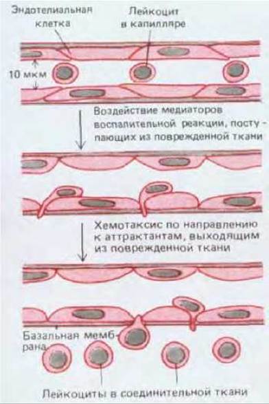
Fig. 17-28. Migration of leukocytes from the bloodstream into damaged or infected tissue during an inflammatory response. This reaction is triggered by various signaling molecules locally released by cells (primarily in Connective Tissue) or generated by complement activation. Some of these mediators act on capillary endothelial cells, weakening their junctions with neighboring cells and thereby increasing capillary permeability. Alterations in the endothelial cell surface also promote blood Cell Adhesion. Other mediators act as chemoattractants, inducing the attached leukocytes to squeeze between capillary endothelial cells into the tissue.
Other signaling molecules generated during the inflammatory response enter the bloodstream and stimulate the bone marrow to step up leukocyte production and release them into Circulation. The bone marrow serves as the primary target for this regulation because, in adult mammals, blood cells—with the exception of lymphocytes and certain macrophages—are produced exclusively in the bone marrow. This regulation is more or less specific to particular cell types: for example, certain bacterial infections lead to a selective increase in the number of neutrophils, whereas infections by protozoa and other parasites result in an accumulation of eosinophils (which is why physicians routinely use differential leukocyte counts to diagnose infectious and other Inflammatory Diseases).
In other instances, the number of erythrocytes increases selectively, such as in humans at high altitudes where oxygen is scarce. Thus, blood Cell Formation (hematopoiesis) is necessarily subject to complex control, in which the number of each cell type is regulated individually in accordance with changing physiological demands. Understanding how these regulatory mechanisms operate is a task of paramount importance for medicine.
Studying hematopoiesis in intact animals is more difficult than studying cell turnover in tissues such as the epidermis. The epidermis exhibits a simple, regular spatial Organization that makes it easy to monitor renewal processes and locate stem cells. This is not as straightforward in hematopoietic tissues. On the other hand, hematopoietic cells live like nomads, which makes them convenient subjects for a different kind of experiment. Dispersed hematopoietic cells can be readily and non-destructively transferred from one Organism to another; furthermore, the proliferation and differentiation of individual cells and their progeny can be observed and studied in culture. For this reason, more is known about the molecules that regulate blood cell formation than about analogous regulatory molecules in other mammalian tissues. Even so, our knowledge regarding blood cells remains far from complete.
17.5.3. Bone Marrow Contains Hematopoietic Stem Cells [23, 26]
The various types of blood cells and their immediate precursors in the bone marrow can be identified by their appearance (Fig. 17-29). They are intermixed with one another, as well as with fat cells and fibroblasts that form a delicate supporting network of Collagen fibers and other Extracellular matrix components. In addition, the entire tissue is permeated by thin-walled blood vessels (blood sinuses) into which newly formed blood cells enter. Megakaryocytes are also present; unlike other blood cells, they remain in the bone marrow even after maturation, constituting one of the most prominent histological features of this tissue. They are unusually large (up to 60 µm in diameter) and possess a highly polyploid nucleus. Under normal conditions, megakaryocytes embrace the walls of blood sinuses and extend cytoplasmic processes through apertures in their endothelial lining; platelets pinch off from these processes and are subsequently swept away by the bloodstream (Fig. 17-30).
The apparent lack of organization in the arrangement of various bone marrow cells makes it difficult to identify precursors of mature blood cells other than their immediate progeny. At very early Selection/3.html">Stages of development, when overt differentiation has not yet begun, all precursor cells appear very similar to one another, and primitive stem cells lack any visible markers by which they can be recognized. Identifying and characterizing stem cells requires a functional assay that allows The Fate of individual cell progeny to be traced. As we will see, this can be accomplished simply by studying colonies formed by individual cells in culture. In the hematopoietic system, however, such cell clones can also be identified within the intact animal.
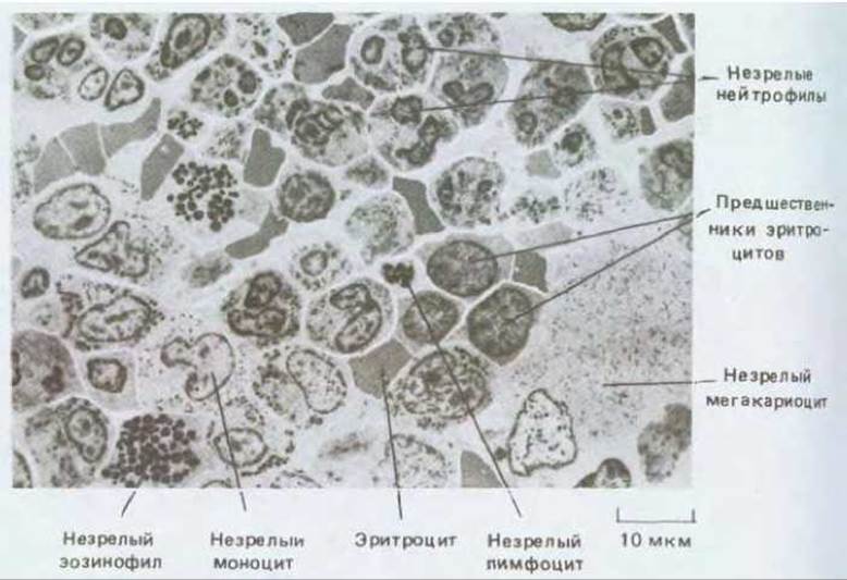
Fig. 17-29. Section of bone marrow (low-magnification electron micrograph). This tissue serves as the primary source of new blood cells (excluding T lymphocytes). Note that immature blood cells of a specific type tend to cluster into "family groups." (J.A.G. Rhodin, Histology: A Text and Atlas. New York: Oxford Univ. Press, 1974.)
If an animal is exposed to a high dose of X-irradiation, its hematopoietic cells are destroyed, and it dies within a few days due to the body's inability to replace lost blood cells. However, an irradiated animal can be saved by injecting cells harvested from the bone marrow of a healthy, immunologically compatible donor. Among these cells, there are evidently some capable of forming colonies in the recipient's body, thereby restoring its hematopoietic tissue. Such colonies develop, in particular, in the Spleen, which acts as an important secondary site of hematopoiesis in normal mice. When the spleen of an irradiated mouse is examined a week or two after the administration of cells from a healthy donor, distinct nodules can be observed, each containing a colony of myeloid cells (Fig. 17-31); after two weeks, some colonies may contain over a million cells. The discrete nature of these nodules suggests that each of them, much like a bacterial colony on an Agar plate, originates from a clone derived from a single ancestral cell—a hypothesis confirmed by genetic marker experiments.

Fig. 17-30. A. A megakaryocyte among other bone marrow cells (diagram). The enormous size of the megakaryocyte is due to its highly polyploid nucleus. A single megakaryocyte produces about 10,000 platelets, which pinch off from long cytoplasmic extensions protruding through Pores in the walls of adjacent blood sinusoids. B. The interior of such a bone marrow sinusoid (scanning electron micrograph). Megakaryocyte extensions are clearly visible. (B - from R.G. Kessel, R.H. Kardon, Tissues and Organs: A Text-Atlas of Scanning Electron Microscopy. San Francisco: Freeman, 1979.)
The founding cell of such a colony is termed a colony-forming unit (CFU). Colony-forming cells are heterogeneous: some give rise to only a single type of myeloid cell, whereas others generate several types. Certain cells undergo numerous division cycles to form large colonies, whereas others divide less frequently and form small colonies. Most colonies eventually die off after producing a limited number of terminally differentiated blood cells. However, some colonies possess a high capacity for self-renewal, generating both terminally differentiated blood cells and new colony-forming cells. These self-renewing colonies are thought to originate from hematopoietic stem cells derived from the transplanted bone marrow.
17.5.4. The Pluripotent Stem Cell Gives Rise to All Classes of Blood Cells [27]
Often, a single splenic colony derived from a single stem cell contains myeloid cells of all types. Thus, the hematopoietic stem cell is pluripotent: it can give rise to many distinct cell types. Although splenic colonies apparently do not contain lymphocytes, other experiments demonstrate that lymphocytes derive from the same stem cell that generates all myeloid cells. This is shown by genetic marker experiments, which make it possible to identify cells belonging to a single clone even after they have entered the bloodstream. Several clonal markers have been employed, but specially engineered Retroviruses have proven to be the most effective. Like all retroviruses, a marker virus can integrate its genome into the chromosome of the infected cell, but it lacks the genes required to produce infectious Viral Particles. Consequently, the marker is present exclusively in the progeny of cells initially infected. The progeny of one such cell differ from those of another solely in the chromosomal site of viral integration. To analyze the progeny of hematopoietic cells, bone marrow cells are first infected with a retrovirus in vitro (Section 5.5.8) and then introduced into lethally irradiated recipients; DNA probes can subsequently be used to trace the progeny of individual infected cells across various hematopoietic and lymphoid tissues.
These experiments not only confirm that all classes of blood cells—both myeloid and lymphoid—originate from a common stem cell (Fig. 17-32), but also allow the "lineages" of these blood cells to be tracked over extended periods. One to two months after bone marrow transplantation, the majority of blood cells in an irradiated recipient mouse are the progeny of no more than a half-dozen pluripotent stem cells; the same holds true a few weeks later, though the blood cells then are the progeny of a different set of stem cells. These observations suggest that an individual stem cell has a very low probability of initiating a clone of differentiated progeny at any given time, and that numerous cell divisions occur between the initial event and terminal differentiation, resulting in a very large final progeny clone comprising up to several million cells. Despite the relative rarity and "quantal" Nature of the initial events, differentiated cells are produced at an overall continuous and steady rate; this is governed by regulatory mechanisms operating at intermediate stages of differentiation that help control the final population size of each cell type.

Each nodule contains a clone of hematopoietic cells derived from one of the injected bone marrow cells
Fig. 17-31. Experimental scheme in which the spleen of a heavily irradiated animal is repopulated with hematopoietic bone marrow cells from a healthy donor. This experiment provided the first means of analyzing individual myeloid progenitor cells, thereby dramatically expanding The Study of hematopoiesis.

Fig. 17-32. Hypothesized scheme of hematopoiesis. Under normal conditions, a pluripotent stem cell occasionally divides to yield either identical pluripotent stem cells (self-renewal) or committed progenitor cells (CFUs — colony-forming units), which are irreversibly determined to form only a restricted number of blood cell types. The proliferation of progenitor cells is stimulated by specific growth factors, but these cells gradually lose their capacity to divide and mature into terminally differentiated blood cells that typically survive for only a few days or weeks. In adult mammals, all the cells shown here develop primarily within the bone marrow. The exceptions are T lymphocytes, which mature in the Thymus, and macrophages, which differentiate from monocytes in most tissues. The most controversial branching point in this scheme is the initial split: it remains unclear whether there exist stem cells restricted to forming solely T AND B lymphocytes alongside stem cells giving rise to all other cell types (myeloid cells). It is quite possible that primary pluripotent stem cells can also differentiate into various tissue cells not shown in this diagram, such as natural killer (NC) cells, mast cells, osteoclasts, and diverse antigen-presenting cells (Section 18.6.10), though these pathways have not yet been definitively established.
17.5.5. The Number of Diverse Blood Cells Increases through the Division of Committed Progenitor Cells [23, 28]
Once a cell has differentiated into an erythrocyte, granulocyte, etc., there is no turning back: the state of differentiation is irreversible. Therefore, at some stage in its development, the progeny of a pluripotent stem cell must definitively and irrevocably commit to a specific pathway of differentiation. Simple microscopic examination of the bone marrow reveals that this commitment occurs long before the final division cycle that yields mature, differentiated cells: specialized progenitor cells can be recognized that still undergo division while already displaying signs of ongoing differentiation. Thus, once a cell enters a specific pathway, it undergoes a series of divisions that expand the population of that specialized cell type.
It follows that the hematopoietic system can be viewed as a cellular hierarchy. Pluripotent stem cells give rise to committed progenitor cells, which have already irreversibly determined to act as precursors for one or more blood cell types. Committed cells are thought to divide rapidly, but for a limited number of times. At the end of this division series, they become terminally differentiated cells that generally cease dividing and die off within a few days or weeks. In vitro studies provide insight into how these cellular processes are regulated.
17.5.6. Factors Regulating Hematopoiesis Can Be Studied Using Cell Cultures [29]
Hematopoietic cells will survive, proliferate, and differentiate in culture only if supplied with specific growth factors or co-cultured with cells that produce these factors. Long-term proliferation of pluripotent stem cells is possible, for example, when they are grown in dispersion over a layer of bone marrow stromal cells, which presumably mimics the microenvironment of intact bone marrow; all types of myeloid cells can be generated in such cultures. Dispersed bone marrow hematopoietic cells can also be cultured in a semisolid agar or methylcellulose medium supplemented with factors extracted from other cells. In such a semisolid matrix, the progeny of each individual cell remain localized, forming a readily identifiable colony. A single committed cell, such as a neutrophil precursor, can give rise to a clone of thousands of neutrophils. Such culture systems enable the assay of activities that support hematopoiesis, facilitating their isolation and the study of their Mechanisms of action. These substances have been identified as Glycoproteins, commonly referred to as colony-stimulating factors (CSFs). Among the growing number of CSFs that have been isolated and purified, some circulate in the blood and act as Hormones, whereas others function as local chemical mediators (Section 12.1). Of the hormonal-type CSFs, the best-characterized is the glycoprotein Erythropoietin, which is produced by the Kidneys and regulates erythropoiesis (red blood cell production).

Fig. 17-33. Stages in erythroblast (red blood cell precursor) development. The erythroblast expels its nucleus to become an immature red blood cell (reticulocyte) shortly before leaving the bone marrow and entering the bloodstream. Within 1 to 2 days, the reticulocyte loses its Mitochondria and Ribosomes to become a mature erythrocyte. Erythrocyte clones develop within the bone marrow On the surface of macrophages, which phagocytose and digest the nuclei discarded by erythroblasts.
17.5.7 Erythropoiesis Depends on the Hormone Erythropoietin [30]
Erythrocytes constitute the bulk of cells circulating in the blood (see Table 17-1). A mature erythrocyte is packed with hemoglobin and virtually devoid of conventional cellular organelles. In adult mammalian erythrocytes, even The Nucleus, Endoplasmic reticulum, mitochondria, and ribosomes are absent—having been extruded from The Cell during development (Fig. 17-33). Consequently, an erythrocyte cannot grow or divide; stem cells represent the sole possible source for generating new erythrocytes. Furthermore, the lifespan of erythrocytes is relatively short—about 120 days in humans and 55 days in mice. Worn-out erythrocytes are engulfed and digested by macrophages in The Liver and spleen.
Oxygen deficiency or a shortage of red blood cells stimulates Kidney cells to synthesize and release elevated levels of erythropoietin into the blood, which in turn drives red blood cell production. Because an accelerated release of new erythrocytes into the circulation is detectable within just a day or two following a rise in blood erythropoietin concentration, the hormone must act on cells that are very close precursors to mature erythrocytes.
Cells responsive to erythropoietin can be identified by adding the hormone to bone marrow cell cultures maintained in semisolid media. After several days, colonies consisting of approximately 60 erythrocytes appear, each originating from a single committed erythroid progenitor cell. Such a cell is termed a colony-forming unit-erythroid, or CFU-E, and it gives rise to mature erythrocytes after approximately six or even fewer division cycles. CFU-Es do not yet contain hemoglobin; they are derived from earlier progenitor cell types whose proliferation is independent of erythropoietin.

Fig. 17-34. Pedigree showing the relationship between the pluripotent stem cell, BFU-E, CFU-E, and mature erythrocytes. BFU-E and CFU-E cells are committed erythroid progenitor cells. BFU-E cells respond to interleukin-3 but do not respond to erythropoietin, whereas CFU-E cells respond well to erythropoietin. A series of cell divisions driven by erythropoietin provides an efficient way to regulate erythropoiesis without disrupting the production of other blood cells.
The second colony-stimulating factor, interleukin-3 (IL-3), is responsible for the survival and proliferation of pluripotent stem cells and the majority of their committed progeny. In its presence, cultured bone marrow cells develop into much larger erythroid colonies of approximately 5,000 erythrocytes each. These colonies originate from erythroid cells called burst-forming units-erythroid (BFU-E). BFU-E differs from the pluripotent stem cell in having a limited capacity for proliferation and giving rise exclusively to erythroid colonies, even under culture conditions that allow other cells to generate alternative types of differentiated blood cells. It differs from CFU-E in that BFU-E are insensitive to erythropoietin and are separated from mature erythrocytes by as many as 12 cell divisions that already require erythropoietin. (These cells also differ from CFU-E in size and can be separated from the latter by centrifugation.) Thus, BFU-E are considered progenitor cells committed to erythroid differentiation and are early precursors of CFU-E (Fig. 17-34).
17.5.8. The production of neutrophils and macrophages is influenced by several colony-stimulating factors (CSFs) [29,31]
The Two Types of "professional phagocytes"—neutrophils and macrophages—develop from the same cells, known as granulocyte-macrophage (GM) progenitors. Like other granulocytes (eosinophils and basophils), neutrophils circulate in the blood for only a few hours before migrating from capillaries into Connective Tissues or other specific sites, where they live for a few days and then die. In contrast, macrophages can reside outside the bloodstream for months, or perhaps even years, where they are capable of renewing proliferation in response to local signals.
Four different CSFs have been identified that stimulate The formation of neutrophil and macrophage colonies in culture. It is believed that in vivo they act in various combinations to regulate the selective production of specific cells. These CSFs are synthesized by various cell types, including endothelial cells, fibroblasts, macrophages, and lymphocytes; during bacterial infection in a given tissue, their concentration in the blood rapidly increases, leading to an accelerated influx of phagocytic cells from the bone marrow into the bloodstream. Of these four factors, IL-3 is the least specific: it acts on pluripotent stem cells and most committed cells, including GM progenitors. The other three factors act more selectively on committed GM progenitors and their differentiated progeny (Table 17-2), although at high concentrations some of them also affect other lineages.
Like erythropoietin, all these CSFs are glycoproteins that act at low concentrations (10-12 M) by binding to specific cell-surface receptors. Their effect on progenitor cells involves not only triggering The Mechanism of differentiated colony formation, but also activating specialized functions (such as phagocytosis and target cell killing) in terminally differentiated cells. Proteins synthesized using cloned genes for these factors serve as potent stimulators of hematopoiesis in experimental animals, suggesting their potential clinical application for stimulating hematopoietic tissue recovery and treating infections.
Table 17-2. Some colony-stimulating factors (CSFs) affecting blood cell production
|
Factor |
Mol. weight |
Target cells |
|
|
Erythropoietin |
51000 |
CFU-E |
Kidney cells |
|
Interleukin-3 |
25000 |
Pluripotent stem cells, most progenitor cells, many terminally differentiated cells |
T lymphocytes, epidermal cells |
|
GM-CSF 1 |
23000 |
GM progenitors 4 |
T lymphocytes, endothelial cells, fibroblasts |
|
G-CSF 2 |
25000 |
GM progenitors 4 and neutrophils |
Macrophages, fibroblasts |
|
M-CSF 3 |
70000(dimer) |
GM progenitors 4 and macrophages |
Fibroblasts, macrophages, endothelial cells |
1 Granulocyte-macrophage colony-stimulating factor (GM-CSF).
2 Granulocyte colony-stimulating factor (G-CSF).
3 Macrophage colony-stimulating factor (M-CSF).
4 Granulocyte-macrophage progenitors.
Factors that specifically stimulate the development of other myeloid lineages, such as megakaryocytes, basophils, and eosinophils, have also been described, but they are not as well characterized as the CSFs discussed above. There is evidence that, alongside soluble CSFs—which include locally secreted products of bone marrow stromal cells—signaling molecules associated with cells and the extracellular matrix also participate in The regulation of hematopoiesis.
17.5.9. Hematopoietic cell behavior is partly stochastic [29, 32]
CSFs are defined as factors that promote the formation of differentiated blood cell colonies. It is difficult to precisely determine The Effect of a CSF on an individual hematopoietic cell. Such a factor could increase the probability of cell survival; it could regulate The rate of Cell Division or the number of divisions required by a progenitor cell prior to differentiation. It could act at late stages of differentiation to facilitate it; or, conversely, it could act at early stages and influence commitment (Fig. 17-35). By tracking the fate of individual isolated hematopoietic cells in culture, one can establish how a specific CSF, such as GM-CSF, can produce all these diverse effects. Nevertheless, it remains unclear which of these effects are significant in vivo. The behavior of pluripotent stem cells is particularly enigmatic: these crucial cells are sparse and rare—accounting for less than one in a thousand bone marrow cells—making them very difficult to identify with absolute certainty.

Fig. 17-35. Parameters through which the production of a specific type of blood cell could be regulated. In vitro studies indicate that colony-stimulating factors (CSFs) can influence all these aspects of hematopoiesis.
Furthermore, in vitro studies show that There is a strong stochastic element in the pathway choice made by a hematopoietic cell. CSFs do not directly dictate what a cell should do, but rather alter the probability of a particular behavior. In cultures of hematopoietic cells, even when these cells have been selected for maximal population homogeneity, they vary greatly in size and frequently in The Nature of the colonies they generate. Moreover, if two sister cells are isolated immediately after division and cultured separately under identical conditions, they will frequently give rise to colonies containing different cell types, or the same cell types in different proportions. Thus, both the programming of cell divisions and the channeling of cells into specific differentiation pathways (commitment) appear to involve stochastic events at the single-cell level, even though the behavior of the multicellular system as a whole is reliably regulated. The question of the molecular mechanisms underlying these processes is the most fundamental unresolved problem in hematopoiesis.
Summary
All numerous types of blood cells originate from a common pluripotent stem cell. In the adult organism, stem cells reside primarily in the bone marrow, where they normally divide rather infrequently to produce new stem cells (self-renewal) or various committed progenitor cells, each capable of giving rise to one or more cell types. Committed cells divide extensively under METABOLISM/18.html">The Influence of signaling glycoprotein molecules (called colony-stimulating factors, or CSFs) and then differentiate into mature blood cells, which typically live for only a few days or weeks. The study of hematopoiesis is greatly facilitated by in vitro culture experiments in which stem cells or committed progenitor cells form clonal colonies when grown in a semisolid medium. However, pluripotent stem cells are rare and difficult to identify, and it remains unclear how they choose their pathway among various developmental options.
Last update: 12/08/2026
Editorial and Educational Adaptation: This material has been compiled based on the primary/original source text. The project team performed an editorial review, corrected technical inaccuracies, structured sections, and adapted the content for an educational format.
What was processed:
- elimination of formatting defects (OCR errors, structural breaks, corrupted characters);
- editorial organization of content;
- standardization of terminology in accordance with academic sources;
- verification of factual statements against the original source text.
All mentions of the author, publication year, and origin of the primary text have been preserved in accordance with the source.