Review of Medical Physiology - William F. Ganong 2002
Blood Circulation
Regulation of the Cardiovascular System
Mechanism of Systemic Nervous Regulation
Nervous Control Mechanisms
Although arterioles and other resistance vessels possess the densest innervation, all Blood Vessels except capillaries and venules contain smooth Muscle and are innervated by motor fibers from the Sympathetic division of the Autonomic Nervous system. Fibers supplying resistance vessels regulate tissue blood flow and ARTERIAL BLOOD PRESSURE, whereas those supplying capacitance vessels regulate the volume of blood pooled in the Veins. Most veins have sparse innervation, but VEINS OF THE Internal Organs are heavily innervated. Venoconstriction is triggered by the same stimuli that activate arteriolar vasoconstrictor nerves. As a result, venous capacitance decreases, the return of blood to The Heart increases, and blood is redistributed to the arterial side of the Circulatory system.
Innervation of Blood Vessels
Noradrenergic fibers terminate in the vessels of all body regions (Fig. 31-5). Functionally, they are vasoconstrictor. In addition to vasoconstrictor innervation, the resistance vessels of skeletal Muscles receive vasodilator fibers which, although coursing with sympathetic nerves, are cholinergic (sympathetic vasodilator system). Evidence also indicates the presence of cholinergic innervation in the Blood vessels of the heart, Lungs, Kidneys, and Uterus. Bundles of noradrenergic and cholinergic fibers form plexuses in the adventitia of the arteriolar wall. Fibers with numerous varicosities extend from these plexuses into the media and terminate on the outer surface of the smooth muscle Cells without penetrating them. Transmitters diffuse into the inner Regions of the media, and action potentials propagate from one smooth muscle Cell to another via Gap Junctions.
Class="center">
Fig. 31-5. Noradrenergic nerve fibers innervating blood vessels in the rat mesentery. Arrows indicate the direction of blood flow (reproduced with permission from Furness JB, Marshall JM: Correlation of the directly observed responses of mesenteric Vessels of the rat to nerve stimulation and noradrenaline with the distribution of adrenergic nerves. J Physiol 1974;239:75-89).
Tonic impulses are not characteristic of vasodilator fibers, whereas the vasoconstrictor fibers of most vascular beds exhibit a degree of tonic activity. Transection of sympathetic nerves (sympathectomy) results in vasodilation of blood vessels. In most Tissues, this vasodilation is caused by a reduction in the tonic activity of vasoconstrictor nerves. However, in skeletal muscles, vessels can also dilate as a result of the activation of the sympathetic vasodilator system (Table 31-3).
Many blood vessels are innervated by fibers that synthesize Polypeptides. Cholinergic nerves also contain VIP, which induces vasodilation. Noradrenergic postganglionic sympathetic fibers synthesize neuropeptide Y, which acts as a vasoconstrictor. Sensory nerve endings near blood vessels contain substance P and CGRP, which cause vasodilation.
Table 31-3. Factors affecting arteriolar diameter
Constriction |
Dilation |
Local factors Local decrease in Temperature Autoregulation |
Increased CO2 and decreased O2 Increased K+, adenosine, lactate, etc. Local decrease in pH Local increase in temperature |
Endothelial factors Endothelin-1 Locally released platelet serotonin Thromboxane A2 |
NO Kinin Prostacyclin |
Circulating Hormones Epinephrine (except in Skeletal Muscle and Liver) Norepinephrine Arginine vasopressin (AVP) Angiotensin II Circulating Na+-K+-ATPase inhibitor Neuropeptide Y |
Epinephrine in skeletal muscle and liver CGRP Substance P Histamine ANP VIP |
Neural factors Increased firing of noradrenergic vasomotor nerves |
Decreased firing of noradrenergic vasomotor nerves Activation of cholinergic vasodilator fibers supplying skeletal muscle |
Afferent impulses in sensory nerves from the Skin travel antidromically down the branches of sensory nerves that innervate blood vessels. These impulses trigger the release of substance P from nerve terminals, which causes vasodilation and increased capillary permeability. This local neural mechanism is known as the axon reflex (see Fig. 32-17). Other cardiovascular Reflexes are mediated by the Central Nervous System.
Innervation of the Heart
Impulses arriving at the heart via noradrenergic nerves lead to an increase in heart rate (chronotropic effect) and myocardial contractility (inotropic effect). They also inhibit the effects of vagal stimulation, possibly through the release of neuropeptide Y, which acts as a cotransmitter in sympathetic nerve endings. Impulses reaching the heart via cholinergic fibers of the vagus nerves decrease heart rate. At rest, the sympathetic nerves to the heart exhibit moderate tonic activity. In humans and large animals, this is counterbalanced by a more pronounced tonic vagal discharge (vagal tone). In experimental animals with transected vagus nerves, heart rate increases. Following the administration of parasympatholytic drugs such as atropine, human heart rate increases from a normal resting value of 70 to 150-180 beats/min. This occurs because parasympathetic tone no longer counteracts sympathetic activity. In humans following simultaneous blockade of both noradrenergic and cholinergic systems, the heart rate is approximately 100 beats/min.
Vasomotor Control
Sympathetic nerves that cause constriction of arterioles and veins, as well as increase stroke volume, exhibit tonic activity (a continuous baseline level of nerve impulses). The regulation of arterial blood pressure occurs through adjustments in this tonic activity (Fig. 31-6). Although arterial pressure is influenced by spinal reflex activity, it is primarily controlled by a group of Neurons in the Medulla Oblongata known as the vasomotor area or vasomotor center.
Neurons that mediate enhanced sympathetic influences on blood vessels and the heart (Fig. 31-7) project to sympathetic preganglionic neurons within the intermediolateral (IML) cell Column of the Spinal Cord. The Cell bodies of these neurons are located bilaterally in the rostroventrolateral medulla (RVLM). Their axons project dorsally and medially, and then descend within the lateral funiculus of the spinal cord to the IML column.
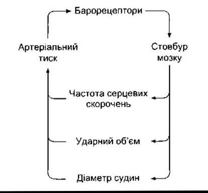
Fig. 31-6. Feedback control of arterial blood pressure.
Impulses originating from the medulla oblongata also influence heart rate via the vagus nerves. The neurons of origin for vagal fibers are located in the dorsal motor Nucleus of the vagus and The Nucleus ambiguus (Fig. 31-8).
An increase in vasoconstrictor activity results in arteriolar constriction and a rise in arterial blood pressure. These changes are usually accompanied by venoconstriction and a decrease in pooled venous blood volume, although changes in capacitance vessels do not always mirror those in resistance vessels. Heart rate and stroke volume also increase with enhanced sympathetic activation, leading to a concurrent rise in Cardiac Output. This is typically accompanied by a reduction in the tonic activity of cardiac vagal fibers. Conversely, a decrease in vasomotor center activity leads to vasodilation, a fall in arterial blood pressure, and increased blood pooling in capacitance vessels. This is generally accompanied by a decrease in heart rate, an effect mediated primarily by enhanced vagal influence on the heart.
Afferent Pathways to the Vasomotor Center
Afferent fibers converging on the vasomotor center are listed in Table 31-4. These include critically important fibers from arterial and venous baroreceptors, as well as inputs from other regions of The Nervous System and from carotid and aortic chemoreceptors. In addition, certain stimuli act directly on the vasomotor center.
Descending pathways to the vasomotor center originate in the Cerebral Cortex (particularly the limbic cortex) and project to the Hypothalamus. These fibers mediate increases in blood pressure and tachycardia in response to emotional factors such as sexual arousal and anger. The connections between the hypothalamus and the vasomotor center are reciprocal, and afferent impulses from the Brainstem complete the loop.

Fig. 31-7. Main pathways involved in medullary regulation of arterial blood pressure. Efferent vagal pathways that slow the heart rate are shown. Putative Neurotransmitters of the Neural Pathways are indicated in parentheses; Glu, glutamate; GABA, gamma-aminobutyric acid; ACh, acetylcholine; NE, norepinephrine; IML, intermediolateral cell column; NTS, nucleus tractus solitarius; CVLM, PVLM, RVLM, caudal, intermediate, and rostral ventrolateral medullary areas; IX and X, glossopharyngeal and vagus nerves.
Lung inflation leads to vasodilation and a decrease in blood pressure. This response is mediated by afferent vagal fibers running from the lungs, which suppress vasomotor center activity. Pain typically induces an elevation in blood pressure via afferent impulses passing from the reticular formation to the vasomotor center. However, prolonged pain can cause vasodilation and dizziness.
Somatosympathetic Reflex
Pain causes an increase in blood pressure. Afferent impulses from working skeletal muscles likely produce a similar pressor effect. This reaction involves C1 neurons in the rostral ventrolateral medulla. Vasoconstriction in response to The stimulation of somatic afferent nerves is termed the somatosympathetic reflex. The neural pathways involved in this response are illustrated in Fig. 31-9.
Baroreceptors
Baroreceptors are receptors that are stimulated by the stretching of the walls of The Heart and blood vessels. The carotid sinus and aortic arch receptors participate in the regulation of arterial Blood Circulation. Baroreceptors are also located in the walls of the right and left atria at the entry points of the superior and inferior venae cavae and Pulmonary veins, as well as in the pulmonary vessels; in the low-pressure sections of the lesser circulation, they are called cardiopulmonary receptors. Because baroreceptor stimulation occurs when the regions where they are localized are stretched, an increase in pressure within these structures leads to heightened firing from the baroreceptors. Impulses from the baroreceptors travel via afferent fibers of the glossopharyngeal and vagus nerves to the medulla oblongata. Most of these fibers terminate in the nucleus of the solitary tract (NST), with glutamate likely acting as their excitatory neurotransmitter. Excitatory glutamatergic fibers project from the NST to the caudal and intermediate ventrolateral areas of the medulla oblongata, where they stimulate GABAergic inhibitory neurons. These neurons project to the rostral ventrolateral area. There are also excitatory, possibly multineuronal, pathways from the NST to the vagal motor neurons in the dorsal motor nucleus and the ambiguous nucleus. Therefore, increased firing from baroreceptors inhibits the tonic activity of vasoconstrictor sympathetic nerves and stimulates vagal effects on the heart, along with arteriolar and venous dilation, resulting in a reduced cardiac output and lowered blood pressure.

Fig. 31-8. Main pathways involved in medullary Regulation of Heart rate via vagus nerves. NST neurons (dashed line) project to preganglionic parasympathetic neurons of the dorsal motor nucleus of the vagus and the ambiguous nucleus. Postganglionic cholinergic neurons innervate the atria and ventricles; Pyr, pyramid; XII, hypoglossal nucleus (modified from Standish R, Enquist LH, Schwaber JS: Innervation of the heart and its central medullary origin defined by viral tracing. Science 1994;263:232).
Table 31-4. Factors influencing the activity of the vasomotor region of the medulla oblongata


Fig. 31-9. Somatosympathetic reflex. Impulses from skin and muscle receptors travel to the RVLM of the medulla oblongata, with axons of RVLM cells projecting directly to the intermediolateral cell column (IML), where they terminate on preganglionic sympathetic neurons; ICP, inferior cerebellar peduncle; ION, inferior olivary nucleus; MLF, medial longitudinal fasciculus; NA, nucleus ambiguus; NTS, nucleus of the tractus solitarius; Py, pyramid; Sp5, spinal trigeminal nucleus (reprinted with permission from Reis DJ, Ruggerio DA, Morrison SF: The C1 area of the rostral ventrolateral medulla oblongata—A critical brainstem region for control of resting and reflex integration of arterial pressure. Am J Hypertens 1989;2:363S).
Carotid Sinus and Aortic Arch
The carotid sinus is a slight dilation of the Internal Carotid Artery located immediately above the bifurcation of the common carotid artery into its external and internal branches (Fig. 31-10). Baroreceptors are found within this dilated region, as well as in the wall of the aortic arch. The receptors are situated in the outer layer of the vascular wall. They are highly branched, bulbous, convoluted endings of Cytology/practical/65.html">Myelinated nerve fibers that resemble Golgi tendon organs. In some species, similar receptors are found in various areas of the large Arteries of the Thorax and neck. Afferent fibers running from the carotid sinus and carotid body form a distinct branch of the Glossopharyngeal nerve—the carotid sinus nerve. However, fibers from the aortic arch form a separate branch of the Vagus nerve only in rabbits. The carotid sinus nerves and the vagal branches from the aortic arch are often referred to as buffer nerves.
Activity of Buffer Nerves
At normal blood pressure, buffer nerve fibers exhibit a low basal firing rate (Fig. 31-11). An increase in pressure within the carotid sinus and aortic arch leads to an increased firing rate, whereas a decrease results in its reduction.
If one carotid sinus of a monkey is isolated and perfused while other baroreceptors are simultaneously denervated, perfusion pressures up to 30 mmHg fail to elicit impulses in the afferent fibers from the carotid sinus, and neither blood pressure nor heart rate drops. At perfusion pressures of 70-110 mmHg, There is a linear relationship between the perfusion pressure and the reduction in blood pressure and heart rate. When the perfusion pressure exceeds 150 mmHg, no further significant enhancement of the response occurs (Fig. 31-12), presumably because baroreceptor discharge and vasomotor center inhibition have reached their maximum levels.

Fig. 31-10. Baroreceptor areas of the carotid sinus and aortic arch. Receptor locations are indicated by X.
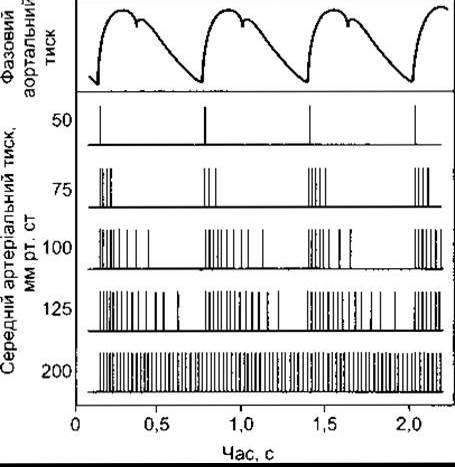
Fig. 31-11. Impulses (vertical lines) in a single afferent nerve originating from the carotid sinus at various arterial pressures, correlated with temporal changes in aortic pressure (reprinted with permission from Berne RM, Levy MN: Cardiovascular Physiology, 3rd ed. Mosby, 1977).
Carotid sinus receptors respond to both static (mean) and pulsatile pressure. A decrease in pulse pressure in the carotid arteries without any change in mean pressure leads to reduced baroreceptor firing, triggering an increase in blood pressure and tachycardia. The receptors also respond to both changing and steady pressures. During pressure fluctuations, impulses sometimes occur during the rising phase and cease during the falling phase (see Fig. 31-11). In contrast, an unfluctuating mean pressure would produce a continuous train of impulses.
Aortic arch receptors have been less thoroughly studied, but there is no reason to believe their response differs significantly from that of carotid sinus receptors.
From the foregoing, it is evident that the baroreceptors of the arterial circulation, their afferent connections with the vasomotor and cardioinhibitory centers, and the efferent pathways from these centers form a negative feedback reflex mechanism aimed at stabilizing blood pressure and heart rate. Any drop in systemic arterial pressure weakens the inhibitory discharge in the buffer nerves, eliciting a compensatory increase in blood pressure and cardiac output. Conversely, any increase in pressure causes arteriolar dilation and a reduction in cardiac output until blood pressure returns to normal levels.
Resetting of Baroreceptors
In chronic Hypertension, the baroreceptor reflex resets to maintain an elevated pressure rather than a normal one. Perfusion studies in experimental animals with hypertension have revealed that raising the pressure within an isolated carotid sinus leads to a drop in the elevated systemic arterial pressure. Conversely, lowering the perfusion pressure causes the elevated pressure to rise further (Fig. 31-12). This resetting occurs rapidly in experimental animals, though little is known about how and why it happens. Both experimentally and clinically, this is a reversible process.

Fig. 31-12. Solid line: decrease in systemic arterial blood pressure in response to an increased pressure within the isolated carotid sinus of a normal monkey. Dashed line: response of a hypertensive monkey, indicating baroreceptor resetting (arrow).
Consequences of Carotid Artery Occlusion and Buffer Nerve Transection
Bilateral occlusion of the common carotid arteries near the carotid sinuses causes an elevation in arterial blood pressure and heart rate due to the resulting decrease in intrasinusoidal pressure. Transection of the carotid sinus nerves on both sides leads to a similar effect. In both cases, the rise in blood pressure is moderate because the aortic arch baroreceptors continue to function normally. If the afferent fibers running from the baroreceptors within the vagus nerve are also severed, arterial pressure surges to 300/200 mmHg or higher and becomes unstable. Bilateral damage to the NTS, the termination site of baroreceptor afferent fibers, results in severe hypertension that can be fatal. These forms of experimental hypertension are referred to as neurogenic hypertension.
Atrial Stretch Receptors
There are two types of atrial stretch receptors: type A, in which impulses fire during atrial systole, and type B, which fire at the end of diastole, coinciding with maximal atrial filling. Impulse generation from type B baroreceptors increases with greater venous return and diminishes under positive pressure Respiration. Consequently, these baroreceptors primarily respond to the distension of the atrial walls. An increase in impulse traffic from most (if not all) of these receptors triggers reflex circulatory changes, specifically vasodilation and a drop in arterial blood pressure. However, in this instance, heart rate does not increase; rather, it decreases.
Role of Baroreceptors in the Hormonal Regulation of ECF Volume
As ECF volume declines, central venous pressure drops, and reduced afferent signaling from atrial baroreceptors triggers an enhanced release of vasopressin (see Chapter 14). Concurrently, sympathetic nervous activity increases, leading to elevated renin production. The release of renin ultimately drives aldosterone synthesis. When ECF volume depletion is substantial, arterial blood pressure falls, and afferent input from the carotid sinus and aortic arch baroreceptors decreases, further promoting hormone secretion. The characteristics of other factors that enhance vasopressin and renin production are detailed in Chapters 20, 24, and 39. The ultimate outcome of these described processes is the retention of Water and sodium, aimed at restoring ECF volume.
Bainbridge Reflex
Rapid infusion of blood or saline into anesthetized animals occasionally accelerates the heart rate, provided the initial heart rate was low. This phenomenon was described by Bainbridge in 1915 and has since been known as the Bainbridge reflex. It is considered a true reflex rather than a local stretch response, as it is abolished by bilateral vagotomy. Infusing fluids into animals with a transplanted heart increases the contraction rate of the recipient's atrial remnant, yet has no effect on The rate of the transplanted heart itself. This response likely involves the atrial receptors that elicit tachycardia (described earlier). This reflex counteracts the baroreceptor-mediated bradycardia in response to volume expansion. The reflex is attenuated or entirely absent if the baseline heart rate is high. Although the physiological importance of this reflex has been widely debated, its definitive role remains unclear.
Left Ventricular Receptors
Distension of the left ventricle in experimental animals leads to a reduction in systemic arterial blood pressure and heart rate. Because the required ventricular stretch for this response is substantial, its definitive physiological significance remains incompletely understood. It is possible that left ventricular stretch receptors contribute to maintaining the vagal tone responsible for low resting heart rates.
When substances such as serotonin, veratridine, capsaicin, phenylbiguanide, and others are injected into the coronary arteries supplying the left ventricle, animals experience transient apnea followed by rapid breathing, hypotension, and bradycardia (the coronary chemoreflex, or Bezold-Jarisch reflex). The receptors appear to be C-fiber endings, with efferent pathways traveling via the vagus nerve. This response does not occur following injections into vessels supplying the atria and the right ventricle. Its ultimate physiological role remains unclear. It is hypothesized that in patients with myocardial infarction, substances released from necrotic tissue stimulate ventricular receptors, contributing to the hypotension that frequently complicates this condition.
Pulmonary Receptors
Administration of serotonin, capsaicin, veratridine, or related compounds into the pulmonary artery activates C-fiber endings located near the pulmonary capillaries, resulting in apnea followed by rapid breathing, hypotension, and bradycardia (the pulmonary chemoreflex). This response (described in Chapter 36) is blocked by vagotomy. Mechanistically, it is analogous to the coronary chemoreflex elicited by injecting substances into the left Coronary Circulation. The reaction develops extremely rapidly—before the substances can reach the left ventricular receptors.
Clinical Studies and Stimulation
Fluctuations in pulse rate and arterial pressure observed in humans upon standing up or lying down (see Chapter 33) are mediated primarily by baroreceptor reflexes. The functional role of these receptors is demonstrated by heart rate changes in response to blood pressure elevations induced by the alpha-adrenergic agonist phenylephrine. The normal physiological response is illustrated in Fig. 31-13. At systolic pressures ranging from 120–150 mmHg, there is a linear relationship between pressure levels and the reduction in heart rate (prolongation of the RR interval). Receptor function can also be assessed by examining pulse and blood pressure alterations during brief periods of strain (forced expiration against a closed glottis: the Valsalva maneuver). At the onset of the maneuver, arterial pressure rises (Fig. 31-14) because increased intrathoracic pressure is transmitted to the aorta. Arterial pressure subsequently falls as high intrathoracic pressure constricts veins, reducing venous return to the heart and decreasing cardiac output. This drop in arterial and pulse pressure weakens baroreceptor signaling, triggering reflex tachycardia and an increase in peripheral vascular resistance. When the glottis is opened, intrathoracic pressure normalizes, cardiac output recovers, but peripheral vessels remain constricted. Consequently, arterial pressure overshoots baseline values, stimulating baroreceptors and causing bradycardia that returns blood pressure to normal levels.
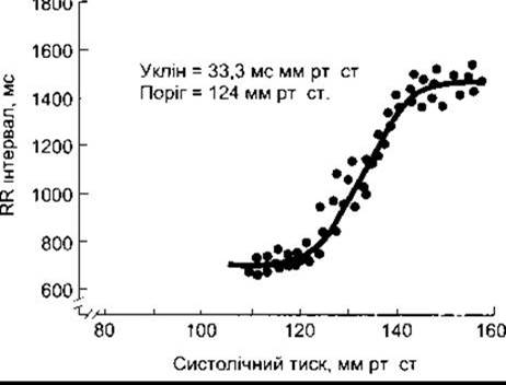
Fig. 31-13. Baroreflex-mediated decrease in heart rate following phenylephrine administration in humans. Note that the ECG RR interval values on the vertical axis are inversely proportional to heart rate (reproduced with permission from Kotrly K et al: Effects of fentanyl-diazepam-nitrous oxide anaesthesia on arterial baroreflex control of heart rate in man. Br J Anaesth 1986; 58:406).
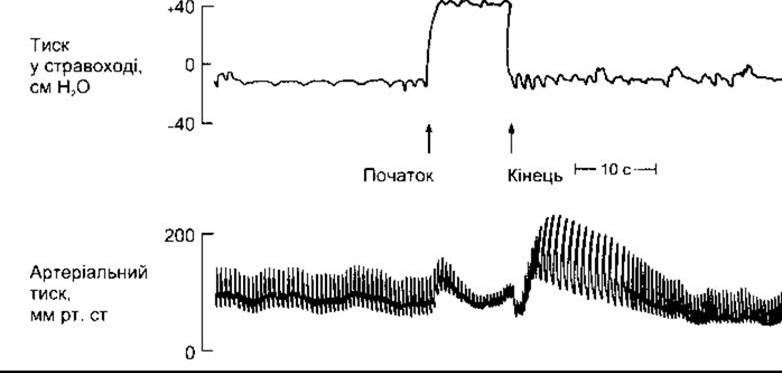
Fig. 31-14. Tracing of the response to the Valsalva maneuver in a healthy subject, recorded via a needle inserted into the brachial artery (courtesy of Dr. J. McIlroy).
The heart rate changes described above are also observed in sympathectomized patients, as their baroreceptors and vagus nerves remain intact. However, in patients with autonomic failure—a syndrome characterized by severe impairment of autonomic nervous system function—heart rate remains unchanged. For reasons that are not entirely clear, neither heart rate nor blood pressure changes in response to normalized intrathoracic pressure in patients with primary hyperaldosteronism. Their response to the Valsalva maneuver normalizes following the surgical removal of the aldosterone-producing tumor.
Effects of Chemoreceptor Stimulation on the Vasomotor Center
Afferent fibers originating from the carotid and aortic bodies are primarily involved in the Regulation of Respiration, a function detailed in Chapter 36. However, these fibers also project to the vasomotor center. The cardiovascular response to chemoreceptor stimulation consists of peripheral vasoconstriction and bradycardia. Additionally, Hypoxia induces tachypnea (hyperpnea) and enhanced release of catecholamines from the Adrenal Glands—responses that respectively drive tachycardia and an increase in cardiac output. During Hemorrhage, hypotension develops, which in turn stimulates the chemoreceptors. This effect is attributed to sluggish Blood flow through chemoreceptor regions, leading to stagnant anoxia in these tissues (see Chapter 37). In hypotensive animals, baroreceptor input is diminished (see below), and transection of the glossopharyngeal and vagus nerves results in a fall in arterial pressure rather than a rise. This occurs because the stimulatory Influence of the chemoreceptors on the vasomotor center is removed. Baroreceptor signaling is also believed to play a role in the generation of Mayer waves. These waves should not be confused with Traube-Hering waves, which are blood pressure oscillations synchronous with respiration. Mayer waves are slow, regular blood pressure oscillations occurring at a frequency of one every 20–40 seconds during states of hypotension. Under these conditions, hypoxia stimulates the chemoreceptors, leading to a compensatory rise in arterial pressure. The increased pressure enhances blood flow through the chemoreceptor zones, subsequently reducing their afferent output. Once pressure drops, a new cycle begins. Mayer waves are attenuated but do not completely disappear following chemoreceptor denervation. They are occasionally observed in vertebrates. Thus, spinal vasopressor reflexes also contribute to the genesis of these waves.
Direct Influences on the Vasomotor Center
Both hypoxia and hypercapnia stimulate the vasomotor center by acting directly on the RVLM, although the direct effect of hypoxia results in a slight increase in intracranial pressure and Impaired blood supply to the vasomotor center, whereas local hypoxia and hypercapnia enhance impulse generation within it. The resulting rise in blood pressure (Cushing's reflex) aims to restore blood flow to the medulla oblongata. This triggers a reflex decrease in heart rate via arterial baroreceptors (see below). Consequently, bradycardia rather than tachycardia is a hallmark symptom in patients with elevated intracranial pressure.
Table 31-5. Factors affecting heart rate1
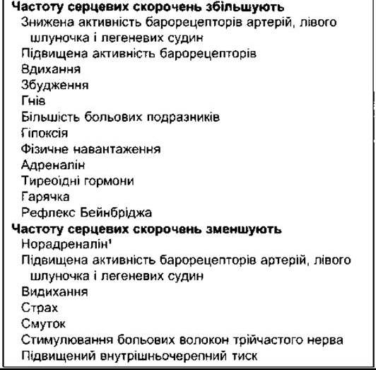
1 Norepinephrine exerts a direct chronotropic effect on the heart. However, in intact animals, its vasopressor action stimulates baroreceptors and triggers a reflex increase in vagal tone, which is sufficient to override the direct effect and cause bradycardia.
An increase in arterial PCO2 stimulates the vasomotor center, whereas the direct peripheral effect of hypercapnia is vasodilation. Thus, the peripheral and central effects counteract each other. Moderate hyperventilation, which significantly reduces blood CO2 tension, causes vasoconstriction in the skin and Brain, leaving arterial pressure virtually unchanged. High concentrations of CO2 lead to pronounced vasodilation in the skin and brain, while vessels in other regions constrict, resulting in a slight increase in arterial pressure.
Sympathetic Vasodilator System
Cholinergic sympathetic vasodilator fibers are part of a regulatory system originating in the cerebral cortex, extending to the hypothalamus and Midbrain, and subsequently passing uninterrupted through the medulla oblongata to the IML of the spinal cord Gray matter. Preganglionic neurons of this system activate postganglionic neurons that supply skeletal muscles and, although anatomically classified as sympathetic, release acetylcholine. Stimulation of this system causes vasodilation in skeletal muscles. However, the resulting increase in blood flow is accompanied by a decrease rather than an increase in O2 consumption, indicating that blood bypasses the capillaries. Stimulation of this system also enhances the release of epinephrine and norepinephrine from The adrenal medulla. The released epinephrine further enhances skeletal muscle vasodilation. In dogs and cats, this system is activated by emotional factors such as fear, anxiety, and rage. Its role in humans has not been thoroughly investigated. It is believed that the sympathetic vasodilator system is involved in emotional fainting. There is direct evidence of cholinergically mediated vasodilation in skeletal muscles during or even at the onset of exercise (see Chapter 33), although pre-exercise vasodilation is known to be relatively mild and inconsistent.
Regulation of Heart Rate
Sympathetic and parasympathetic innervation of the heart, as well as baroreceptor-mediated reflex changes, have been described in detail in previous chapters. However, Table 31-5 summarizes and categorizes the principal factors affecting heart rate. In general, stimuli that increase heart rate concurrently elevate blood pressure.
Factors that decrease heart rate generally lower blood pressure, although there are exceptions, such as hypotension and tachycardia induced by atrial baroreceptor stimulation, or hypertension and bradycardia associated with elevated intracranial pressure (see above).
Last update: 10/08/2026
Editorial and Educational Adaptation: This material has been compiled based on the primary/original source text. The project team performed an editorial review, corrected technical inaccuracies, structured sections, and adapted the content for an educational format.
What was processed:
- elimination of formatting defects (OCR errors, structural breaks, corrupted characters);
- editorial organization of content;
- standardization of terminology in accordance with academic sources;
- verification of factual statements against the original source text.
All mentions of the author, publication year, and origin of the primary text have been preserved in accordance with the source.