Review of Medical Physiology - William F. Ganong 2002
Circulation
Regulation of the Cardiovascular System
Systemic Regulation via Hormones
Many Hormones circulating in the Blood act on The Vascular System. Vasodilator hormones include kinins, VIP, and ANP. Hormones that promote vasoconstriction include vasopressin, noradrenaline, adrenaline, and angiotensin II.
Kinins
The body contains two related Peptides known as kinins. One of them is the nonapeptide bradykinin, and the other is the decapeptide lysylbradykinin, also known as kallidin (Fig. 31-3). Lysylbradykinin can be converted into bradykinin by the action of aminopeptidase. Both peptides are metabolized into inactive fragments by the action of kininase I (a carboxypeptidase that cleaves C-terminal Arg residues). In addition, dipeptidyl carboxypeptidase—kininase II—inactivates bradykinin and lysylbradykinin by cleaving Phe-Arg from the C-terminus. Kininase II is the exact same enzyme as angiotensin II-converting enzyme (see Chapter 24), which cleaves His-Leu from the C-terminus of angiotensin I.
Class="center">Table 31-2. BIOLOGICAL EFFECTS OF endothelins1
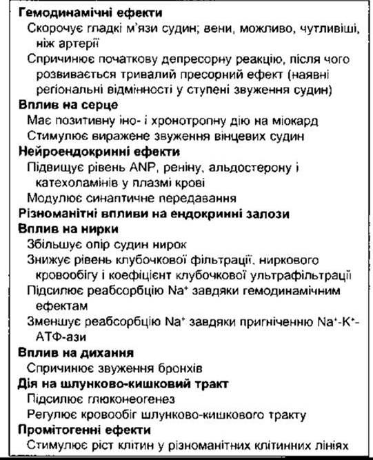
1 Modified from Thomas CP, Simonson MS, Dunn MJ: Endothelin: Receptors and transmembrane signals. News Physiol Sci 1992;7:207.
Bradykinin and lysylbradykinin are formed from two precursor Proteins—high-molecular-weight and low-molecular-weight kininogens (Fig. 31-4). They are generated through Alternative Splicing of a single Gene located on chromosome 3. The biological activity of bradykinin and lysylbradykinin is identical, and it remains unclear why two distinct kinins are produced.
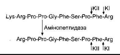
Fig. 31-3. Kinins. Lysylbradykinin (top) is converted to bradykinin (bottom) by aminopeptidase. The peptidases are inactivated by kininase I (KI) or kininase II (KII) at the sites indicated by the short arrows.
The generation of peptides from their precursors occurs via proteases known as kallikreins. In humans, The production of kallikreins is encoded by a family of three genes located on chromosome 19. There are two types of kallikreins: plasma kallikrein, which circulates in an inactive form, and tissue kallikrein, which is localized primarily on the apical membrane of Cells important for electrolyte transport. Tissue kallikrein is found in numerous Tissues, including sweat and Salivary Glands, the Pancreas, the Prostate Gland, the intestine, and the Kidneys. Tissue kallikrein acts on high-molecular-weight and low-molecular-weight kininogens to produce lysylbradykinin. Upon activation, plasma kallikrein acts on high-molecular-weight kininogen to generate bradykinin.
The conversion of inactive plasma kallikrein (prekallikrein) into its active form—kallikrein—is triggered by active factor XII, which initiates the intrinsic pathway of Blood Coagulation. Kallikrein, in turn, activates factor XII via a positive feedback mechanism. High-molecular-weight kininogen also possesses The ability to activate factor XII (see Fig. 27-25).
The action of kinins is similar to that of histamine. They function primarily as tissue hormones, although small amounts circulate in the blood. Kinins induce the contraction of visceral smooth Muscle. They relax vascular smooth muscle by acting indirectly via NO, thereby lowering blood pressure. Kinins also increase capillary permeability, attract leukocytes, and cause pain at the site of subcutaneous injection. They are released during active secretion in the Sweat Glands, salivary glands, and the Exocrine Pancreas (see Chapter 26). Furthermore, kinins ensure increased BLOOD FLOW IN these tissues during active secretion. Kinins are also present in the kidneys, where their exact function remains to be fully elucidated.
Two Types of bradykinin receptors have been identified: B1 and B2. Their Amino acid sequences are 36% identical. Both belong to the family of serpentine G protein-coupled receptors. B1 receptors may be involved in mediating the pain-producing effects of kinins, but their distribution and function are poorly understood. B2 receptors closely resemble H2 receptors and are found in numerous tissues.
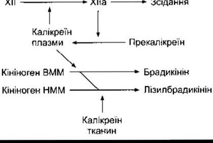
Fig. 31-4. Generation of kinins from high-molecular-weight (HMW) and low-molecular-weight (LMW) kininogens.
Adrenomedullin
Adrenomedullin (AM) is a depressor polypeptide first isolated from pheochromocytoma cells. Its prohormone gives rise to another depressor polypeptide known as proadrenomedullin N-terminal 20 peptide (PAMP). AM also inhibits aldosterone secretion in sodium chloride-depleted animals and exerts a depressor effect through the enhanced production of PAMP acts by suppressing peripheral sympathetic nerve activity.
In addition to The adrenal medulla, AM and PAMP are found in plasma and many other tissues, including the kidneys and the Brain. However, the precise role of AM and PAMP (if any) in cardiovascular regulation remains unproven.
Natriuretic Hormones
Atrial natriuretic peptide (ANP) is synthesized in The Heart (see Chapter 24); it counteracts the effects of vasoconstrictors and lowers blood pressure. Nevertheless, its exact role in circulatory regulation is not yet fully understood. The Na+-K+-ATPase natriuretic inhibitor, now considered to be endogenous ouabain (strophanthidin), does not lower blood pressure; rather, it increases it.
Circulating Vasoconstrictors
Vasopressin is a potent vasoconstrictor, yet when administered to healthy individuals, it does not significantly alter blood pressure levels due to a compensatory decrease in stroke volume. Its role in Blood Pressure Regulation is described in Chapter 14.
Noradrenaline exerts a systemic vasoconstrictor effect, whereas adrenaline dilates the Blood Vessels of Skeletal Muscle and the Liver. Chapter 20, which provides a detailed Description of the effects of catecholamines on The Cardiovascular system, highlights the relatively minor role of circulating noradrenaline compared to noradrenaline released by vasomotor nerves.
The octapeptide angiotensin II exerts a systemic vasoconstrictor effect. It is formed from angiotensin I, which is cleaved from circulating angiotensinogen by the action of renin (see Chapter 24). The production of angiotensin II is stimulated by enhanced renin release triggered by a drop in blood pressure or a reduction in extracellular fluid volume. This mechanism helps maintain blood pressure levels. Angiotensin II also promotes Water retention and stimulates aldosterone secretion. The increased generation of angiotensin II is part of a homeostatic mechanism aimed at maintaining extracellular fluid volume (see Chapter 20). Additionally, renin-angiotensin systems exist in many other Organs, and one may reside within the walls of blood vessels. Angiotensin II generated locally in the vascular wall may play an important role in certain forms of Hypertension.
Urotensin-II is a polypeptide first isolated from the Spinal Cord of fish, which is also present in The Heart and vascular wall. It is the most potent vasoconstrictor known to date in mammals. However, its physiological role remains to be conclusively determined.
Last update: 10/08/2026
Editorial and Educational Adaptation: This material has been compiled based on the primary/original source text. The project team performed an editorial review, corrected technical inaccuracies, structured sections, and adapted the content for an educational format.
What was processed:
- elimination of formatting defects (OCR errors, structural breaks, corrupted characters);
- editorial organization of content;
- standardization of terminology in accordance with academic sources;
- verification of factual statements against the original source text.
All mentions of the author, publication year, and origin of the primary text have been preserved in accordance with the source.