Review of Medical Physiology - William F. Ganong 2002
Circulation
Regulation of the Cardiovascular System
Endothelium-Derived Factors
Endothelial Cells
As noted in Chapter 30, endothelial cells form a system of critical importance. These cells secrete numerous growth factors and vasoactive substances. Vasoactive compounds include Prostaglandins and thromboxanes, nitric oxide, and endothelins.
Prostacyclin and Thromboxane A2
Prostacyclin is produced by endothelial cells, whereas thromboxane A2 is produced by platelets. Both substances are derived from arachidonic acid via the cyclooxygenase pathway (see Fig. 17-33). Thromboxane A2 promotes platelet aggregation and vasoconstriction, whereas prostacyclin inhibits platelet aggregation and causes vasodilation. Through the combined action of thromboxane A2 and prostacyclin, local platelet aggregation and subsequent thrombus formation are regulated (see Chapter 27), which prevents excessive systemic thrombosis while maintaining Blood FLOW IN adjacent vascular beds.
The balance between thromboxane A2 and prostacyclin can be shifted toward prostacyclin by the administration of low-dose aspirin. Aspirin causes irreversible inhibition of cyclooxygenase by acetylating a Serine residue in the Active Site of the enzyme, thereby reducing The production of both thromboxane and prostacyclin. However, within several hours, endothelial cells synthesize new cyclooxygenase. In contrast, platelets are incapable of synthesizing new Enzymes, and platelet cyclooxygenase levels increase only with the appearance of newly formed platelets. This process is slow, as the half-life of platelets is approximately four days. Consequently, long-term low-dose aspirin therapy suppresses thrombosis and is recommended for the Prevention of myocardial infarction, unstable angina pectoris, transient ischemic attacks, and stroke.
Endothelium-Derived Relaxing Factor
Approximately 20 years ago, it was established that the endothelium plays a vital role in vasodilation. In response to various stimuli, endothelial cells can produce endothelium-derived relaxing factor (EDRF), which has been identified as nitric oxide (NO). NO is synthesized from Arginine (Fig. 31-1) in a reaction catalyzed by nitric oxide synthase (NOS). Three isoforms of NOS have been identified: NOS 1 in The Nervous system, NOS 2 in macrophages and other immunocompetent cells, and NOS 3 in endothelial cells. NOS 1 and NOS 3 are activated by factors that increase intracellular Ca2+ concentrations, including the vasodilators acetylcholine and bradykinin. Conversely, NOS in immunocompetent cells is activated by cytokines rather than Ca2+. Once produced by the endothelium, NO diffuses into vascular smooth Muscle cells, where it activates soluble guanylyl cyclase, leading to The formation of cGMP (see Fig. 31-1), which subsequently induces vascular smooth muscle relaxation.
Class="center">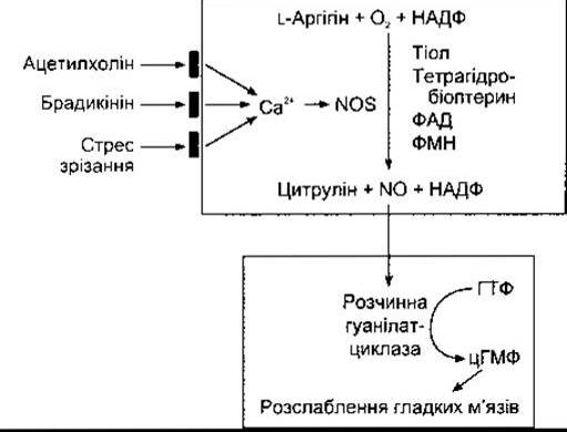
Fig. 31-1. Synthesis of NO from arginine in endothelial cells and its action, mediated by The stimulation of soluble guanylyl cyclase and cGMP formation, leading to the relaxation of vascular smooth muscle cells. Activation of the endothelial form of nitric oxide synthase (NOS) occurs in response to an increase in intracellular Ca2+ concentration, which may be triggered by the action of acetylcholine (ACh), bradykinin, or shear stress on The Cell membrane. Tetrahydrobiopterin, FAD, and FMN serve as Cofactors.
Hemoglobin serves to inactivate NO.
Adenosine, ANP, and histamine act on H2 receptors to induce endothelium-independent relaxation of vascular smooth Muscles. However, the effects of acetylcholine, bradykinin, VIP, substance P, and certain other Polypeptides are endothelium-dependent. Various vasoconstrictor substances that act directly on vascular smooth muscles would induce even greater vasoconstriction if they did not simultaneously stimulate NO release. When tissue blood flow suddenly increases due to arteriolar dilation, the large feeding Arteries supplying the tissue also dilate. This flow-mediated vasodilation is similarly driven by the local production of NO. Substances released during platelet aggregation also trigger NO production. NO-mediated vasodilation helps maintain blood flow in vessels with an intact endothelium. Conversely, endothelial injury leads to platelet aggregation and vasoconstriction (see Chapter 27).
Further evidence for The Physiological Role of NO comes from the finding that administration of arginine derivatives that inhibit nitric oxide synthase to laboratory animals results in a transient increase in blood pressure. This indicates that NO release is essential for maintaining normal blood pressure.
In addition, NO is involved in vascular remodeling and angiogenesis, and plays a role in the Pathogenesis of atherosclerosis. In this context, it is noteworthy that some patients develop a progressive form of graft vasculopathy following Heart transplantation, which is believed to be linked to endothelial injury.
Nitroglycerin and other nitrovasodilators, which are widely used in the Treatment of angina pectoris, stimulate guanylyl cyclase through a mechanism similar to that of NO.
It has also been established that penile erection is mediated by the release of NO, which leads to vasodilation and engorgement of the corpora cavernosa (see Chapter 23).
It has become a recurring pattern in physiology that a substance is initially discovered for its crucial role in The Cardiovascular system, only to be later found synthesized in other systems where it performs entirely different Functions. This was the case, for example, with angiotensin II (see Chapter 24) and endothelins (see below), and the same holds true for NO. Nitric oxide is present in the Brain, where it plays a vital role in neural function via cGMP signaling (see Chapter 4). It is also required for the cytotoxic activity of macrophages, including their ability to induce tumor cell death. In the gastrointestinal tract, NO acts as an important factor in smooth muscle relaxation. Other functions of NO are discussed in various sections of this book.
Endothelins
Endothelial cells also produce endothelin-1, one of the most potent vasoconstricting substances known. Endothelin-1 (ET-1), endothelin-2 (ET-2), and endothelin-3 (ET-3) belong to a family of three homologous polypeptides containing 21 amino acid residues (Fig. 31-2). Each endothelin is encoded by a distinct Gene. The unique Structure of endothelins bears a striking resemblance to sarafotoxins, polypeptides found in snake venom.
Endothelin-1
In endothelial cells, post-translational Processing initially yields a 39-amino-acid prohormone known as big endothelin-1, which possesses approximately 1% of the biological activity of mature endothelin-1. Subsequently, endothelin-converting enzyme cleaves the Trp-Val bond to generate endothelin-1. A family of endothelin-converting enzymes exists, responsible for processing big endothelin-1, big endothelin-2, and big endothelin-3. Although small amounts of big endothelin-1 and endothelin-1 enter the bloodstream, they are primarily released into the tunica media of Blood Vessels, where they act as paracrine agents.
Two distinct types of endothelin receptors have been cloned, both of which are G protein-coupled and signal via phospholipase C (see Chapter 1). ETA receptors are specific for endothelin-1, are distributed across numerous Tissues, and mediate endothelin-1-induced vasoconstriction. ETB receptors bind all three isoforms of endothelins and are coupled to Gi Proteins; they are involved in vasodilation and mediate various endothelin effects (see below).

Fig. 31-2. Structure of human endothelins and a snake venom sarafotoxin. Amino acid residues that differ from those in endothelin-1 are highlighted in color.
Regulation of Secretion
Endothelin-1 is not stored in secretory granules; rather, most regulatory factors directly affect METABOLISM/31.html">Transcription of its gene, resulting in immediate changes in secretion levels. Factors that stimulate and inhibit gene transcription are summarized in Table 31-1.
Effects on the Cardiovascular System
As noted above, endothelin-1 functions primarily as a local paracrine regulator of vascular tone. Both big endothelin-1 and endothelin-1 are present in the Circulation, although their concentrations do not rise in Hypertension. Mice with a heterozygous knockout of the endothelin-1 gene show a greater tendency toward elevated blood pressure than toward hypotension. Circulating levels of endothelin-1 are elevated in congestive Heart Failure and following myocardial infarction, indicating that endothelin-1 plays a significant role in the pathogenesis of these conditions.
Other Effects of Endothelins
In addition to being synthesized by endothelial cells, endothelin-1 is also found in the brain and Kidneys. Endothelin-2 is produced predominantly in nerves and the intestine. Endothelin-3 is present in the blood and occurs in high concentrations in the brain, as well as in the kidneys and gastrointestinal tract. The effects listed in Table 31-2 highlight the physiological roles of endothelins across various tissues. Endothelins are abundant in the brain, where early in life they are produced by both astrocytes and Neurons. They are detected in sensory ROOT ganglia, anterior horn cells, the Cerebral Cortex, the Hypothalamus, and cerebellar Purkinje cells, and they also participate in regulating Transport Across the blood-brain barrier. Endothelin receptors are located on mesangial cells (see Chapter 38), where this polypeptide is thought to decrease the Glomerular Filtration rate.
Table 31-1. Transcriptional Regulation of Endothelin-1 Secretion

Mice homozygous for an endothelin-1 gene knockout exhibit severe craniofacial malformations and die at birth due to respiratory failure. They also present with megacolon (Hirschsprung's disease), presumably because the precursor cells that normally form the myenteric plexus fail to migrate into the distal bowel. Furthermore, endothelins play a crucial role in the closure of the ductus arteriosus at birth.
Last update: 10/08/2026
Editorial and Educational Adaptation: This material has been compiled based on the primary/original source text. The project team performed an editorial review, corrected technical inaccuracies, structured sections, and adapted the content for an educational format.
What was processed:
- elimination of formatting defects (OCR errors, structural breaks, corrupted characters);
- editorial organization of content;
- standardization of terminology in accordance with academic sources;
- verification of factual statements against the original source text.
All mentions of the author, publication year, and origin of the primary text have been preserved in accordance with the source.