BIOLOGY Volume 2 - A Guide to General Biology - 2004
14. TRANSPORT IN ANIMALS
14.7. The Heart
14.7.4. Regulation of Heart Rate
As previously noted, the intrinsic Heart rate is set by the rhythmic activation of the SA node (pacemaker). Even after being removed from the body and placed in an artificial medium, The Heart continues to beat rhythmically, albeit more slowly. However, the body places constantly changing demands on The Cardiovascular system, and the heart rate varies accordingly. This is achieved through two regulatory systems: nervous and endocrine (hormonal, chemical). These are homeostatic responses whose function is to maintain constant internal environmental conditions (Homeostasis) amidst continuously changing external conditions.
The volume of Blood pumped out of the heart over a given period of time (usually measured per minute) is called the Cardiac Output. It depends on the volume of blood ejected during each contraction (the stroke volume) and the heart rate:
Class="center">Cardiac output = Stroke volume × Heart rate
Cardiac output is a critical variable in supplying blood to the body, and altering the heart rate is one of the primary ways it is regulated.
Nervous Regulation of Heart Rate
The Medulla Oblongata (part of the Hindbrain) contains centers that regulate cardiovascular activity, including heart rate. Appropriate nerves run from these centers to the heart (Fig. 14.24). The Nervous system (NS) includes the Autonomic nervous system, which operates automatically without conscious control. The autonomic NS is further divided into the sympathetic nervous system (SNS) and the parasympathetic nervous system (PNS). The SNS generally excites Organs and stimulates their activity, whereas the PNS inhibits their activity. Both systems are involved in regulating heart rate.
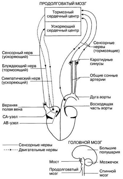
Fig. 14.24. Nervous regulation of heart rate. Neurons connect the heart to the cardioinhibitory and cardioacceleratory centers located in the medulla oblongata.
The medulla oblongata contains two regions that influence heart rate: the cardioinhibitory center, which decreases heart rate, and the cardioacceleratory center, which increases it. Like other structures of the medulla oblongata, these are referred to as medullary centers. A pair of vagus nerves, containing parasympathetic fibers, originates symmetrically from the cardioinhibitory center and runs down both sides of the Trachea to the heart (only one is shown in Fig. 14.24). Within the heart, these nerve fibers lead to the SA node, the AV node, and the bundle of His, and the impulses they carry decrease the heart rate. NERVES OF THE sympathetic nervous system originate from the cardioacceleratory center in the medulla oblongata. Running along the spine, these nerves reach the SA node. The impulses they carry increase the heart rate. It is the coordinated activity of both medullary centers that regulates the heart rate.
Sensory nerve fibers from stretch receptors located in the walls of the aortic arch, carotid sinuses, and SUPERIOR VENA CAVA lead to the cardiovascular centers of the medulla oblongata. Impulses from the aorta and carotid sinuses stimulate the cardioinhibitory center and slow down the heart, whereas signals from the superior vena cava, reaching the cardioacceleratory center, speed it up. When the blood volume increases in any of these vessels, their walls stretch, resulting in an increased rate of impulses sent from them to the cardiovascular centers of the medulla oblongata.
For example, during intense Physical Exercise, Muscles contract strongly, which accelerates the return of blood to the heart via the Veins. The influx of a large volume of blood into the superior vena cava causes its walls to stretch, leading to enhanced heart activity. At the same time, the increased blood flow to the heart raises the internal pressure on its walls. The heart responds to this automatically (without nerve intervention) with a more forceful systolic contraction, meaning it ejects more blood into the Arteries (in other words, the stroke volume increases). This correlation between the volume of blood entering The Heart and the stroke volume is known as Starling's law, named after the English physiologist who discovered this relationship.
The increase in stroke volume leads to the stretching of the aorta and carotid sinuses and the generation of impulses that travel to the cardioinhibitory center, causing the heart rate to slow down. Thus, there is an automatic self-regulatory mechanism that prevents excessively rapid beats and allows the heart's activity to adjust so that it can effectively cope with the incoming blood volume at any given moment.
Hormonal Regulation of Heart Rate
Heart rate is influenced directly or indirectly by A number of Hormones. Below we examine those that have a direct effect.
The most important direct-acting hormone is adrenaline (epinephrine), secreted by The adrenal medulla. Smaller amounts of noradrenaline (norepinephrine), which has a similar effect, are produced there as well. Both hormones stimulate heart activity, although adrenaline is more potent in this regard. Under their influence, the heart rate increases, leading to an elevated cardiac output and higher blood pressure. These hormones also trigger other effects that prepare the body for rapid action in stressful situations (the "fight-or-flight" response), as described in Section 17.1.2.
Thyroxine, synthesized by The Thyroid Gland, increases the basal metabolic rate (Section 17.6.4). This increases the oxygen demand of Tissues and leads to the release of large amounts of heat. As a result, Blood Vessels dilate (vasodilatation) and blood flow increases, which in turn leads to a higher cardiac output. Additionally, thyroxine directly increases the heart rate.
Other Factors Regulating Heart Activity
There are a number of other factors that directly affect the heart Muscle or the sinoatrial node. These factors are listed in Table 14.4.
Table 14.4. Humoral and other factors affecting heart rate
Stimulus |
Effect on heart rhythm |
Increase in pH |
Slows down |
Decrease in pH (e.g., during high |
Speeds up |
CO2 content in the blood, as in physical exercise) |
|
Low Temperature |
Slows down |
High temperature |
Speeds up |
Inorganic ions |
Affect directly or indirectly |
The cardiovascular centers of the medulla oblongata are influenced in one way or another by many factors, including emotions associated with anger or The impact of auditory and visual stimuli, which manifest as blushing or pallor. In such cases, sensory impulses are transmitted to the Brain and, via intracerebral connections, to the medullary cardiovascular centers, which respond accordingly.
Last update: 06/08/2026
Editorial and Educational Adaptation: This material has been compiled based on the primary/original source text. The project team performed an editorial review, corrected technical inaccuracies, structured sections, and adapted the content for an educational format.
What was processed:
- elimination of formatting defects (OCR errors, structural breaks, corrupted characters);
- editorial organization of content;
- standardization of terminology in accordance with academic sources;
- verification of factual statements against the original source text.
All mentions of the author, publication year, and origin of the primary text have been preserved in accordance with the source.