Human Anatomy - Kotsan I. Ya. 2009
Autonomic (vegetative) nervous system
Segmental autonomic centers
Segmental autonomic centers comprise Neurons that function as interneurons within the reflex arc. Unlike suprasegmental centers, segmental autonomic centers are subdivided into distinct sympathetic and parasympathetic components.
Topographically, segmental autonomic centers are classified into cerebral (cranial) and spinal centers.
The cranial centers are further divided into mesencephalic (Midbrain) and bulbar (Brainstem) centers, whereas the spinal centers comprise the thoracolumbar and sacral divisions.
The mesencephalic (midbrain) segmental autonomic center is represented by the parasympathetic Nucleus of the Oculomotor nerve (paired accessory oculomotor nucleus), which innervates two Muscles of the Eyeball: the ciliary Muscle and the sphincter pupillae.
The bulbar segmental autonomic center is located in the Pons and Medulla Oblongata. It is represented by the parasympathetic nuclei of the facial (superior salivatory nucleus), glossopharyngeal (inferior salivatory nucleus), and vagus (dorsal nucleus) nerves, which provide innervation to the lacrimal and Salivary Glands, the Glands of the nasal and oral cavities, as well as the Organs of the neck, Thorax, and Abdominal cavity.
The thoracolumbar sympathetic segmental center is located in the lateral horns of the eighth cervical (VIII), all thoracic, and the first to second (I–II) lumbar segments of the Spinal Cord (represented by the intermediolateral sympathetic nuclei).
The sacral parasympathetic segmental autonomic center is situated in the lateral horns of the second to fourth (II–IV) sacral segments of the spinal cord (represented by parasympathetic nuclei).
Thus, the mesencephalic and bulbar centers (of the Brain) and the sacral center (of the spinal cord) belong to the parasympathetic Autonomic Nervous system, whereas the thoracolumbar center (of the spinal cord) belongs to the sympathetic division.
The Peripheral Division of the Autonomic Nervous System includes: 1) autonomic nerves, branches, and nerve fibers emerging from the BRAIN AND SPINAL cord; 2) Autonomic plexuses; 3) autonomic ganglia; 4) the Sympathetic trunk (right and left) with its ganglia, interganglionic and communicating branches, and sympathetic nerves (Fig. 263).
In the somatic nervous system, nerve fibers are classified According to the direction of impulse conduction into afferent (sensory) and efferent (motor) fibers. In the autonomic nervous system, alongside centrally originating afferent and efferent fibers, There are also local afferent and efferent pathways. Nerve fibers of central origin are the processes of neurons located in the autonomic nuclei of the spinal cord and brain, as well as neurons in sensory spinal ganglia and cranial nerve sensory ganglia. Nerve fibers of local origin are the processes of afferent and efferent neurons situated within autonomic ganglia.
The sources of afferent nerve fibers of the autonomic nervous system are:
1) neurons of spinal ganglia;
2) neurons of sensory ganglia of Cranial Nerves;
3) intrinsic sensory neurons of the autonomic nervous system (Dogiel type II Cells).
Class="center">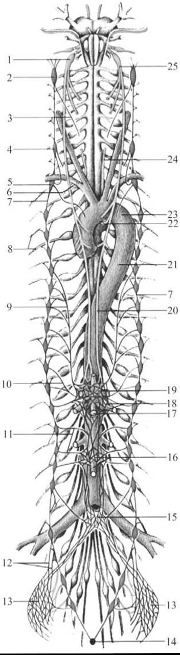
Fig. 263. Diagram of The Structure of the autonomic nervous system
1 — right Vagus nerve; 2 — superior cervical ganglion of the sympathetic trunk; 3 — superior cardiac branch of the sympathetic trunk; 4 — middle cervical ganglion of the sympathetic trunk; 5 — inferior cervical ganglion of the sympathetic trunk; 6 — right recurrent laryngeal nerve; 7 — sympathetic trunk; 8 — intercostal nerve; 9 — greater splanchnic nerve; 10 — lesser splanchnic nerve; 11 — lumbar splanchnic nerves; 12 — sacral splanchnic nerves; 13 — inferior hypogastric ganglion and plexus; 14 — ganglion impar; 15 — superior hypogastric plexus and ganglion; 16 — inferior mesenteric ganglion and artery; 17 — superior mesenteric artery and ganglion; 18 — aortico-renal plexus and renal artery; 19 — celiac trunk with celiac ganglion; 20 — left vagus nerve; 21 — Thoracic Aorta; 22 — left recurrent laryngeal nerve; 23 — aortic arch; 24 — left recurrent laryngeal nerve; 25 — left vagus nerve
Afferent fibers originating from spinal ganglia and sensory ganglia of cranial nerves possess a well-developed myelin sheath. Their Nerve Impulse Conduction velocity ranges from 12 to 120 m/s. Afferent fibers belonging to intrinsic sensory neurons of the autonomic nervous system (processes of Dogiel type II cells) are unmyelinated. Their conduction velocity is significantly lower, ranging from 1 to 7 m/s.
Efferent fibers are subdivided into preganglionic and postganglionic fibers.
Preganglionic fibers are the processes of neurons located in the autonomic nuclei of the spinal cord and the autonomic nuclei of cranial nerves. They are covered with a myelin sheath, which gives them a whitish appearance. The conduction velocity of excitation in sympathetic preganglionic fibers is 1.5–4 m/s, and in parasympathetic fibers, it is 10–20 m/s. Preganglionic fibers terminate in synapses on neurons within autonomic ganglia. The length of preganglionic fibers varies depending on the distance to the autonomic ganglia where these fibers terminate. Parasympathetic preganglionic fibers are typically long because they terminate on neurons within organ plexuses. The length of sympathetic preganglionic fibers depends on which autonomic ganglia (first- or second-order) they terminate upon.
Postganglionic fibers are the processes (axons) of efferent neurons located in autonomic ganglia. They have a smaller diameter and lack a myelin sheath. Their nerve impulse conduction velocity is slow (~1 m/s). Postganglionic fibers represent the final link of the autonomic reflex arc, transmitting nerve impulses from ganglia to smooth muscle, glands, and Tissues. The length of postganglionic fibers depends on the Location OF THE Cell bodies of the neurons from which they originate.
There is an inverse relationship between the lengths of pre- and postganglionic fibers within the same reflex arc: the longer the preganglionic fiber, the shorter the postganglionic fiber, and conversely, the shorter the preganglionic fiber, the longer the postganglionic fiber.
Autonomic ganglia are complex peripheral structures. Based on their topography (distance from the Central Nervous System), they are classified into first-, second-, and third-order ganglia. First-order ganglia (paravertebral) lie alongside THE Vertebral Column and collectively form the sympathetic trunks. Second-order ganglia (prevertebral) are located anterior to the vertebral column within prevertebral or vascular plexuses. Third-order ganglia (terminal or organ-associated) are situated either within the walls of organs (intraorgan) or adjacent to them (extraorgan). First- and second-order ganglia are generally classified as part of the Sympathetic division of the autonomic nervous system, whereas third-order ganglia predominantly contain parasympathetic neurons, though a certain number of parasympathetic neurons are also found in sympathetic ganglia.
An autonomic ganglion is a distinct organ characterized by a specific location, shape, size, Blood supply, and innervation.
The size of autonomic ganglia is primarily determined by the number of Nerve Cells they contain, ranging from just a few (3–5) to many thousands per ganglion. The cells comprising a ganglion are subdivided into motor (Dogiel type I cells), sensory (Dogiel type II cells), and associative neurons.
Each autonomic ganglion has a Connective Tissue sheath, or capsule, whose extensions penetrate into the ganglion, dividing it into lobules or sectors. Furthermore, each neuron within the ganglion possesses its own thin capsule, the outer layer of which is formed by connective tissue and the inner layer by flat endothelial cells.
The Blood supply to autonomic ganglia is provided by branches from adjacent Arteries. Blood and Lymphatic vessels spread through the connective tissue septa, which together constitute the stroma of the ganglion. Neurons are nourished through the direct contact of their cell membranes with blood capillaries mediated by glial satellite cells.
Autonomic ganglia have a complex innervation. Sensory endings, formed by afferent neurons from spinal ganglia and intrinsic afferent autonomic neurons, are found on the neurons, connective tissue, and Blood Vessels of autonomic ganglia. This demonstrates that the state and activity of autonomic ganglia are subject to continuous neural control, including input from the central nervous system.
Autonomic ganglia perform the following Functions: 1) reflex functions, acting as peculiar peripheral centers where the arcs of true peripheral Reflexes are closed; 2) integrative-coordinative function, integrating two streams of impulses—from the center and the periphery; 3) receptor function, through which the central nervous system automatically regulates the Functions of the autonomic ganglia themselves.
All this provides grounds to believe that autonomic ganglia possess an integrative-coordinative function. As coordination centers, they are tuned to the automatic regulation of internal organ activity.
Autonomic (vegetative, visceral) plexuses (plexus autonomici (viscerales)) are located in the Body Cavities, predominantly anterior to the aorta and its branches, surrounding Internal Organs (extraorgan plexuses) and within the walls of internal organs (intraorgan plexuses).
These plexuses consist of autonomic plexus ganglia and internodal nerve fibers. Within the plexus ganglia, preganglionic autonomic fibers synapse onto postganglionic fibers. Sympathetic postganglionic fibers, afferent (sensory) fibers, and some parasympathetic preganglionic fibers do not synapse within the plexuses and pass through them in transit. The number of neurons in the body cavity autonomic plexuses exceeds the number of neurons in the spinal cord, which has led some scientists to group them under the term "third brain," serving as the phylogenetic prototype of the diffuse nervous system of Coelenterates. Physiological studies of the "third brain" demonstrate that it is capable of storing information, influencing human emotions, and so forth.
Autonomic plexuses are divided into 2 groups: Primary and secondary.
Primary, or vascular, plexuses are sympathetic in function. Their structure invariably includes second-order ganglia (prevertebral ganglia).
Secondary, or organ, plexuses are mixed in function (sympathetic and parasympathetic). Sympathetic organ plexuses are derivatives of primary (vascular) plexuses and are formed by postganglionic fibers. Parasympathetic organ plexuses are formed by pre- and postganglionic fibers and contain third-order (organ) ganglia. Secondary (organ) plexuses are, in turn, subdivided into submucosal, subserosal, and intermuscular plexuses.
The Separation of the Autonomic (vegetative) nervous system from the unified nervous system is due to A number of features that distinguish it from the somatic (animal) nervous system.
1. The functions of the autonomic nervous system (with some exceptions) are not under conscious control.
2. Somatic nerves emerge from the brainstem and spinal cord segmentally along their entire length, and this segmentality is partially preserved at the periphery as well. In contrast, autonomic nerves emerge from several distinct centers (foci) of the central nervous system.
3. The STRUCTURE OF THE autonomic reflex arc differs from the reflex arc of the somatic division of The Nervous System. In the reflex arc of the autonomic division, the efferent link consists not of a single neuron, but of two. Overall, a simple autonomic reflex arc is represented by three neurons. The first link of the reflex arc is a sensory neuron, whose cell body is located in the spinal ganglia or sensory ganglia of the cranial nerves. The peripheral process of such a neuron, bearing a sensory ending—a receptor—originates in organs and tissues. Its central process runs within the posterior roots of the spinal cord or the sensory roots of the cranial nerves to the corresponding nuclei in the spinal cord or brain. The second link of the reflex arc is efferent, as it carries impulses from the spinal cord or brain to the effector organ. This efferent pathway of the autonomic reflex arc is represented by two neurons. The first of these neurons, which is the second neuron in sequence within a simple autonomic reflex arc, is located in the autonomic nuclei of the central nervous system. It can be called an interneuron, as it lies between the sensory (afferent) link of the reflex arc and the second (effector) neuron of the efferent pathway. The effector neuron represents the third neuron of the autonomic reflex arc. The Cell bodies of the effector (third) neurons lie in the peripheral ganglia of the autonomic nervous system (sympathetic trunk, autonomic ganglia of cranial nerves, ganglia of autonomic plexuses). The processes of these neurons run to organs and tissues as part of organ-specific autonomic or mixed nerves. Postganglionic nerve fibers terminate on smooth muscles, glands, and other tissues via corresponding terminal apparatuses.
4. The presence of ganglia in the efferent part of the reflex arc is also a characteristic feature of the autonomic nervous system that distinguishes it from the animal nervous system.
5. There are also certain differences in the nerves. The Afferent Pathways of the autonomic nervous system do not appear as microscopically visible nerves; their fibers run within the composition of other nerves (greater and lesser splanchnic nerves, vagus nerve, etc.). Moreover, the sympathetic division is characterized by the fact that the sensory innervation associated with it can extend over a considerable distance and, accordingly, the sympathetic division can be considered a system of collateral innervation. For example, afferent spinal nerve fibers that participate in The formation of the celiac plexus, which innervates the abdominal organs, originate from multiple spinal ganglia (CV-LIII). This circumstance determines the multiplicity and multisegmental Nature of the pathways and sources of afferent innervation of abdominal organs. This also explains why Pain Sensation from internal organs can be transmitted via both autonomic and somatic nerves.
As for the Efferent Pathways of the autonomic nervous system, they form distinct nerves and ganglia. Therefore, one can speak of two centrifugal PATHWAYS OF THE unified nervous system: one pathway consists of animal, somatic, motor nerves, and the second consists of autonomic nerves.
6. Autonomic nerves form plexuses around blood vessels, alongside which they approach and enter organs. The presence of perivascular plexuses is also a characteristic feature of the autonomic nervous system that distinguishes it from the somatic system.
7. There is also a difference in the diameter of nerve fibers: somatic, especially efferent nerve fibers, have a larger diameter and are covered with a thick myelin sheath.
Autonomic efferent (postganglionic) nerve fibers are almost unmyelinated (gray), and since the conduction velocity of excitation along nerve fibers depends on their diameter and the presence of a myelin sheath, the conduction velocity of a nerve impulse along somatic motor fibers is much higher (100—120 m/s), whereas along autonomic fibers it is much lower (2—10 m/s).
8. There is also a major difference in the innervation territories of the autonomic and somatic nervous systems. The autonomic nervous system is distributed throughout the entire Organism; it has a wide region of efferent innervation that encompasses all organs and Tissues of the body, not excluding skeletal musculature (which it tonifies). In contrast to the autonomic system, the somatic nervous system innervates only skeletal musculature via centrifugal fibers, meaning it has a limited innervation area.
Based on the topography of autonomic nuclei and ganglia, differences in the length of the First and Second neurons of the efferent pathway, as well as functional specificities, the autonomic nervous system is divided into two parts: the sympathetic and the parasympathetic.
Last update: 08/08/2026
Editorial and Educational Adaptation: This material has been compiled based on the primary/original source text. The project team performed an editorial review, corrected technical inaccuracies, structured sections, and adapted the content for an educational format.
What was processed:
- elimination of formatting defects (OCR errors, structural breaks, corrupted characters);
- editorial organization of content;
- standardization of terminology in accordance with academic sources;
- verification of factual statements against the original source text.
All mentions of the author, publication year, and origin of the primary text have been preserved in accordance with the source.