Human Anatomy - Kotsan I. Y. 2009
Autonomic Nervous System
Sympathetic part of the nervous system
The term "sympathetic Nervous system" was introduced in 1732 by Winslow, who believed that these specific structures were responsible for human sympathy, empathy, and physiological harmony. Modern physiologists have proven that pleasant sensations are primarily associated with the parasympathetic division, whereas unpleasant ones are mediated by the sympathetic division.
The Sympathetic part of the Autonomic nervous system comprises central and peripheral divisions (Fig. 264).
In addition to common suprasegmental centers, the central division of the sympathetic nervous system features a segmental thoracolumbar sympathetic center, represented by a group of ganglionic Cells within the Gray matter of the Spinal Cord. These cells form the so-called lateral intermediate (sympathetic) nuclei—right and left—located in the lateral horns of the spinal cord extending from the eighth cervical to the second or third lumbar segments (CVIII-ThI-LIII).
The peripheral division of the sympathetic nervous system consists of the right and left sympathetic trunks with their constituent ganglia, the nerves branching from these trunks, as well as the nerves and ganglia forming Autonomic plexuses situated along the Blood Vessels either outside or inside the Organs.
The Sympathetic trunk (truncus sympatheticus) is a paired Structure composed of a chain of 17–22 ganglia (ganglia sympathica) connected by interganglionic branches.
The interganglionic branches (rami interganglionares) are small nerve trunks that connect adjacent ganglia of the sympathetic trunk. They contain both myelinated preganglionic fibers and unmyelinated postganglionic fibers.
Preganglionic sympathetic fibers typically enter the sympathetic trunk segmentally; originating from the cellular elements of the central Divisions of the sympathetic nervous system, they form white communicating branches (rami communicantes albi). In most cases, the sympathetic fibers of these branches either synapse within the ganglia of the sympathetic trunk or continue onward within sympathetic nerves to terminate in one of the prevertebral ganglia.
Class="center">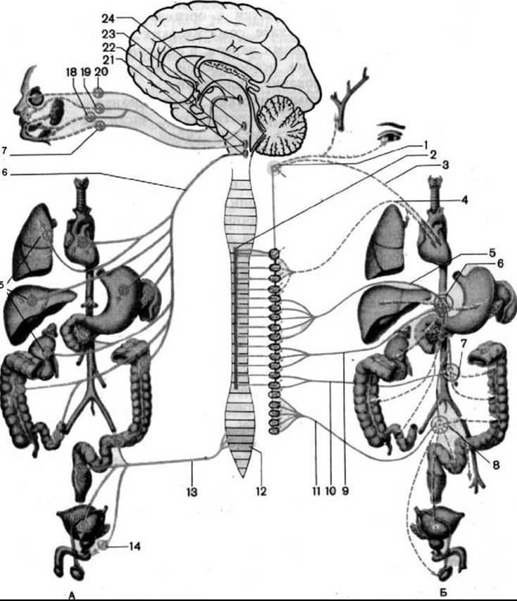
Fig. 264. Diagram of the autonomic innervation of Internal Organs (according to M. R. Sapin; arrows indicate the pathways of nerve impulses to organs, showing the parasympathetic (A) and sympathetic (B) divisions)
1 — superior cervical ganglion of the sympathetic trunk; 2 — lateral horn of the spinal cord; 3 — superior cervical cardiac nerve; 4 — thoracic cardiac and pulmonary nerves; 5 — greater splanchnic nerve; 6 — celiac plexus; 7 — inferior mesenteric plexus; 8 — superior and inferior hypogastric plexuses; 9 — lesser splanchnic nerve; 10 — lumbar splanchnic nerves; 11 — sacral splanchnic nerves; 12 — sacral (parasympathetic) nuclei; 13 — pelvic splanchnic nerves; 14 — pelvic (parasympathetic) ganglia; 15 — parasympathetic ganglia (within organ plexuses); 16 — Vagus nerve; 17 — otic (parasympathetic) ganglion; 18 — submandibular (parasympathetic) ganglion; 19 — pterygopalatine (parasympathetic) ganglion; 20 — ciliary (parasympathetic) ganglion; 21 — dorsal Nucleus of the vagus nerve; 22 — inferior salivatory nucleus; 23 — superior salivatory nucleus; 24 — accessory oculomotor nucleus
Postganglionic sympathetic fibers emerge from each ganglion of the sympathetic trunk. Some of these fibers enter the Spinal Nerves as gray communicating branches (rami communicantes grisei), while others participate in The formation of sympathetic nerves and autonomic plexuses.
Topographically, the sympathetic trunk is divided into four regions: cervical, thoracic, lumbar, and sacral.
The cervical portion of the sympathetic trunk extends from the Base of the Skull to the neck of the first rib. It is located posterior to the carotid Arteries on the Deep Muscles of the Neck and comprises three cervical sympathetic ganglia: superior, middle, and inferior.
1. The superior cervical ganglion (ganglion cervicale superius) is the largest ganglion of the sympathetic trunk. It is spindle-shaped, measuring up to 2 cm or more in length and 0.5 cm in thickness. It lies at the level of the second and part of the third cervical vertebrae, posterior to the Internal Carotid Artery and medial to the vagus nerve. Interganglionic connecting fibers approach the ganglion, while postganglionic fibers emerge from it to form the following nerves and branches.
The superior cervical cardiac nerve (n. cardiacus cervicalis superior) descends toward The Heart parallel to the sympathetic trunk, anterior to the prevertebral layer of the Cervical fascia. In the thoracic cavity, the right superior cervical cardiac nerve passes anterior to the brachiocephalic trunk, whereas the left nerve of the same name runs anterior to the common carotid artery; both nerves then course along the anterior surface of the aortic arch to enter the cardiac plexus.
The internal carotid nerve (n. caroticus internus) extends from the upper pole of the superior cervical ganglion to the corresponding artery, forming the internal carotid plexus (plexus caroticus internus) along its course within the carotid canal. Upon exiting the carotid canal, the deep petrosal nerve (n. petrosus profundus) branches off from the internal carotid plexus, passing through the foramen lacerum to the pterygoid canal where, upon joining the greater petrosal nerve (a branch of the Facial Nerve), it forms the nerve of the pterygoid canal (Vidian nerve). Through the nerve of the pterygoid canal, sympathetic fibers pass via the pterygoid canal to reach the mucous membranes, blood vessels, glands, and Tissues of the Oral Cavity, Nasal cavity, and Orbit. The portion of the internal carotid plexus surrounding the cavernous part of the internal carotid artery is frequently designated as the cavernous plexus (plexus cavernosus). Sympathetic fibers extend from the cavernous plexus alongside all Branches of the internal carotid artery, forming periarterial plexuses that reach the cerebral and orbital blood vessels. Within the Eyeball, sympathetic fibers innervate the dilator pupillae Muscle, the vascular tunic (uvea), and the ocular blood vessels.
The external carotid nerves (nn. carotici externi), numbering 2 to 3, run toward the External Carotid Artery and, enveloping it, form the external carotid plexus (plexus caroticus externus). The external carotid plexus extends along the branches of the corresponding artery, providing sympathetic innervation to the blood vessels, glands, smooth muscle elements, and tissues of the HEAD organs. The internal and external carotid plexuses interconnect on the common carotid artery to form the common carotid plexus (plexus caroticus communis). Branches of the glossopharyngeal and vagus nerves join the common carotid plexus, jointly participating in the Formation of the carotid body (glomus caroticus).
The jugular nerve (n. jugularis) ascends along the wall of the Internal jugular vein to the jugular foramen, where it divides into branches directed to the superior and inferior ganglia of the vagus nerve, the inferior ganglion of the Glossopharyngeal nerve, and the Hypoglossal nerve. Through these connections, sympathetic fibers are distributed within the branches of the 9th, 10th, and 12th pairs of Cranial Nerves.
The gray communicating branches (rami communicantes grisei) connect the superior cervical ganglion with the first three (and occasionally four) cervical spinal nerves, the inferior ganglion of the vagus nerve, as well as the hypoglossal and phrenic nerves.
The laryngopharyngeal branches (rami laryngopharyngeales) participate in the formation of the laryngopharyngeal plexus and innervate the blood vessels, muscles, and mucous membrane of the Pharynx and Larynx.
II. The middle cervical ganglion (ganglion cervicale medium) is inconstant, located at the level of the transverse process of the sixth cervical vertebra, adjacent to the inferior thyroid artery. It has an oval or triangular shape and small dimensions. Interganglionic connecting fibers also reach this ganglion, and the following branches and nerves arise from it.
Gray communicating branches (rami communicantes grisei) pass to the 4th through 6th (and occasionally 7th) cervical spinal nerves, the common carotid plexus, the plexus of the inferior thyroid artery, and to the thyroid and Parathyroid glands.
The middle cervical cardiac nerve (n. cardiacus cervicalis medius) descends lateral to the superior cervical cardiac nerve and reaches the cardiac plexus. Along its path, the middle cardiac nerve gives off small branches to the common carotid artery and the inferior thyroid artery, contributing to the formation of their respective plexuses.
III. The inferior cervical ganglion (ganglion cervicale inferius) is quite large and is situated posterior to the Water/144.html">Origin of the vertebral artery. It frequently fuses with the first, and occasionally the second, thoracic ganglion to form a combined cervicothoracic (stellate) ganglion (ganglion cervicothoracicum (stellatum)). The stellate ganglion has an irregular shape with transverse dimensions of up to 8 mm. A white communicating branch and interganglionic fibers approach the ganglion, and a distinct group of nerves and branches originates from it.
The inferior cervical cardiac nerve (n. cardiacus cervicalis inferior) descends posterior to the brachiocephalic trunk (right nerve) and posterior to the aorta (left nerve) toward the cardiac plexus. Along its course, the inferior cervical cardiac nerve gives off communicating branches to the middle cardiac nerve.
The vertebral nerve (n. vertebralis) approaches the posterior wall of the vertebral artery and participates in the formation of the sympathetic vertebral plexus (plexus vertebralis).
The subclavian loop (ansa subclavia) loops anterior to the Subclavian Artery. The sympathetic fibers of the loop form the subclavian plexus (plexus subclavius) on the wall of the subclavian artery, and then continue onto the branches of the subclavian artery, reaching the organs and tissues of the neck, thoracic cavity, and upper limb.
The gray communicating branches (rr. communicantes grisei) connect to the VI, VII, and VIII cervical spinal nerves. These branches, like those from the middle cervical sympathetic ganglion, represent postganglionic fibers that branch out within the peripheral NERVES OF THE Brachial Plexus in the upper extremity, innervating vascular walls, Skin glands, and arrector pili muscles.
The thoracic part of the sympathetic trunk consists of 11–12 thoracic ganglia. They have a somewhat flattened spindle-like or triangular shape, with an average size of 3–5 mm. The ganglia are located anterior to the heads of the Ribs on the lateral surface of the vertebral bodies, posterior to the endothoracic fascia and parietal Pleura. White communicating branches containing preganglionic fibers approach the thoracic ganglia of the sympathetic trunk from all thoracic spinal nerves. The ganglia are interconnected by interganglionic fibers. In turn, several types of branches and nerves depart from the thoracic ganglia of the sympathetic trunk.
The gray communicating branches (rr. communicantes grisei) contain postganglionic fibers and join adjacent spinal nerves.
The thoracic cardiac branches (rr. cardiaci thoracici) originate from the II–IV thoracic ganglia, run anteriorly and medially, and take part in the formation of the cardiac plexus.
The thoracic pulmonary branches (rr. pulmonales thoracici) are formed by fibers from the II–IV thoracic ganglia and participate in the formation of the pulmonary plexus.
The esophageal branches (rr. oesophageales) stem from the II–V thoracic ganglia and contribute to the formation of the esophageal plexus.
The greater splanchnic nerve (n. splanchnicus major) is formed by several branches originating from the V–IX thoracic ganglia of the sympathetic trunk and consists predominantly of preganglionic fibers. On the lateral surface of the thoracic vertebral bodies, these branches unite into a common nerve trunk that runs inferiorly and medially, penetrates the Abdominal cavity between the muscular bundles of the lumbar part of the Diaphragm alongside the azygos vein on the right and the hemiazygos vein on the left, and terminates in the ganglia of the celiac plexus.
The lesser splanchnic nerve (n. splanchnicus minor) arises from the tenth and eleventh thoracic ganglia of the sympathetic trunk, runs inferiorly along with the greater splanchnic nerve, and reaches the celiac plexus. The lesser splanchnic nerve also consists mainly of preganglionic fibers. A significant portion of the fibers of the lesser splanchnic nerve forms the renal branch (r. renalis), which heads toward the renal plexus, bypassing the celiac plexus.
The lowest splanchnic nerve (n. splanchnicus imus) originates from the twelfth (occasionally eleventh) thoracic ganglion of the sympathetic trunk and terminates in the renal plexus. All three splanchnic nerves are part of the plexuses that participate in the Innervation of the abdominal organs, as well as the blood vessels and Lymph Nodes of the thoracic and abdominal cavities.
The lumbar, or abdominal, part of the sympathetic trunk consists of three to five lumbar ganglia. The sympathetic trunks in the lumbar region are located slightly closer to each other than in the thoracic cavity.
The lumbar ganglia are spindle-shaped, and their dimensions do not exceed 6 mm. The ganglia are located on the anterolateral surface of the lumbar vertebral bodies, medial to the psoas major muscle, and are covered by the retroperitoneal fascia. The lumbar ganglia of the right and left sympathetic trunks are connected by transversely oriented communicating branches that lie on the anterior surface of the lumbar vertebrae, posterior to the aorta and the INFERIOR VENA CAVA. White communicating branches (from the upper 2–3 lumbar spinal nerves) approach only the top two lumbar ganglia. The lower lumbar ganglia lack white communicating branches and are instead interconnected by interganglionic fibers.
Two Types of branches emerge from each lumbar ganglion: gray communicating branches (rr. communicantes grisei), which contain postganglionic fibers and run to the lumbar spinal nerves, and lumbar splanchnic nerves (nn. splanchnici lumbales), which head toward the celiac plexus and visceral autonomic plexuses (celiac, aortic, renal, suprarenal) and contain both preganglionic and postganglionic fibers.
The sacral, or pelvic, part of the sympathetic trunk is typically formed by four spindle-shaped ganglia, each measuring about 5 mm. The ganglia of the pelvic part, like those of the lumbar part, are interconnected not only by longitudinal (interganglionic) branches but also by transverse branches—commissures. The sacral ganglia lie on the pelvic surface of the sacrum, medial to the pelvic sacral foramina. Inferiorly, the right and left sympathetic trunks converge and finally merge into the ganglion impar (ganglion impar), which lies on the anterior surface of the first coccygeal vertebra.
Gray communicating branches (rr. communicantes grisei) extend from the sacral ganglia to the sacral and coccygeal spinal nerves; via these branches, postganglionic sympathetic fibers travel to innervate blood vessels, glands, organs, and tissues in the areas where the somatic sacral nerves branch out. Additionally, sacral splanchnic nerves (nn. splanchnici sacrales) run to the superior and inferior hypogastric plexuses.
Last update: 08/08/2026
Editorial and Educational Adaptation: This material has been compiled based on the primary/original source text. The project team performed an editorial review, corrected technical inaccuracies, structured sections, and adapted the content for an educational format.
What was processed:
- elimination of formatting defects (OCR errors, structural breaks, corrupted characters);
- editorial organization of content;
- standardization of terminology in accordance with academic sources;
- verification of factual statements against the original source text.
All mentions of the author, publication year, and origin of the primary text have been preserved in accordance with the source.