HUMAN MEDICAL BIOLOGY, ANATOMY, PHYSIOLOGY, AND PATHOLOGY - Ya.I. Fedonyuk 2010
BIOLOGY
CHAPTER 1. BIOLOGICAL BASES OF HUMAN VITAL ACTIVITY
1.2. THE CELL. TISSUES. CONCEPT OF THE ORGAN, ORGAN SYSTEM, ORGANISM
Fundamentals of General Cytology
Cytology (from Greek cytos - Cell, logos - science) is the science of The Cell. Cells are divided into prokaryotic (lacking a morphologically distinct nucleus) and eukaryotic (possessing a morphologically distinct nucleus). Accordingly, There are two groups of organisms - prokaryotes (pre-nuclear) and eukaryotes (nuclear). Humans belong to eukaryotes. The human Organism is multicellular. The cell is the fundamental Structural and functional unit of The Human Body. Non-cellular structures (symplasts, syncytia) are cell derivatives. Tissues are formed from cells, Organs from tissues, and Organ Systems from organs.
Cells of various human tissues and organs are highly diverse in shape, size, Structure, chemical composition, and metabolic profile (Fig. 1.1). The human body contains 200 cell types, with a total population ranging from 1014 to 1015. All of them possess a nucleus, with the sole exception of erythrocytes, which lose their nucleus during development.
Class="center">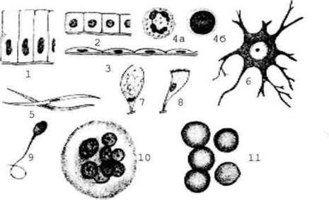
Fig. 1.1. General Structure and various cell shapes: 1 - cylindrical epithelial Cells of the intestine; 2 - cubic cells of the renal tubules; 3 - squamous cells of the peritoneal mesothelium; 4 - round Blood Cells: a - with a segmented nucleus (neutrophilic leukocyte); b - with a rounded nucleus (lymphocyte); 5 - spindle-shaped cell with a rod-like nucleus (smooth Muscle cell); 6 - branched cell (nerve cell); 7 - goblet cell; 8 - ciliated cell; 9 - flagellated cell (spermatozoon); 10 - megakaryocyte (multinucleated cell); 11 - erythrocytes (anucleated cells).
Cell shape is inextricably linked to its function. For instance, Nerve Cells, which conduct impulses, possess branching processes.
Despite their considerable diversity, all Eukaryotic cells consist of three main parts: The cell membrane (Plasmalemma), Cytoplasm, and nucleus (Fig. 1.2). The cytoplasm contains hyaloplasm (cytoplasmic matrix); Organelles, which are permanent structures with characteristic features and specific cellular Functions; and inclusions, which are temporary structures resulting from cellular activity. The cytoplasm is separated from the surrounding environment and adjacent cells by the plasmalemma, the outer cell membrane.
The cytoplasm includes the hyaloplasm, mandatory cellular elements known as organelles, as well as various non-permanent structures called inclusions.
The hyaloplasm is the internal environment of the cell where intermediary metabolic reactions take place. It is the most fluid part of the cytoplasm, housing organelles and inclusions. It comprises the Cytosol (Water with dissolved inorganic and organic substances) and the cytomatrix (a network of protein microfilaments).
Organelles are divided into general-purpose and special-purpose organelles. General-purpose organelles include Mitochondria, Ribosomes, The Endoplasmic reticulum, the Golgi apparatus, Lysosomes, Peroxisomes, and the centrosome. These organelles are present in all cells. Special-purpose organelles are found only in specific cells, ensuring their specialized functions. These include flagella, cilia, neurofibrils, and myofibrils.

Fig. 1.2. General layout of Cell Structure.
A ribosome is a submicroscopic, non-membranous, general-purpose organelle—a ribonucleoprotein granule consisting of two subunits, large and small. The function of ribosomes is Protein Synthesis. Typically, Proteins are synthesized not by a single ribosome, but simultaneously by a group of ribosomes (a polysome) bound together by a Messenger RNA (mRNA) molecule. Ribosomes and Polysomes may lie free in the cytoplasm, producing proteins for the cell's own needs, or be attached to the membranes of the endoplasmic reticulum to synthesize secretory proteins exported from the cell.
The endoplasmic reticulum is a submicroscopic, membranous, general-purpose organelle forming a system of tubules and flattened cisternae. The endoplasmic reticulum with ribosomes attached to the outer surface of its membranes is termed the granular (rough) endoplasmic reticulum, whereas the ribosome-free version is termed the agranular (smooth) endoplasmic reticulum.
One of the primary Functions of the endoplasmic reticulum is the Transport of substances. In addition, the granular endoplasmic reticulum is characterized by protein synthesis, while the agranular reticulum is involved in Glycogen Synthesis and Breakdown, and Lipid METABOLISM (particularly steroid hormone synthesis). In the Liver, it participates in the detoxification of hypnotics, carcinogens, and other substances.
Mitochondria are microscopic, membranous, general-purpose organelles that appear in light Microscopy as short rods and filaments. The primary function of mitochondria is to supply the cell with energy. Mitochondria also participate in the synthesis of A number of proteins and contain the necessary components for this process: Mitochondrial DNA, small ribosomes, transfer and messenger RNA, and Enzymes.
The Golgi apparatus is a microscopic, membranous, general-purpose organelle that, in silver nitrate-stained preparations, appears as a network of interwoven dark threads. Submicroscopically, the Golgi apparatus represents a collection of interconnected cisternae, flattened in the central region and expanded at the periphery. Small vesicles and vacuoles are located around the cisternae within the organelle. The functions of the Golgi apparatus include the packaging, Condensation, and export of protein secretions, participation in carbohydrate synthesis and their attachment to polypeptide chains during glycoprotein synthesis, and The formation of lysosomes.
Lysosomes are submicroscopic, membranous, general-purpose organelles essential for the intracellular enzymatic degradation of both exogenous substances (internalized via endocytosis) and endogenous Materials—eliminating organelles and inclusions during normal turnover or in response to pathological changes.
Peroxisomes are submicroscopic, membranous, general-purpose organelles resembling lysosomes, but lacking the hydrolytic enzymes characteristic of the latter. They contain amino acid oxidases and catalase, which destroys peroxides. Peroxisomal catalase can play a protective role by breaking down hydrogen peroxide, which is toxic to cells.
The centrosome (cell center) is a microscopic, non-membranous, general-purpose organelle located within the cell in pairs (diplosome). These are cylinders positioned at right angles to each other, whose walls consist of nine triplets of microtubules.
During cell mitosis, the centrosome participates in the Formation of the mitotic spindle, as well as the microtubules of the cell's motile apparatus—Cilia and flagella. The latter are cytoplasmic extensions containing a microtubule system in their core, consisting of two central filaments and nine peripheral doublets. The base of a cilium or flagellum is anchored by a basal body, which is a modified centriole.
The function of microtubules is associated with maintaining and altering cell shape. Microtubules are composed of the protein tubulin.
Inclusions are non-essential cell components that appear and disappear depending on the cell's functional state. They can be composed of chemical substances of diverse origin: Lipids, CARBOHYDRATES, proteins, and Vitamins. Inclusions are classified into trophic, secretory, excretory, and pigment inclusions. Trophic inclusions are subdivided, based on The Nature of the accumulated substances, into lipid, carbohydrate, and protein inclusions.
The Nucleus is an essential cell component containing genetic material—the Chromosomes (Fig. 1.3).
Most often, the nucleus has a spherical shape, but it may conform to the shape of the cell. The size of the nucleus depends on the cell type and its functional state.
The nucleus of a non-dividing cell (interphase nucleus) is surrounded by a nuclear envelope (karyotheca), which consists of outer and inner nuclear membranes and a perinuclear space located between them. The nucleus contains the nucleolus, Chromatin, and karyoplasm. The nuclear envelope contains pores that facilitate metabolic processes between the karyoplasm and cytoplasm. The outer membrane of the nuclear envelope features a small number of ribosomes on its surface and is connected to the tubules of the granular endoplasmic reticulum. The nuclear envelope performs a barrier function, which consists in separating the nuclear contents from the cytoplasm.
Karyoplasm is the fluid component of the nucleus that forms the microenvironment for nuclear structures. It is the analogue of hyaloplasm in the cytoplasmic part of the cell.
Chromatin is the main structural component of the interphase nucleus, serving as the analogue of chromosomes in a fixed and stained cell. Due to its chemical composition (DNA, histone proteins, RNA), chromatin readily absorbs basic stains, producing a chromatin pattern specific to each cell type. Two Types of chromatin are distinguished: euchromatin and heterochromatin. Euchromatin consists of decondensed, poorly staining chromosome regions (functionally active). Heterochromatin consists of condensed, well-staining chromosome regions (functionally inactive).
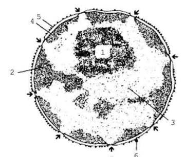
Fig. 1.3. Cell Nucleus - general view:
1 - nucleolus; 2 - heterochromatin; 3 - euchromatin; 4 - inner membrane; 5 - outer membrane; 6 - perinuclear space; 7 - nuclear pore.
The nucleolus is the densest, round, well-stained STRUCTURE OF THE nucleus, which originates from the chromosomes. The nucleolus ensures the formation of Ribosomal RNAs and ribosomes.
Chromosomes are dense rod- or thread-like structures that stain well and are revealed in the cell nucleus during mitotic division. Each species of PLANT AND ANIMAL organisms has a specific number, size, and structure of chromosomes—its karyotype. The Human Karyotype is characterized by the presence of 23 pairs of chromosomes, which include twenty-two pairs of autosomes and one pair of sex chromosomes. The latter comprise X and Y chromosomes. The number of chromosome sets in a cell is denoted by the term ploidy and the letter n. Somatic cells have a diploid set of chromosomes (2n), while Germ Cells have a haploid set (n).
The capacity for self-reproduction is a defining feature of living systems. Cell reproduction in a multicellular organism occurs through the division of a parent cell. The entire period of a cell's existence from division to division or from division to death is called the Cell Cycle. Cells of various organs and tissues have different division capacities and, consequently, different cell cycles. Cell Division is preceded by the duplication of its chromosome set, which occurs during a precisely defined period of interphase. Only after this process does cell division begin. There are two types of cell division: indirect (mitosis) and direct (amitosis). Meiosis is a specialized variant of mitosis.
Mitosis is the universal mode of cell reproduction. It comprises four phases: prophase, metaphase, anaphase, and telophase. During these phases, As a result of euchromatin condensation, the duplicated chromosomes become visible in the nucleus, and the mitotic spindle forms, which participates in transporting chromosomes to the opposite poles of the cell, ultimately leading to the division of the cell in half (cytotomy, cytokinesis). Mitosis is characteristic of somatic cell division, which possess a double (diploid) set of chromosomes (2n).
Amitosis is a direct cell division in which the nucleus remains in the interphase state. In this process, chromosome condensation does not occur, and a mitotic spindle is not formed. Amitosis results in nuclear division and the appearance of bi- or multinucleated cells. Division of the Cell Cytoplasm occurs less frequently.
Meiosis is a specialized form of cell reproduction characteristic of the formation of Gametes (germ cells). During meiosis, two consecutive Divisions of the genetic material take place, resulting in a haploid set of chromosomes remaining in the Nucleus of the germ cell.
A more detailed characterization of eukaryotic cells is provided below in sections 1.3.2, 1.3.3, and 1.3.4.
Last update: 08/08/2026
Editorial and Educational Adaptation: This material has been compiled based on the primary/original source text. The project team performed an editorial review, corrected technical inaccuracies, structured sections, and adapted the content for an educational format.
What was processed:
- elimination of formatting defects (OCR errors, structural breaks, corrupted characters);
- editorial organization of content;
- standardization of terminology in accordance with academic sources;
- verification of factual statements against the original source text.
All mentions of the author, publication year, and origin of the primary text have been preserved in accordance with the source.