FUNDAMENTALS OF MEDICAL BIOLOGY - 2012
The Population-Statistical Method. Medical Genetic Counseling
The population-statistical (population-genetic) method allows for the investigation of the genetic composition of human populations, the frequency of normal and pathological genes and genotypes across various populations (heterozygotes, dominant and recessive homozygotes), and the frequency of normal and pathological phenotypes. The frequency of a specific genotype (allele) is the relative number of individuals in a population possessing that genotype (allele). Frequency is expressed as a percentage or a fraction of one. For example, the frequency of blue-eyed individuals in a population is 49%, or 0.49. This method is based on observing hereditary traits in large population groups. Researchers either conduct direct selective studies on a portion of the population, examine archives of hospitals and other medical institutions, or survey the population using questionnaires. The choice of method depends on the research objectives. The final stage involves the mathematical Analysis of the obtained data.
One of the simplest and most universal Methods for analyzing genotype and Gene frequencies in a population was proposed by G. Hardy and W. Weinberg. In 1908, mathematician G. Hardy in England and physician-anthropologist W. Weinberg in Germany formulated the law maintaining genetic
equilibrium in an ideal population. They suggested using the binomial expansion formula to represent the distribution of genotypes in a panmictic population: (a + b) = a2 + 2ab + b2.
The frequency of genotypes and phenotypes is calculated using the Hardy-Weinberg formula:
Class="center">p2 + 2pq + q = (p + q) = 1 (or 100 %),
where p is the frequency of the dominant allele (A); q is the frequency of the recessive allele (a); p is the frequency of homozygotes for the dominant allele (AA); q is the frequency of homozygotes for the recessive allele (aa); 2pq is the frequency of heterozygotes (Aa).
Using this equation, one can determine the genetic Structure of a population without conducting special studies. To do this, one only needs to know the frequency of recessive homozygotes (aa) - q . This is the only genotype that can be determined directly from the phenotype.
Example. In a population, the frequency of Rh-negative individuals (aa) is 15%. Determine the frequency of Rh-positive homozygotes (AA) and Rh-positive heterozygotes (Aa) in the population.
Solution. The frequency of the aa genotype (q2) = 15 % = 0.15. Hence, the frequency of allele a (q) = √0.15 = 0.387. The Frequency of Gene A (p) = 1 — q = 1 - 0.387 = 0.613. The frequency of genotype AA (p2) = 0.6132 = 0.376. The frequency of genotype Aa (2pq) = 2 x 0.613 x 0.387 = 0.474. Therefore, Rh-negative individuals in the population account for 15%, and Rh-positive individuals account for 85% (among them, homozygotes make up 37.6%, and heterozygotes make up 47.4%).
A number of other specialized mathematical methods have been developed to analyze the genetic structure of populations and the processes occurring at this level of life. The studied populations differ in biological traits, geographical living conditions, and economic status. The Study of gene distribution across specific territories indicates that they can be categorized as follows:
1) universally distributed genes that occur in all regions and populations, albeit with varying frequencies (this includes most known genes, such as those for phenylketonuria, galactosemia, hemophilia, and certain forms of mental retardation). For instance, the phenylketonuria gene occurs with a frequency of 1% in the European population, while the color blindness gene has a frequency of 7% in males and 13% in females (in the heterozygous state).
2) locally distributed genes found predominantly in specific regions and populations; for example, the Sickle-Cell Anemia gene, which is prevalent in African and Mediterranean countries where malaria is endemic.
The population-statistical method is used to study:
1) gene frequencies in populations, including the frequency of Hereditary diseases;
2) the Mutational Process;
3) the roles of HEREDITY AND ENVIRONMENT in the onset of diseases, particularly those with a hereditary predisposition;
4) the roles of heredity and environment in shaping human phenotypic polymorphism regarding normal traits;
5) The Significance of genetic factors in Human Evolution (anthropogenesis), particularly in racial differentiation (N.P. Bochkov, 2001).
Knowledge of the genetic composition of human populations is of great importance for medicine, particularly for social hygiene, medical genetics, and MEDICAL Genetic Counseling.
Medico-GENETIC ASPECTS OF the Family
In humans, the allelic composition of offspring genotypes depends on the mating system. A distinction is made between random and selective matings, outbreeding, Inbreeding, and incestuous marriages. In random matings (panmixia), any individual has an equal probability of mating with any individual of the opposite sex. Selective (assortative) matings occur between individuals with similar phenotypes. Positive and negative assortative matings are distinguished. In the first case, phenotypically similar individuals mate more frequently, while In the second case, they mate less frequently. For example, marriages between deaf-mute individuals, as well as between people of similar height or intellectual ability, are observed more often than would be expected under panmixia. Conversely, red-haired men and women avoid marrying one another. Outbreeding refers to non-related matings, while inbreeding refers to consanguineous matings. Incestuous (prohibited) marriages are a form of inbreeding involving first-degree relatives (father-daughter, brother-sister). Historically, this form of inbreeding existed as a system within the Ptolemaic dynasty that ruled Egypt from 305–30 BC. Today, such marriages are prohibited almost everywhere. In many states, marriages between an uncle/aunt and a niece/nephew are forbidden. In the United States, more than a third of states prohibit marriages between first cousins. In high-altitude Swiss villages and Jewish communities in many German cities, the frequency of marriages between first cousins reaches 6–12%. A high frequency of consanguineous marriages is typical for small groups of people isolated by geographical, economic, religious, national, and racial factors.
Medico-genetic significance of various Mating Systems: 1) inbreeding increases homozygosity, including for pathological genes; close consanguineous marriages lead to increased infant mortality and a higher probability of hereditary diseases with an Autosomal Recessive Inheritance pattern; 2) outbreeding maintains a high level of heterozygosity, which can enhance vitality.
Medico-genetic aspects of the family. In recent years, The structure of population morbidity has changed significantly. Congenital Malformations and hereditary diseases have become predominant, occupying the top place in the structure of infant mortality. About 5% of children are born with various genetic defects. Ideally, every couple should undergo a complete medical genetic evaluation prior to conception before planning to have children. Through this process, guided by a physician, couples develop a proper attitude toward Contraception, Pregnancy, family life, and childcare. At the present stage, In vitro Fertilization with subsequent embryo implantation into the Uterus is possible, which increases reproductive success.
Medical genetic counseling
Medical genetic counseling (MGC) is a specialized healthcare service aimed primarily at preventing the birth of children with Hereditary and Congenital disorders. MGC dates back to the 1920s and is associated with the name of the Russian geneticist and neuropathologist S.M. Davidenkov. Later, in the 1940s, the American scientist D. Nelson established the first medical genetic counseling clinic.
The MAIN OBJECTIVES OF MGC are: 1) establishing an accurate Diagnosis of a genetic disease; 2) determining the mode of Inheritance of the disorder in a given family; 3) calculating the risk of recurrence of the genetic disease in the family; 4) determining the most effective Prevention strategy; 5) explaining the collected information, genetic prognosis, and prevention methods to the counselees.
MGC is conducted in four stages: 1) diagnosis establishment; 2) prognosis formulation; 3) Conclusions or evaluation; 4) counseling the family on preventing the birth of an affected child.
At The First stage, the clinical diagnosis is refined using Genetic Methods. At the Second Stage, the probability of having an affected child (genetic risk) is calculated. The prognosis depends on the mode of inheritance. For Mendelian (monogenic) diseases, genetic risk assessment is performed through calculations based on Mendel's Laws; for polygenic diseases, it is primarily based on empirical data, i.e., actual observations. At the Third Stage, the medical geneticist provides a Conclusion with recommendations regarding the degree of genetic risk. A genetic risk of up to 5% is considered low and is not a contraindication for childbearing in the family. A risk ranging from 6% to 20% is considered moderate. In this case, recommendations depend on the magnitude of the genetic risk, as well as the medical and social consequences of the condition and the availability of prenatal diagnosis. If the genetic risk exceeds 20% and prenatal diagnostic Methods for the condition are unavailable, further childbearing is not recommended. At the Fourth Stage, the medical geneticist provides advice on preventing the birth of an affected child.
A fundamentally important task of MGC is to find simple and accessible methods for detecting heterozygous carriers of mutant genes, and to perform Differential diagnosis with phenocopies and new Mutations. Practically every couple should undergo MGC prior to planning a pregnancy (prospective) and mandatory after the birth of an affected child (retrospective). Mandatory MGC should be carried out in families at risk based on the following indications:
1) birth of a child with a congenital malformation;
2) established hereditary disease or suspicion thereof in the family:
a) one of the parents is a carrier of a balanced chromosomal translocation;
b) the mother is a carrier of an X-linked pathological gene;
c) children have congenital malformations or inborn errors of METABOLISM;
3) physical developmental delay or mental retardation in a child;
4) recurrent spontaneous abortions, miscarriages, or stillbirths of unexplained Etiology;
5) consanguineous marriages;
6) exposure to teratogens or suspected teratogenic factors During the first trimester of pregnancy;
7) adverse pregnancy course;
8) maternal age over 35 years;
9) ultrasound suspicion of a congenital fetal malformation.
To provide medical genetic counseling services in Ukraine, a specialized structural subsystem exists within the healthcare system (operating out of polyclinics and hospitals) known as the medical genetic service. It comprises 53 interdistrict medical genetic offices (IMGO) and one municipal office, 18 regional medical genetic consultations (RMGC), and 7 interregional medical genetic centers (IRMGC) located in Kyiv, Lviv, Kharkiv, Kryvyi Rih, Crimea, Donetsk, and Odesa. In 1992, the Ukrainian Scientific Center of Medical Genetics was established under the Ministry of Health and the Academy of Medical Sciences of Ukraine. It, along with the Lviv Research Institute of Hereditary Pathology, was entrusted with coordinating all Branches of the medical genetic service in Ukraine. The entire medical genetic service is supervised by the Coordination Council for Medical Genetics under the Ministry of Health of Ukraine and the chief specialist of the Ministry of Health of Ukraine. Specialized care for patients with hereditary diseases is also provided by specialists from research institutes of the Ministry of Health of Ukraine, departments of higher medical educational institutions, and postgraduate medical institutes. All Methods of Medical genetic care are regulated by Ministry of Health Order No. 203 dated October 18, 1988, "On measures to improve medical genetic care in Ukraine," and Ministry of Health Order No. 503 dated December 28, 2002, "On improving outpatient care for the population of Ukraine."
Prenatal Diagnosis of hereditary pathology
Prenatal diagnosis enables the detection of congenital malformations or Genetic Disorders in the fetus during early Selection/3.html">Stages of development. Early diagnosis helps either in making a decision regarding pregnancy termination or in preparing the family for the birth of an affected child. Prenatal diagnosis is indicated in the following cases:
1. Maternal age of 35 years or older, paternal age over 40 years.
2. A family history of a previous child with chromosomal pathology, including Down syndrome (previous aneuploid).
3. Occurrence of a definitively diagnosed genetic disorder in the family.
4. Parental chromosomal rearrangements.
5. Family history of X-linked inherited disorders.
6. Carrier state of an X-linked recessive pathological gene in the mother;
7. Fragile X syndrome.
8. Hemoglobinopathies.
9. Inborn errors of metabolism.
10. Various hereditary disorders diagnosed via DNA marker linkage analysis.
11. Neural tube defects of unknown etiology, children with multiple congenital malformations, and chromosomal abnormalities.
12. Occurrence of structural chromosomal rearrangements (especially translocations and inversions) in one of the parents.
13. Heterozygosity of both parents for the same pair of alleles in Autosomal Recessive Disorders.
14. Pregnant women from areas of elevated radiation exposure, exposed to teratogenic agents, etc.
15. Presence of polyhydramnios or oligohydramnios during the current pregnancy.
16. Elevated maternal serum alpha-fetoprotein levels exceeding 2.5 MoM or decreased below 0.5 MoM.
17. Ultrasound suspicion of a congenital fetal malformation.
18. History of viral infections during the first trimester of pregnancy.
19. Extragenital disorders in the pregnant woman (Hypertension, Diabetes Mellitus, thyroid disorders, congenital and Acquired Heart defects).
Prenatal diagnostic methods can be divided into three categories: 1) screening; 2) non-invasive, and 3) invasive.
Screening methods. These methods help identify women at an increased risk of giving birth to a child with a hereditary or congenital disorder. They include laboratory assays for alpha-fetoprotein, human chorionic gonadotropin, and unconjugated estriol levels. Alpha-fetoprotein (AFP) is a protein of fetal origin. It is initially produced by the yolk sac and, starting from the end of the first trimester, by the fetal Liver. Its concentration varies throughout pregnancy and also depends on environmental factors. AFP levels are measured in maternal serum and Amniotic Fluid between 16 and 18 weeks of gestation. An elevated AFP level may indicate neural tube defects, abdominal wall defects, or malformations of other systems. Decreased AFP levels are observed in chromosomal abnormalities (Down syndrome, Edwards syndrome) and fetal demise. Measuring human chorionic gonadotropin levels in maternal serum is a highly effective method for diagnosing Down syndrome. After the first trimester of pregnancy, its level should decline. When a mother carries a fetus with a chromosomal abnormality, the concentration of this hormone remains unchanged. In Down syndrome, maternal serum levels of unconjugated estriol are significantly lower.
Non-Invasive Methods are Procedures that do not require surgical intervention. Ultrasonography is the most commonly used approach.
Ultrasonography (ultrasound scan, echography, ultrasonography). This method is based on transmitting ultrasound waves through the Abdominal cavity. Upon reflecting off fetal tissue surfaces, these waves are captured and amplified, producing an image of the placental contours and fetal Organs. The transducer acts as both the source and receiver of the ultrasound waves. During an uncomplicated pregnancy, ultrasound examinations are performed twice: at 20-24 weeks and 28-32 weeks. Ultrasonography detects fetal malformations as well as Central Nervous system pathologies, such as hydrocephaly (Water on the Brain), microcephaly (abnormally small brain), and anencephaly (absence of a brain); gastrointestinal and renal disorders, including agenesis (absence) or hypoplasia (underdevelopment), Hydronephrosis, and Polycystic Kidney Disease; respiratory and cardiovascular anomalies, skeletal defects, fetal tumors, etc. There are certain precautions regarding The Use of ultrasound in early pregnancy, as neither harmful nor entirely harmless effects of ultrasound on the developing fetus have been conclusively proven.
Invasive methods. Invasive prenatal diagnostic procedures for hereditary diseases include chorionic villus sampling, amniocentesis, cordocentesis, fetal tissue biopsy, and fetoscopy.
Chorionic villus sampling and trophoblast testing are performed during the first trimester of pregnancy (9–11 weeks) under ultrasound guidance. The Procedure is carried out via a transabdominal or transcervical approach. The retrieved chorionic Cells are analyzed to determine the karyotype, DNA profile, enzyme activity, fetal sex, and presence of hemoglobinopathies (Sickle cell anemia, β-thalassemia).
Amniocentesis is the extraction of amniotic fluid using a fine needle to obtain and analyze fetal cells contained within it (fig.). The transabdominal method (through the abdominal wall) is the most common. It is performed under ultrasound guidance between 12 and 18 weeks of gestation. Fetal cells are either examined immediately or cultured for 2-4 weeks for subsequent cytogenetic, molecular-genetic, and biochemical analyses. This method enables the detection of chromosomal abnormalities and Metabolic Disorders.
Cordocentesis is the puncture of the fetal umbilical cord Blood Vessels to collect a blood sample. It is performed under ultrasound guidance. Blood samples are used for the same purposes as in amniocentesis. However, cordocentesis has the advantage that blood is a more convenient specimen for research than amniotic fluid cells. Blood Cells can be cultured more rapidly (within 2-3 days) and reliably than amniocytes.
Fetal tissue biopsy is performed in the 2nd trimester of pregnancy under ultrasound guidance without fetoscopy. A Skin biopsy is performed to diagnose inherited skin disorders (ichthyosis, epidermolysis bullosa). The sample is examined under a light or Electron microscope. To diagnose Duchenne muscular dystrophy, a Muscle biopsy is performed followed by immunofluorescence assay, which detects the dystrophin protein. Affected individuals lack this protein because the normal gene responsible for its production is non-functional.
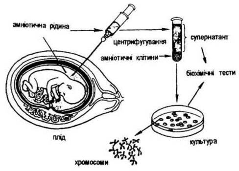
Fig. 36. Amniocentesis is a method of prenatal diagnosis of hereditary diseases.
Fetoscopy is a visual examination of the fetus using an endoscope (fetoscope). Inserted into the amniotic cavity (via the anterior abdominal wall or the posterior fornix), the endoscope allows for visual inspection of fetal parts, blood sampling, and skin biopsy for subsequent analysis. This method makes it possible to diagnose visible congenital malformations (such as polydactyly and Achondroplasia) and to detect conditions like ichthyosis or epidermolysis bullosa through the analysis of skin biopsy samples. Furthermore, it enables the diagnosis of hereditary hemoglobinopathies, erythrocyte enzymopathies, and immunodeficiency states. It is performed exclusively under specific clinical indications at 18–23 weeks of gestation under ultrasound guidance. Miscarriage rates following fetoscopy have been reported in 7–8% of cases.
In recent years, the hypothesis of preconceptional prevention has been gaining traction. It begins several months before conception and continues through the Cytology/cytology/16.html">Early stages of embryonic development. Preparing the mother's body for conception (through vitamin supplementation, antioxidant therapy, immune system enhancement, and stress reduction) and maintaining these conditions during the early stages of embryonic development (up to 10 weeks) helps reduce the incidence of multifactorial congenital malformations. According to N.P. Bochkov (1997), women who received additional vitamin supplementation experienced a decrease in the recurrence rate of neural tube defects in their offspring from 4.6% to 0.7%.
Last update: 08/08/2026
Editorial and Educational Adaptation: This material has been compiled based on the primary/original source text. The project team performed an editorial review, corrected technical inaccuracies, structured sections, and adapted the content for an educational format.
What was processed:
- elimination of formatting defects (OCR errors, structural breaks, corrupted characters);
- editorial organization of content;
- standardization of terminology in accordance with academic sources;
- verification of factual statements against the original source text.
All mentions of the author, publication year, and origin of the primary text have been preserved in accordance with the source.