Practical Course in Zoology: Study Guide - T. A. Dauda 2014
Metazoa
Chordates
Subphylum Vertebrates, or Craniata - Superclass Fishes - Class Bony Fishes
The Class Bony Fishes (Osteichthyes) encompasses the vast majority of species within the superclass of fishes and about half (more than 20,000) of all vertebrate species. Bony fishes inhabit A wide variety of aquatic environments. The primary Selection/11.html">General features of bony fishes include the following structural characteristics. The Skeleton is ossified to a greater or lesser degree. The interbranchial septa are reduced to varying extents, and the gill filaments lie directly on the gill arches. A bony operculum is invariably present, covering the gill apparatus externally. The vast majority of species possess a swim bladder, an important hydrostatic organ that plays an adaptive role, facilitating the vertical migration of fish through different Water horizons.
In most bony fishes, Fertilization is external; the eggs are small and lack horn-like envelopes. Viviparity occurs in a negligible number of species.
The Classification of bony fishes is extremely difficult, and at present, there is no single consensus on this issue.
To become acquainted with the external Morphology, perform a dissection, and study the internal Structure, one can use the European perch (Perca fluviatilis), which is widely distributed in the freshwaters of Europe and Asia.
European perch (Perca fluviatilis).
The European perch is a predatory fish. It feeds on various invertebrates and other fish, often consuming large quantities of fry, which makes it detrimental in fish-farming areas.
Materials and equipment. For every one or two students, the following are required: a freshly killed perch fixed in formalin; a dissecting tray; a scalpel; scissors; tweezers; dissecting needles; pins (10–15 pcs.); absorbent cotton; gauze pads; a hand lens; watch glasses; Petri dishes; and wall charts: "Internal Structure of the Perch," "Diagram of the Fish Circulatory system," and "STRUCTURE OF THE Fish Brain."
Assignment. Examine the external Features of the perch. Pay attention to the adaptations of fishes to an aquatic lifestyle. Perform a dissection and memorize the Topography of the Internal Organs. Dissect and study The structure of individual organs and Organ Systems. Prepare the following drawings: "Internal Structure," "Structure of the Brain," and "Diagram of the Circulatory System."

Fig. 119 External and internal structure of the perch:
1 — Mouth with Teeth; 2 — operculum; 3 — bony scales; 4 — caudal fin; 5 — dorsal fin; 6 — anal fin; 7 — eye; 8 — nostrils; 9 — lateral line; 10 — anus; 11 — genital opening; 12 — urinary opening; 13 — dissected Stomach; 14 — intestine; 15 — rectum; 16 — Liver; 17 — Gallbladder; 18 — gills; 19 — Spleen; 20 — swim bladder; 21 — Kidney; 22 — Ureter; 23 — Urinary Bladder; 24 — Ovary; 25 — atrium; 26 — ventricle of The Heart; 27 — ventral aorta.
Study of external morphology using a freshly killed fish and preserved specimens. Place the perch in the dissecting tray. Note that its body is streamlined in shape. The body of the perch can be divided into the HEAD, trunk, and tail, although there are no sharply defined boundaries between these regions. The boundary between the head and the trunk is considered to be the posterior edge of the operculum, while the boundary between the trunk and the tail is the anus (Fig. 119). The caudal region, terminating in the caudal fin and accounting for about one-third of the body length, serves as the fish's primary propulsive organ.
Observe the dark coloration of the back, which makes the perch inconspicuous from above. From below, the light coloration of the belly blends with the bright Background of the water. Dark longitudinal stripes along the flanks Complement the protective coloration of the body. Both the perch and the pike lie in wait for prey among aquatic vegetation.
The body of the perch, like that of all vertebrates, is covered by Skin consisting of two layers: the lower layer, or dermis (corium), formed by Connective Tissue, and the upper layer, the epidermis, represented by Stratified Epithelium.
Unicellular glands that abundantly secrete mucus are located in the epidermis. Notice that the body of the perch is covered with mucus. Mucus reduces friction when the fish swims in water and protects the body from microbial invasion and the leaching of salts from the Organism.
Examine the overlapping scales embedded in the skin like roof tiles. They form a flexible and robust protective cover that shields the body from injury and reduces friction. Locate the lateral lines, which run along the sides of the perch's body from the head to the tail as a dashed-like sequence. The lateral line is a specialized sensory organ through which the fish perceives water pressure and current velocity; it Functions as a physiological locator enabling the fish to sense approaching objects in the water.
Examine the fins, which are the organs of locomotion in water. A distinction is made between paired fins—pectoral and pelvic—and unpaired fins—dorsal, caudal, and anal. Draw the outline of the perch's body, the lateral line, and both paired and unpaired fins in your notebook.
Spread out one fin after another using tweezers and examine them against transmitted light. It can be seen that each fin consists of fin rays spanned by a membranous web. The paired fins and the caudal fin (organs of locomotion) feature soft, branched fin rays. In the unpaired dorsal and anal fins, which help the fish maintain an upright position in the water, stiff, spiny fin rays predominate.
Examine the head of the perch: it has a wedge-like shape and is connected directly to the trunk, with no cervical region (neck). Notice that the eyes of the perch, located on the sides of the head, lack eyelids. The nostrils are divided by a skin flap into inhalant and exhalant openings. Verify that the nostrils do not communicate with the Oral Cavity by inserting a dissecting needle into them. Like all fishes, the perch possesses only an Inner ear; external and middle ears are absent in fishes.
Examine the mouth of the perch. Like the pike and other predatory fishes, the perch has a wide mouth; the jaws are lined with sharp, backward-pointing teeth, which serve not for chewing, but for grasping and holding prey.
Insert your finger into the mouth of the perch and run it along the jaws and the palate. In the perch, small conical teeth are found on the jaws, the palate, and the Pharynx. Examine the Tongue on the floor of the fish's oral cavity, which is simply a fold of the mucous membrane lacking musculature.
The gills of the perch are located beneath the opercula on the sides of the head. Due to the rhythmic Movements of the opercula, water enters through the mouth and exits outward from beneath the gill covers. As it washes over the gills, the water delivers oxygen necessary for Respiration into the Blood and carries away carbon dioxide.
Using a dissecting needle, locate the anus just anterior to the anal fin, as well as the genital and urinary openings situated slightly posterior to it on a small papilla.
Pull out a perch scale with tweezers, place it on a Glass slide, and examine it under low magnification of a Microscope. The age of the fish and its living conditions in different years can be determined by the concentric rings lying parallel to the outer margin of the scale. In the perch, the outer edge of the scale bears small sharp denticles; such a scale is termed ctenoid (Fig. 120). Scales with a smooth outer margin are called cycloid.
Make drawings of ctenoid and cycloid scales.
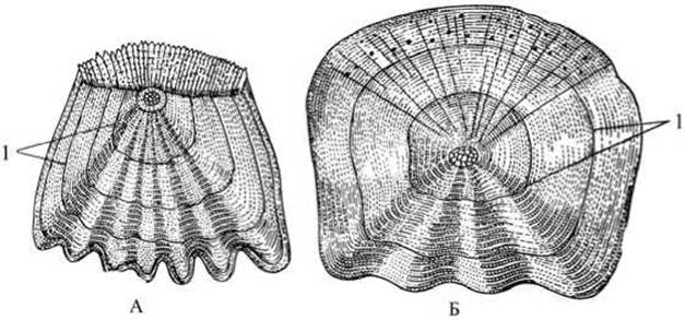
Fig. 120. Scales of bony fish:
A — ctenoid scale (perch); B — cycloid scale (roach); 1 — annual rings.
Dissection. The internal structure of a fish is studied through dissection, supplemented by a preserved specimen of a dissected fish. Hold the perch in your left hand with its belly facing up and its head pointing away from you, and make a small transverse incision in the belly near the anus. Insert the scissors into the incision and carefully, without damaging the internal organs, cut along the midline of the ventral side up to eye level, using the tips of the scissors. The girdles of the paired fins will be cut through in the process.
Grasp the left body wall with forceps and, lifting it up, make an incision from the anus almost up to the lateral line. Turn the scissors and cut parallel to and just below the lateral line toward the posterior corner of the operculum (gill cover). While cutting, lift the body wall upward with forceps, taking care not to damage the swim bladder and other internal organs with the scissors.
Lift the operculum and cut it so that the end of the first incision meets the opercular cut, opening up the gill cavity. Remove the excised piece of the body wall along with the musculature, Ribs, and part of the operculum.
Place the fish in the dissecting tray on its right side and pin down the tip of the tail and the head (the pin should pass through the mouth opening). Pour water over the fish until all dissected organs are submerged. Examine the overall arrangement of the organs.
In the dissected fish, the gills are visible in the anterior part of the body, while the heart lies posterior and ventral to the gill cavity. The entire Abdominal cavity is filled with digestive organs. Posterior to the heart lies the sac-like stomach, partially covered by a large, light brown or reddish liver. A intestine with three pyloric caeca extends from The Stomach. The intestine forms loops and terminates at the anus. The spleen is visible within one of the intestinal loops. The Gonads (reproductive organs) are located in the posterior region of the fish's body. Behind them lies the urinary bladder. A large swim bladder lies dorsal to the digestive and reproductive organs, and the Kidneys are situated above the swim bladder. All internal Organs of the fish are located within the secondary body cavity (coelom).
The gill apparatus is formed by four gill arches on each side of the head. Cut out a gill arch with scissors, place it in a Petri dish with water, and examine its structure using a hand lens while placing the Petri dish on a white sheet of paper. Alternatively, the gill arch can be placed on a small white plate and covered with water.
Examine the convex outer side of the gill arch, which bears fringe-like gill filaments arranged in two rows; these are permeated with Blood Vessels. On the inner concave side, gill rakers are visible, forming a straining apparatus (filter). They trap solid food particles entering the pharynx along with water. The trapped food particles are directed from the pharynx into the Esophagus.
Remove the gills: to do this, cut all the gill arches at the top and bottom and transfer them to a Petri dish using forceps. The heart can now be easily located near the gills within the pericardial cavity. The fish heart consists of two chambers: a sac-like dark red atrium and, located slightly posterior and closer to the ventral side, a pointed, lighter-colored ventricle. Pull the ventricle backward with forceps and locate the bulbus arteriosus, which marks the Origin of the ventral aorta. Examine the sac-like, thin-walled, dark-red sinus venosus located dorsal to the atrium. Having excised The Heart and placed it in a watch glass with water, palpate the walls of the ventricle with forceps — they are dense and muscular. In the same way, verify that the walls of the atrium are thin and soft.
Make a drawing of the internal structure of the perch and proceed to study the individual organ systems.
Circulatory system. Trace The pathway of blood through the fish's body using the Circulation diagram. Upon ventricular contraction, deoxygenated (venous) blood is pumped through the ventral aorta toward the gills. The ventral aorta branches to form 4 pairs of afferent branchial Arteries (corresponding to the number of gill arches). In the gill filaments, where the arteries form a dense capillary network, gas exchange takes place: carbon dioxide passes from the blood into the water, while oxygen passes from the water into the blood. The oxygenated blood enters 4 pairs of efferent branchial arteries, through which it flows into the paired epibranchial vessels — the roots of the dorsal aorta. The carotid arteries branch off from these toward the head. In the posterior region, the roots of the aorta merge to form the dorsal aorta.
The dorsal aorta runs beneath THE Vertebral Column. Arteries branch off from it, distributing blood to organs and Tissues. Within the organs and tissues, the arteries branch into a dense network of capillaries. Gas exchange occurs through their capillary walls: the blood delivers oxygen and nutrients to body Cells and receives carbon dioxide and Metabolic waste products, thereby turning into venous blood. Small Veins carrying venous blood from organs and tissues unite, much like in amphioxus, into larger paired anterior and posterior cardinal veins. From the cardinal veins, blood flows through the Cuvierian ducts into the sinus venosus. Venous blood from the abdominal organs, similarly to amphioxus, passes through the hepatic portal system and also enters the sinus venosus. From the sinus venosus, blood flows into the atrium and then into the ventricle. Thus, only venous blood passes through the heart of the perch.
The CIRCULATORY SYSTEM OF fish is closed, and There is a single circuit of circulation. The structure of the fish circulatory system shows great similarity to that of amphioxus, but fish possess a true heart.
Digestive System. Dissect out the alimentary canal and examine its individual sections. To do this, carefully cut the mesentery supporting the intestinal loops, untangle them, and straighten them out. Locate the gallbladder, which lies on the stomach against the inner surface of the liver. It receives the liver secretion — Bile, which flows through the bile duct into the duodenum. Remove the liver with extreme care, as it tears easily.
Insert a glass rod into the mouth of the perch and, gently advancing it, trace the path of food through the digestive tract. The perch swallows its prey whole. From the oral cavity, food enters the pharynx, which leads directly into the esophagus — a short, thin-walled tube opening into the stomach. The stomach is elongated and sac-like, with its blind end directed backward. The rod inserted through the mouth and pushed along the alimentary canal abuts against the posterior part of the stomach. Upon cutting open the stomach, a whole swallowed fish can frequently be found inside.
Locate the Small Intestine extending from the anterior lower edge of the stomach. Note that at the beginning of the intestine, bordering the stomach, three short blind pyloric caeca branch off; these increase the digestive surface area of the intestine. The small intestine transitions gradually, without clear boundaries, into the hindgut (rectum), which terminates at the anus.
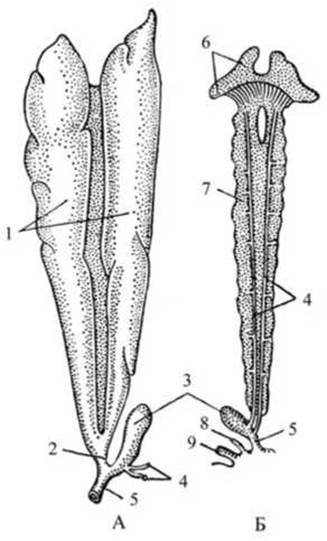
Fig. 121. Excretory and reproductive organs of the perch:
A — Male reproductive organs; B — female kidneys; 1 — Testes; 2 — vas deferens (sperm duct); 3 — urinary bladder; 4 — Ureters; 5 — urogenital opening (Urethra); 6 — head kidney (pronephros); 7 — trunk kidney (mesonephros); 8 — oviduct; 9 — anus.
The Pancreas, consisting of small lobules located within the mesentery, is poorly developed in the perch and difficult to observe.
Cut off the alimentary canal in the region of the esophagus and hindgut, transfer it to the bottom of the dissecting tray, and lay it out for final examination and drawing. Find the reddish spleen — a hemopoietic (blood-forming) organ — nestled in the loop near the duodenum.
Examine the swim bladder located between the Vertebral Column and the intestine. In the perch, it is a closed sac with sealed ends and dense, silvery-shining walls, fused to the body wall of the fish. When attempting to remove it from the body, it most often tears.
The swim bladder plays an important adaptive role in the life of fish in the aquatic environment. It is filled with gas and acts as a hydrostatic organ that enables the fish to submerge or rise.
Reproductive System. Locate the gonads of the perch in the posterior part of the body (Fig. 121). Their size depends on the fish's sexual maturity. Examine the male gonads—paired testes (milt); they have an elongated shape. The common duct of the testes opens onto the urogenital papilla posterior to the anus.
The ovary of the perch is unpaired, yellowish, and has a granular structure. Eggs (roe) are produced within the ovary. Note the almost complete absence of specialized genital ducts. The gonads are elongated, as the Ovaries and, particularly, the testes extend almost to the genital aperture. Fertilization in most bony fish is external.
Excretory system. Remove the swim bladder and reproductive organs. Examine the kidneys of the perch—they appear as long, narrow, reddish-brown ribbons extending along both sides of the vertebral column for almost the entire length of the body. These are the trunk kidneys. The anterior PARTS OF THE kidneys are noticeably expanded, fused along the midline, and enclose the esophagus; this is a rudiment of the head kidney (pronephros).
The ureters are thin tubes running along the inner margin beneath the peritoneal lining of each kidney.
Posteriorly, they merge into a common duct that empties into the urinary bladder. Locate the urinary bladder and its opening, which is situated next to the genital aperture on the urogenital papilla.
Muscular System. Examine the Muscles on the remaining intact right side of the perch. Muscles are easier to observe if the perch is first immersed in boiling water for 1–2 minutes. Note that, similar to amphioxus, the trunk musculature of the fish is arranged metamerically.
Nervous system. Fish possess a BRAIN AND SPINAL cord (Fig. 122). Nerves extend from the brain and Spinal Cord to all organs and body parts. Remove all internal organs from the body cavity of the fish and examine the white thread-like nerves emerging from the spinal cord.
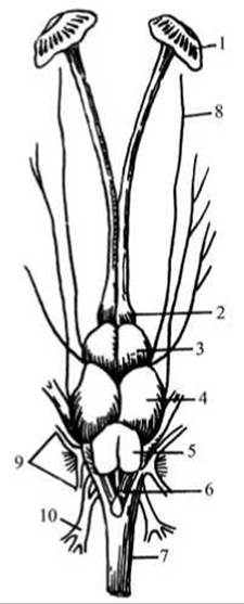
Fig. 122 Brain of the perch:
1 — olfactory capsules; 2 — olfactory lobes; 3 — Telencephalon (Forebrain); 4 — mesencephalon (Midbrain); 5 — Cerebellum; 6 — myelencephalon (Medulla Oblongata); 7 — spinal cord; 8-10 — Cranial Nerves.
Open the Skull of the perch.
To do this, remove the perch from the dissection tray, hold it in your left hand, and use a sharp scalpel to gradually pare away the skull bones (much like sharpening a pencil) from the occiput toward the eyes. Using a pipette, gently wash away the loose fatty tissue covering the brain with a weak stream of water, and carefully remove it with tweezers if necessary. Examine the small forebrain; the olfactory lobes adjoin it anteriorly, giving rise to olfactory nerves that terminate in the olfactory bulbs (capsules). Fish have a well-developed SENSE OF SMELL. The olfactory capsules lie in blind nasal sacs. The Diencephalon adjoins the forebrain posteriorly and is covered from above by the large optic lobes of the midbrain (the diencephalon can only be observed in a wet preparation where it is shown from below).
Locate the midbrain, which is the largest region of the fish brain. Posterior to it lie the cerebellum (Hindbrain) and the medulla oblongata featuring the Rhomboid fossa, which transitions into the spinal cord. The cerebellum in fish is well-developed in connection with high mobility and the coordination of movement.
Make a drawing of the brain structure of fish.
Last update: 13/08/2026
Editorial and Educational Adaptation: This material has been compiled based on the primary/original source text. The project team performed an editorial review, corrected technical inaccuracies, structured sections, and adapted the content for an educational format.
What was processed:
- elimination of formatting defects (OCR errors, structural breaks, corrupted characters);
- editorial organization of content;
- standardization of terminology in accordance with academic sources;
- verification of factual statements against the original source text.
All mentions of the author, publication year, and origin of the primary text have been preserved in accordance with the source.