MICROBIOLOGY - M.H. Serhiichuk - 2008
Chapter 6. PHYSIOLOGICAL CHARACTERISTICS OF ANAEROBIC MICROORGANISMS
Anaerobic Respiration
Anaerobic Respiration is a process of oxidation of organic and inorganic substrates in which the TERMINAL ELECTRON ACCEPTORS are:
- inorganic substances: oxidized anions (SO42-, SO32-, NO3-, NO2-); oxidized metal cations (Fe3+, Mn4+), СО2; oxygen-containing metal anions (selenates, arsenates); radionuclides (U+6, Tc+7); oxidized forms of CSpounds (СO2, S0);
- a range of Organic compounds: sulfur-containing compounds (dimethyl sulfoxide), organic nitrogen oxides (trimethylamine N-oxide), halogenated organic compounds (3-chlorobenzoate, 1,1,2,2-tetrachloroethane), fumarate.
Depending on The Nature of the terminal electron acceptor, anaerobic respiration is classified as nitrate, sulfate, sulfur, iron, fumarate, carbon respiration, etc.
There are two MAIN TYPES OF anaerobic respiration:
- involving acceptors with a positive (high) redox potential (nitrate, nitrite, Fe3+, fumarate, dimethyl sulfoxide, nitrogen oxides). This type of respiration is typical for facultative anaerobes; it competes with aerobic respiration and is inhibited by molecular oxygen;
- involving acceptors with a negative (low) redox potential (sulfur, sulfate, carbonate). This type of respiration has been found exclusively in obligate anaerobes.
The most important electron acceptors and their standard redox potentials (Eo') at pH 7.0 are listed in Table 6.1.
Class="center">Table 6.1. Standard redox potentials of potential electron acceptors in respiration
Redox pair |
Eo', V |
НСО3-/НСО2- |
-0,41 |
Н+/Н2 |
-0,42 |
НСО3-/ацетат |
-0,35 |
НСО3-/СН4 |
-0,33 |
S0/HS- |
-0,27 |
HSO3-/HS- |
-0,12 |
SeO42-/SeO32- |
+0,02 |
Фумарат / сукцинат |
+0,03 |
Триметиламіноксид/триметиламін |
+0,13 |
Диметилсульфоксид / диметилсульфід |
+0,16 |
NO2-/NH4+ |
+0,34 |
NO 2-/ NO |
+0,35 |
NO3-/NO2- |
+0,43 |
Fe3+/Fe2+ |
+0,77 |
O2/H2O |
+0,82 |
The redox potential (RP) of the terminal electron acceptor determines the site where electrons exit the Cell/36.html">Respiratory Chain and, consequently, The amount of ATP generated via Oxidative Phosphorylation. The higher the RP of the acceptor, the more energy the microorganism obtains from respiration. In natural environments, where several potential electron acceptors are usually present simultaneously, facultative anaerobes preferentially utilize the most energetically favorable ones—those with the highest redox potentials—whereas obligate anaerobes rely exclusively on low-potential acceptors. The Sequence of electron acceptor utilization in each microorganism is genetically determined and controlled by complex regulatory mechanisms.
Nitrate respiration is a process in which facultatively anaerobic microorganisms use nitrate (NO3-) as a terminal electron acceptor during anaerobic respiration. Because nitrate is reduced in the process, nitrate respiration is also referred to as dissimilatory nitrate reduction, and the microorganisms performing it are called nitrate reducers or nitrate-reducing Bacteria.
The substrates or electron Donors in nitrate respiration include various organic compounds (glucose, ethanol, succinate, benzoate) and some inorganic substances (molecular hydrogen, reduced sulfur compounds). Thus, nitrate reducers can function as chemoorganotrophs (Pseudomonas aeruginosa), chemolithotrophs (Hydrogenomonas agilis, Thiobacillus denitrificans), or mixotrophs (Micrococcus denitrificans). The Mechanism of organic substrate oxidation generally does not differ from that under aerobic conditions, meaning that nitrate reducers obtain a portion of their ATP through substrate-level phosphorylation.
The reduction of nitrate nitrogen involves changes in its oxidation state and The formation of several intermediate products, as shown in Table 6.2.
Table 6.2. Oxidation states of key nitrogen compounds
Compound |
Oxidation state |
Ammonium (NH4+) |
-3 |
Ammonia (NH3) - gas |
-3 |
Molecular nitrogen (N2) - gas |
0 |
Nitrous oxide (N2O) - gas |
+ 1 (average) |
Nitric oxide (NO) - gas |
+2 |
Nitrite (NО2-) |
+3 |
Nitrate (NО3-) |
+5 |
During nitrate respiration, bacteria reduce nitrate via two pathways (Fig. 6.20):
1. Via nitrite to gaseous products NO, N2O, and N2. Because this process depletes the medium of nitrogen, it is termed denitrification. Denitrification is a widespread phenomenon characteristic of representatives of over 40 bacterial genera and species: Pseudomonas (P. aeruginosa, P. fluorescens, P. stutzeri), Thauera, Alcaligenes, Paracoccus, Hyphomicrobium, Thiobacillus (T. denitrificans), Bacillus, and many others.
2. Via nitrite to ammonium. This process is known as dissimilatory (respiratory) ammonification. It is typical for certain bacilli, enterobacteria, and streptococci. Unlike assimilatory ammonification—an energy-consuming reduction of nitrate to ammonium aimed at incorporating nitrogen into Biosynthesis (see Chapter 5)—dissimilatory ammonification proceeds with the generation of ATP via oxidative phosphorylation. In some microorganisms, dissimilatory ammonification can stall
at the nitrite stage until the nitrate supply is completely exhausted. Nitrite then accumulates in the medium or is subsequently reduced further to ammonium, which is characteristic of Escherichia coli.

Fig. 6.20. Two pathways of nitrate reduction by bacteria during nitrate respiration
The Enzymes catalyzing both nitrate reduction pathways are synthesized exclusively under anaerobic conditions. Molecular oxygen strongly inhibits The activity of enzymes involved in dissimilatory nitrate reduction.
Oxidative phosphorylation during nitrate respiration comprises several steps:
1. Reduction of nitrate to nitrite (this stage is common to both assimilatory ammonification and denitrification).
2. Reduction of nitrite to ammonium (assimilatory ammonification).
3. Reduction of nitrite to gaseous products (denitrification).
The reduction of nitrate to nitrite proceeds According to the equation
NO3- + 2e- + 2H+ → NO2- + H2O
Nitrate is reduced to nitrite along the respiratory chain by means of NADH2 generated during glucose oxidation in Glycolysis. Nitrate reduction is catalyzed by the enzyme dissimilatory nitrate reductase. Bacteria possess Two Types of dissimilatory nitrate reductases: NAR and NAP. NAR is bound to the cytoplasmic membrane on the cytoplasmic side and Functions when sufficient nitrate is present in the medium. NAR is an enzyme complex (sometimes designated as NarGHI) consisting of three subunits: NarI, NarH, and NarG (Fig. 6.21).
The NarI enzyme is a multiheme cytochrome b containing two Hemes. It accepts electrons from a quinone (ubiquinone or menaquinone) and transfers them further to the membrane protein NarH. Protons are simultaneously released into the periplasm, resulting in the generation of a transmembrane electrochemical proton gradient. The NarH protein contains four Fe-S clusters. Its function is to transfer electrons from NarI to NarG. The NarG protein consists of an Fe-S cluster and a molybdenum-containing cofactor, molybdopterin guanine dinucleotide (MGD). Nitrate reduction to nitrite actually takes place on the MGD cofactor as follows (Fig. 6.22):
- the oxo group of oxidized Mo(VI) loses Water upon reduction of the cofactor to Mo(IV);
- nitrate binds to the reduced cofactor, accepts electrons from it, and is reduced to nitrite;
- Mo(IV) is simultaneously oxidized to Mo(VI).
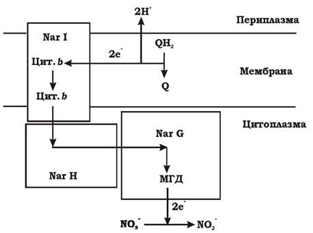
Fig. 6.21. Structure AND FUNCTIONS of nitrate reductase:
Q - ubiquinone; NarI, NarH, NarG - nitrate reductase subunits; Cyt. b - cytochrome b, MGD - molybdopterin guanine dinucleotide
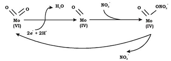
Fig. 6.22. MECHANISM OF ACTION of the NarG MGD cofactor in nitrate reductase
Nitrate is reduced to nitrite in the Cytoplasm; therefore, The Cell maintains specialized mechanisms for transporting nitrate inward and rapidly exporting toxic nitrite out of the cell.
Another type of dissimilatory nitrate reductase, NAP, functions within the cell when nitrate levels in the medium are low. It is localized in the periplasm and consists of two subunits.
The reduction of nitrite to ammonium during dissimilatory ammonification proceeds according to the equation
NO2- + 3 NAD(P)H + 5H+ → NH4++ 3 NAD(P)+ + 2H2O in three stages. At each stage, two electrons are transferred to the acceptor, while intermediates remain enzyme-bound:
1. HNO2 is initially reduced to HNO (N+3 → N+1).
2. HNO is reduced to NH2OH (N+1 → N-1).
3. NH2OH is reduced to NH4+ (N-1 → N-3).
This process is catalyzed by a complex of periplasm-localized or membrane-bound enzymes known as dissimilatory nitrite reductases (which exist in several types). This complex enzyme can accept electrons from NADH2 dehydrogenase and reduced quinone. Nitrite reduction is coupled to the generation of proton-motive force (Δρ), although the mechanism of the proton pump remains poorly understood. Despite the generation of a proton gradient during the reduction of nitrite to ammonium, practically no ATP is synthesized. This is because nitrite is a toxic compound for the cell. Immediately following nitrate reduction, nitrite is exported from the cytoplasm into the periplasm via proton-coupled antiporters. Consequently, the proton gradient generated during nitrite reduction is utilized by the cell not for ATP synthesis, but for nitrite efflux. The reduction of nitrite to ammonium takes place in the periplasm. The synthesis of dissimilatory nitrite reductases is repressed by oxygen.
The reduction of nitrite during denitrification proceeds via a series of gaseous intermediates according to the following equations:
NO2- + e- + 2H+→ NO + H2O (enzyme: NO-forming nitrite reductase)
2NO + 2e- + 2H+ → N2O + H2O (enzyme: NO reductase)
N2O + 2e- + 2H+ → N2 + H2O (enzyme: N2O reductase)
Some denitrifiers reduce nitrite completely to N2, others do not utilize nitrite but instead reduce NO or N2O to molecular nitrogen, some species can reduce nitrite to NO and N2O to N2, whereas others do not reduce NO to N2O.
The reduction of nitrite to NO is catalyzed by periplasmic NO-forming nitrite reductase. There are two forms of this enzyme, which never occur simultaneously in the same Cells:
- 75% of denitrifiers contain the tetraheme cytochrome cd1;
- 25% of bacteria contain a copper-containing nitrite reductase.
Both enzymes receive electrons from Quinones, which is accompanied by the translocation of protons from the cytoplasm.
NO is reduced to N2O by NO reductase, known as NOR. This is a cytochrome bc complex that is synthesized exclusively under anaerobic conditions and is located on the periplasmic side of the cytoplasmic membrane. At this stage of denitrification, protons are not translocated out of the cell, but the N-N bond is formed.
The reduction of N2O to N2 involves the formation of a triple N=N bond. This is an energetically favorable process that results in proton extrusion. All types of N2O reductase enzymes are localized in the cytoplasmic membrane and contain copper, with some also possessing a unique Cu/S center.
Thus, the general metabolic pathway of facultatively anaerobic bacteria performing denitrification is shown in Fig. 6.23.

Fig. 6.23. Electron flow during aerobic respiration and denitrification (FP - flavoprotein)
Anoxic (anaerobic) ammonium oxidation (ANAMMOX) is a microbial process in which ammonium and nitrite are converted into molecular nitrogen under anaerobic conditions (described in 1992).
Bacteria performing anaerobic ammonium oxidation belong to the order Planctomycetales and are identified as a novel, deeply branching group of planctomycetes that includes three genera: Brocadia, Kuenenia, and Scalindua. Known anammox bacterial species include Brocadia anammoxidans, Kuenenia stuttgartiensis, Scalindua wagneri, S. sorokinii, and S. brodae. The first two genera were discovered in wastewater, whereas Scalindua has been found in numerous marine ecosystems.
Anammox bacteria are a fascinating group with many highly unusual characteristics. Their cell walls lack peptidoglycan, they reproduce by budding, and their cytoplasm is divided into compartments separated by membrane partitions. These compartments perform distinct cellular functions. It appears that these ancient microorganisms are positioned
at the ROOT of the evolutionary tree. Other planctomycete species are aerobic chemoorganoheterotrophs and differ significantly from anammox bacteria, which are anaerobic chemolithoautotrophs.
The METABOLISM of anammox bacteria is based on energy generation via anaerobic ammonium oxidation, coupled with the reduction of nitrite as an electron acceptor through intermediates such as hydrazine (N2H4) and hydroxylamine (NH2OH). CO2 serves as the primary carbon source for these bacteria.
Overall equation of the process: NH4+ + NO2- → N2 + 2H2O
The scheme of anaerobic ammonium oxidation is shown in Fig. 6.24.

Fig. 6.24. Scheme of anaerobic ammonium oxidation
One of the Key Enzymes in anaerobic ammonium oxidation is hydroxylamine oxidoreductase. The Importance of this enzyme is underscored by the fact that it accounts for 10% of the total cell protein. It catalyzes The oxidation of both hydrazine and hydroxylamine. Hydroxylamine oxidoreductase is localized exclusively within an organelle-like, membrane-bound structure located in the cytoplasm. This organelle is called the anammoxosome and occupies over 30% of the cell volume (Fig. 6.25).

Fig. 6.25. Cytology/cytology/92.html">SCHEMATIC STRUCTURE OF an anammox bacterium cell
Anammoxosomes are the centers of anaerobic ammonium oxidation. They are enclosed by an extremely dense double membrane containing unique ladderane Lipids: tightly packed, concatenated 4-carbon (cyclobutane) rings. The mechanism of synthesis of these lipid moieties remains unknown. Classic microbial Membrane Lipids contain 3-, 5-, 6-, or even 7-membered aliphatic rings, but 4-membered cyclobutane rings had not been known prior to their discovery in anammox bacteria. From an evolutionary perspective, It is interesting to note that currently only a small number of bacteria (thermophiles and sulfate reducers) are known to possess fragments of such lipids. They can serve as biomarkers for detecting anammox bacteria in the environment.
Membranes containing ladderane lipids exhibit very low permeability to chemical compounds, allowing them to maintain concentration gradients over extended periods. If hydrazine were able to diffuse out of the anammoxosome during the prolonged anammox process, the cells would suffer severe energy losses. Anammoxosome membranes also protect the rest of the cell from toxic intermediates—hydroxylamine and hydrazine.
The anammox process occurs at temperatures ranging from 6 to 43 0C and pH 6.7–8.3. For intensive anammox activity, nitrite must be aerobically produced from ammonium by first-stage nitrifiers. Therefore, the process functions most effectively in a co-culture of aerobic and anaerobic ammonium oxidizers at an oxygen concentration of about 0.5%.
It has been estimated that anaerobic ammonium oxidation accounts for up to 70% of molecular nitrogen production in the world's oceans.
Sulfate respiration (dissimilatory sulfate reduction) is a process in which obligate anaerobic bacteria use sulfate (SO42-) as a terminal electron acceptor during anaerobic respiration.
The ability to perform sulfate reduction has been identified in microorganisms belonging to four phylogenetically distinct groups:
1. The largest group belongs to the γ-subclass of the class Proteobacteria and includes the genera Desulfovibrio, Desulfobulbus, Desulfomicrobium, Desulfobacter, Desulfobacterium, Desulfosarcina, Desulfococcus, Desulfonema, and others.
2. Gram-positive bacteria of the genus Desulfotomaculum.
3. Gram-negative bacteria of the genus Thermodesulfobacterium.
4. Archaea of the genus Archaeoglobus.
Sulfate-reducing bacteria (SRB) can exhibit chemoorganoheterotrophic and chemolithoautotrophic Types of Metabolism. Their electron donors include molecular hydrogen, formate, acetate, lactate, fumarate, succinate, propionate, Fatty acids (C4–C20), methanol, ethanol, and propanol. Some sulfate reducers can also grow on Alanine, glutamate, Choline, glycerol, fructose, nicotinic acid, indole, Hydrocarbons (predominantly C16, but not CH4), starch, Peptides, benzoate, phenylacetate, and phenol.
Autotrophic carbon dioxide assimilation in sulfate reducers proceeds via the reductive acetyl-CoA pathway (see Fig. 5.32).
Based on their carbon metabolism, sulfate reducers are divided into two groups (Table 6.3):
1. Those unable to oxidize acetyl-CoA (they oxidize organic compounds only to acetate and do not use acetate as a source of electrons and carbon—"incomplete oxidizers").
2. Those that oxidize acetyl-CoA and consequently break down organic compounds completely to CO2 (these bacteria are capable of growing on acetate).
Table 6.3. Classification of sulfate reducers based on carbon metabolism
Sulfate reducers oxidizing organic compounds only to acetate |
Sulfate reducers oxidizing organic compounds to CO2 |
Desulfovibrio |
Desulfobacter |
Desulfomicrobium |
Desulfobacterium |
Desulfomonile |
Desulfococcus |
Desulfobotulus |
Desulfonema |
Desulfomonas |
Desulfurella |
Desulfotomaculum (some) |
Desulfosarcina |
Desulfobulbus |
Desulfuromonas |
Thermodesulfobacterium |
Desulfoarculus |
Archaeoglobus |
Desulfotomaculum (some) |
Sulfate reducers capable of complete substrate oxidation and growth on acetate degrade acetyl-CoA via two pathways:
- the oxidative acetyl-CoA pathway (Fig. 6.26);
- the modified Krebs cycle (Fig. 6.27).
The modified Krebs cycle (Fig. 6.27, b), which is used
by sulfate reducers to degrade acetate, differs from the classical cycle by the presence of the enzyme succinyl-CoA:acetate CoA-transferase, through which acetate is activated without the participation of ATP:
Succinyl-CoA- + Acetate- → Succinate2- + Acetyl-CoA
Species of the genus Desulfobacter also contain an additional enzyme, ATP-dependent citrate lyase. In the reaction catalyzed by this enzyme, acetyl-CoA is converted into citrate via acetyl phosphate, resulting in the synthesis of one ATP molecule:
Acetyl-CoA + OAA2- + ADP + HPO42- → Citrate3- + CoASH + ATP
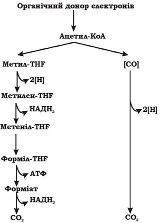
Fig. 6.26. Acetyl-CoA oxidation pathway (Wood-Ljungdahl pathway):
THF - tetrahydrofolic acid
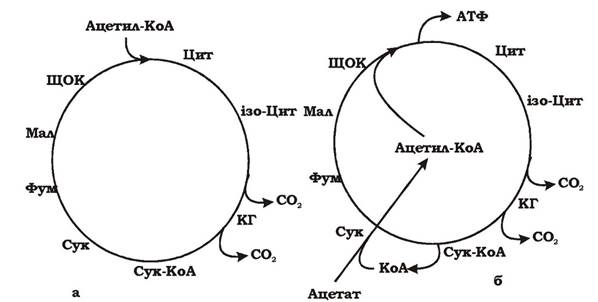
Fig. 6.27. Acetate utilization by sulfate-reducing bacteria via a modified Krebs cycle:
a - conventional Krebs cycle; b - modified Krebs cycle; OAA - oxaloacetate; Cit - citrate; iso-Cit - isocitrate; KG - α-ketoglutaric acid; Suc - succinate; Fum - fumarate; Mal - malate
The electron acceptors in sulfate respiration include sulfate (SO42-), as well as oxidized sulfur compounds such as thiosulfate (S2O32-), tetrathionate (S4O62-), and sulfite (SO32-). Some species of sulfate reducers, such as Desulfovibrio gigas, Desulfovibrio multispirans, and Desulfuromicrobium baculatum, are also capable of reducing elemental sulfur (S0). The end product of the reduction of these compounds is sulfide (S2-), which is excreted from the cell into the environment.
Table 6.4 lists some of the intermediates of sulfate reduction and their oxidation states.
Table 6.4. Oxidation states of key sulfur compounds
Compound |
Oxidation state |
Organic sulfur compounds |
-2 |
Sulfide (H2S) |
-2 |
Elemental sulfur S0 |
0 |
Thiosulfate (S2O32-) |
+2 (average for S) |
Tetrathionate (S4O62-) |
+2 (average for S) |
Sulfur dioxide (SO2) |
+4 |
Sulfite (SO32-) |
+4 |
Sulfur trioxide (SO3) |
+6 |
Sulfate (SO42-) |
+6 |
Depending on the pH of the medium, sulfide can exist in the following forms:
- at low pH values - H2S gas has low solubility in water and is therefore volatile;
- at neutral pH values - HS-;
- at high pH values - S2-.
The latter two compounds are highly soluble in water. Upon contact with heavy metals, they form insoluble, dark-colored precipitates, such as FeS, PbS, and CuS.
All stages of sulfate reduction take place in the cytoplasm. SO42- ions enter the cell via symport with H+ or Na+. The transport of 1 mole of sulfate consumes approximately 1/3 mole of ATP, although this amount depends on the concentration of the acceptor in the environment.
Dissimilatory sulfate reduction to sulfide within the cell can be described by the following equations:
1. ATP + SO42- → APS + PPi (ATP sulfurylase)
2. APS + 2e- → AMP + SO3- (APS reductase)
3. HSO3- + 7H+ + 6e- → H2S + 3H2O (sulfite reductases)
The first step involves an endergonic reaction. Because the redox potential (midpoint potential) of the SO42-/HSO3- couple is very low ($E_0' = -516\text{ mV}$), sulfate is first activated by ATP to form adenosine phosphosulfate (APS) (Fig. 5.17), which has a significantly higher redox potential ($E_0' = -60\text{ mV}$). This reaction is catalyzed by the enzyme ATP sulfurylase.
In the second step, APS is converted into hydrosulfite and AMP by the enzyme APS reductase. The two electrons required for this process are supplied by FADH2.
The third step involves the reduction of sulfite (hydrosulfite) to sulfide. This process requires six electrons and is catalyzed by sulfite reductases. Four types of sulfite reductases have been identified in sulfate-reducing bacteria: desulfoviridin, desulforubidin, desulfofuscidin, and the P-582 cytochrome-like enzyme. There are two models for this process:
- direct six-electron reduction of sulfite without intermediate products;
- the trithionate pathway (Fig. 6.28) of sulfite reduction, resulting in intermediates such as trithionate (S3O62-) and thiosulfate (S2O32-). The trithionate pathway comprises three reactions:
1. 3HSO3- + 2е- + 3Н+ → -O3S-S-SO3- + 3Н2О
2. -O3S-S-SO3- + 2е- + Н+ → -S-SO3-+ HSO3-
3. -S-SO3- + 2е- + 3Н+ → HSO3- + H2S
Balance: HSO3- + 6е- + 7Н+→ H2S + 3Н2О
Initially, three hydrosulfite ions participate in the reaction, which are converted into trithionate upon The addition of two electrons. The latter is reduced to thiosulfate and, finally, to sulfide. The second and third steps involve the regeneration of sulfite groups.
Inorganic pyrophosphate (PPi), released during APS formation, can be cleaved in sulfate-reducing bacteria in two ways:
1. Desulfovibrio hydrolyzes it using inorganic pyrophosphatase without energy accumulation: PPi + Н2О → 2Pi
2. Desulfotomaculum cleaves inorganic pyrophosphate via acetyl phosphate with ATP formation. This reaction is catalyzed by the enzyme acetate kinase (acetate-pyrophosphate kinase):
Acetate + PPi → Acetyl phosphate + Pi
Acetyl phosphate + ADP → Acetate + ATP
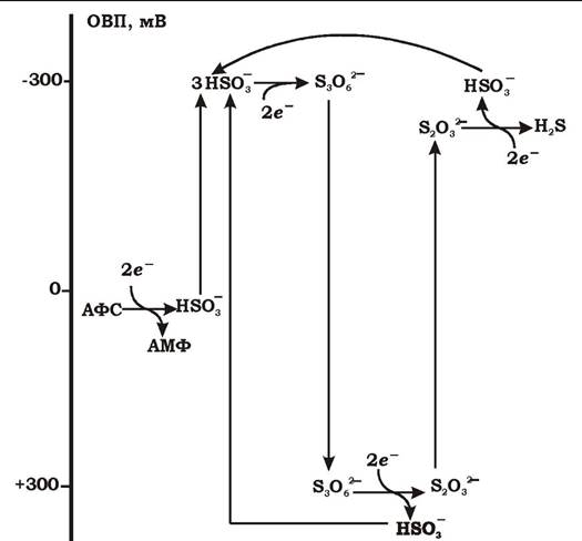
Fig. 6.28. The trithionate pathway of adenosine phosphosulfate and sulfite reduction by sulfate reducers: APS — adenosine phosphosulfate
Dismutation (disproportionation) of sulfur-containing compounds by sulfate-reducing bacteria. Some sulfate reducers possess a unique Energy Metabolism known as the disproportionation or dismutation of sulfite and thiosulfate. This process can be viewed as a type of inorganic Fermentation. The term "disproportionation" refers to the Cleavage of a compound into two components, one of which is more oxidized and the other more reduced than the parent compound. Let us examine this process using the disproportionation of thiosulfate by Desulfovibrio sulfodismutans as an example. Thiosulfate (S2O32-) contains two sulfur atoms with a cumulative oxidation state of +2. The bacterium uses one sulfur atom as an electron donor and the other as an electron acceptor:
S2+ - 4e- → SO42- (oxidation of thiosulfate sulfur to sulfate) S2+ + 4e- → S2- (reduction of thiosulfate sulfur to sulfide)
Overall equation:
S2O32- + Н2О → SO42- + H2S; ΔG0'= - 27.6 kJ/mol.
Sulfate reducers can also disproportionate sulfite:
4S032-+ Н2О → 3SO42-+ H2S; ΔG0' = - 241.3 kJ/mol.
Since the electron donor is an inorganic compound, this process can be regarded as a form of lithotrophy. Under such conditions, microorganisms can grow in the presence of trace amounts of organic compounds in the medium, or even autotrophically, utilizing solely the energy derived from the disproportionation reaction.
The mechanisms of ATP generation in sulfate reducers are not yet fully understood. It is known that they perform both substrate-level and oxidative phosphorylation.
The respiratory chain of sulfate reducers has several distinctive features. It is characterized by the presence of various low- and high-potential carriers, such as ferredoxins (E0'= -130...-440 mV), flavodoxins (E0'= -140...-440 mV), rubredoxins (E0'= +5...-5 mV), rubrerythrins (E0'= +225 mV), and menaquinones. Sulfate reducers feature unique c3-type Cytochromes, which exhibit the lowest redox potential among all known cytochromes (E0'= -260 mV). These cytochromes are localized in the periplasm and possess a multiheme structure. Tri-, tetra-, and octaheme c3-type cytochromes are known. Fatty acid-oxidizing bacteria contain b-type cytochromes, which also exist in several variants. Unlike oxygen-respiring bacteria, which possess a single cytoplasmic membrane-bound terminal oxidase, sulfate reducers contain multiple soluble terminal oxidases: APS reductase, sulfite reductase, trithionate reductase, and thiosulfate reductase. In addition, periplasmic Hydrogenase enzymes are involved in proton gradient generation by sulfate reducers. Sulfate reducers harbor Three types of hydrogenases: [Fe]-, [NiFe]-, and [NiFeSe]-hydrogenase.
The Mechanism of Oxidative phosphorylation in sulfate reducers is illustrated in Fig. 6.29.
Regardless of the Nature of the electron donor (organic substances or H2), hydrogen diffuses across the cytoplasmic membrane into the periplasm, where it is oxidized by the hydrogenase and cytochrome c3 enzyme complex: 4Н2 → 8Н+ + 8е-. Eight electrons return to the cell, reducing the activated sulfate in the form of APS to sulfide. Eight protons (8Н+) remain in the periplasm, establishing a proton concentration gradient between the interior and exterior of the cell. ATP is synthesized via ATP synthase according to Mitchell's chemiosmotic hypothesis. The mechanism of proton gradient generation, driven by the simple charge Separation of hydrogen atoms via hydrogenase coupled with the return of electrons to the cytoplasm, is referred to as vectorial electron transport.

Fig. 6.29. Putative mechanism of oxidative phosphorylation in sulfate-reducing bacteria
Sulfur respiration is a process in which bacteria utilize elemental sulfur (S0) as a terminal electron acceptor during anaerobic respiration. This pathway is found in such obligately anaerobic eubacteria as Desulfuromonas acetoxidans, Desulfurella acetivorans, Wolinella succinogenes, Desulfovibrio gigas, and Desulfomicrobium sp. Additionally, two aerobic bacteria, Pseudomonas mendonica and Alteromonas putrefaciens, are capable of respiring on sulfur in the absence of oxygen. Sulfur respiration is also observed in certain thermophilic anaerobic archaea, including Acidianus infernus, Desulfolobus ambivalens, Pyrobaculum islandicum, Pyrodictium occultum, and Thermoproteus neutrophilus. Among sulfur reducers, both chemoorganotrophs and chemolithotrophs are represented.
The Reduction of Elemental sulfur by certain bacteria can occur not only via anaerobic respiration but also as a secondary process during fermentation. However, in the latter case, Oxidative phosphorylation is absent (no ATP is generated), and sulfur merely serves as an auxiliary electron acceptor—a "sink for excess hydrogen." For instance, the thermophilic eubacteria Thermosipho africanus and Thermotoga sp. reduce sulfur to hydrogen sulfide concurrently with The breakdown of sugars and other organic substrates.
Iron and manganese respiration. The reduction of iron(III) to iron(II) via microbial respiration is an ancient evolutionary process. It is hypothesized to have occurred in Earth's hyperthermal environments more than 3.5 billion years ago. At that time, the Earth was rich in Fe3+, which was generated from Fe2+ through photochemical reactions in primordial oceans and hydrothermal vents.
The Redox Potential of the Fe3+/Fe2+ couple is exceptionally high (+770 mV), closely approaching that of oxygen (+810 mV); consequently, the utilization of Fe3+ as an electron acceptor by microorganisms is energetically highly advantageous. Iron respiration is characteristic of two major microbial groups: hyperthermophilic archaea and mesophilic eubacteria.
Several types of dissimilatory iron and manganese reduction are distinguished:
1. During the fermentation of organic compounds (sugars, Amino Acids) by cultures of Escherichia coli, Bacillus polymyxa, Clostridium pasteurianum, Lactobacillus lactis, and Vibrio spp., minor REDUCTION OF Fe(III) and Mn(IV) may occur alongside traditional fermentation products. Under these conditions, only 0.03–5% of the reducing equivalents are channeled into metal reduction. Thus, dissimilatory metal reduction in this context acts merely as a minor auxiliary catabolic pathway, serving primarily to remove excess reducing agents generated during fermentation.
2. The reduction of Fe(III) and Mn(IV) can be carried out by the bacteria Thiobacillus thiooxidans, Thiobacillus ferrooxidans, and Sulfolobus acidocaldarius during the oxidation of elemental sulfur as an electron donor:
S0+ 6Fe(III) + 4H2O → HSO4- + 6Fe(II) + 7H+
However, The energy released in this reaction is insufficient to sustain bacterial growth.
3. The reduction of Fe(III) and Mn(IV) can proceed via the chemolithotrophic OXIDATION OF MOLECULAR hydrogen. Pseudomonas sp. and Shewanella putrefaciens oxidize hydrogen using Fe(III) as the sole electron acceptor. Shewanella putrefaciens is also capable of growth via manganese respiration:
H2+ 2Fe(III) → 2H++ 2Fe(II)
H2+ Mn(IV) → 2H+ + Mn(II)
4. Shewanella putrefaciens and several unidentified bacteria can utilize iron and manganese respiration as their sole energy-yielding metabolism when growing on organic acids (acetate, lactate, Pyruvate, formate) according to the following equations:
Форміат- + 2Fe(III) + Н2О → НСО3- + 2Fe(II) + 2H+
Лактат- + 4Fe(III) + 2Н2О → Ацетат- + НСО3- + 4Fe(II) + 5H+
Піруват- + 2Fe(III) + 2Н2О → Ацетат- + НСО3- + 2Fe(II) + 3H+
Форміат- + Mn(IV) + Н2О → НСО3- + Mn(II) + 2H+
Лактат- + 2Mn(IV) + 2Н2О → Ацетат- + НСО3- + 2Mn(II) + 5H+
Піруват- + Mn(IV) + 2Н2О → Ацетат- + НСО3- + Mn(II) + 3H+
5. In microbial communities, primary dissimilatory iron(III) reduction is mediated by members of the family Geobacteraceae (Geobacter chapelleii, G. hydrogenophilus, G. metalliraducens, G. sulfurreducens, Geobacter spp., Geothrix fermentans, Geovibrio ferrireducens).
The primary challenge in utilizing Fe(III) for respiration stems from the insolubility of its compounds at neutral pH (~7), which prevents their uptake into the cell. Gram-negative bacteria, such as Shewanella, overcome this obstacle through the mechanism illustrated in Fig. 6.30. Their cells produce abundant tetra- and decahaem c-type cytochromes that form a multihaem "conduit" spanning the inner and outer membranes. Through this conduit, electrons derived from cytoplasmic carbon metabolism are transferred across both membranes and the periplasm to the cell exterior, where the actual reduction of insoluble iron(III) compounds takes place. Examples of such multihaem cytochromes identified in Shewanella frigidimarina include:
- CymA – a tetrahaem cytochrome anchored to the membrane quinol dehydrogenase;
- Pcc35 – a periplasmic decahaem cytochrome;
- Ifc3 – an iron-induced flavocytochrome c3;
- OmcA: an outer-membrane decaheme lipoprotein.
The genes responsible for the synthesis of these cytochromes are present in The Genome in multiple copies, enabling the cell to synthesize them in large quantities when needed. It has been found that when Shewanella frigidimarina is cultured under anaerobic conditions with iron(III), the number of cytochromes in its cells increases dramatically.
The multiheme iron(III) reductase system is non-specific. It is also capable of reducing other extracellular compounds, such as Mn(IV) and insoluble forms of sulfur.
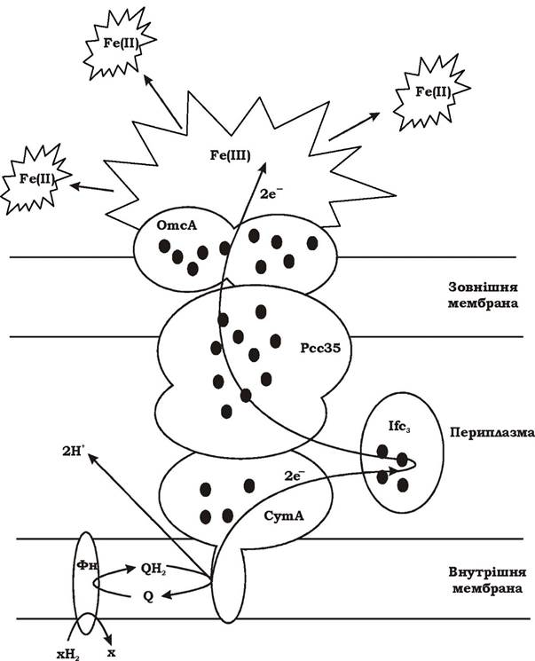
Fig. 6.30. Hypothetical scheme of the involvement of the inner and outer membranes, periplasm, and multiheme cytochromes in iron respiration of Shewanella frigidimarina: CymA - tetraheme cytochrome associated with membrane quinol dehydrogenase; Pcc35 - periplasmic decaheme cytochrome; Ifc3 - iron-induced flavocytochrome c3; OmcA - outer-membrane decaheme lipoprotein; Фн — flavoprotein
Fumarate respiration is an anaerobic process in which an organic compound, fumaric acid, serves as the electron acceptor. Upon reduction, fumarate is converted into succinate. The redox potential (RP) of the fumarate/succinate couple is relatively high, standing at +30 mV. Electron donors in fumarate respiration include formic acid, molecular hydrogen, sulfide, and NADH2 generated during the Catabolism of organic compounds. Fumarate respiration is characteristic of many facultatively anaerobic bacteria, such as E. coli, Proteus rettgeri, Salmonella, and Klebsiella. It can also be performed by obligate anaerobes: Clostridium formoaceticum, Desulfovibrio gigas, Propionibacterium, Vibrio succinogenes, Bacteroides fragilis, and Wolinella succinogenes.
Fumarate can be reduced As a result of NADH2 oxidation. The transfer of electrons from NADH2 to fumarate occurs via a series of respiratory chain carriers, the specific ones being:
- menaquinone (short for methylnaphthoquinone);
- type b cytochromes;
- fumarate reductase.
Menaquinone is a lipophilic substance with a redox potential of ~ -74 mV, enabling it to transfer electrons to fumarate. Since the redox potential of ubiquinone is +133 mV, this quinone does not participate in fumarate respiration.
Bacteria that respire via fumarate typically contain several types of type b cytochromes with different redox potential values: -200 mV and -20 mV.
Fumarate reductase is an iron-sulfur flavoprotein with a covalently bound FAD prosthetic group. This enzyme is tightly bound to the membrane and consists of two subunits. Fumarate reductase is synthesized in the cell only under specific conditions, as it is an inducible enzyme.
The General scheme of fumarate respiration is shown in Fig. 6.31.
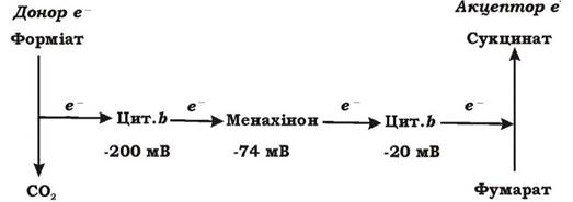
Fig. 6.31. General scheme of fumarate respiration
Carbon dioxide respiration is characteristic of homoacetogenic bacteria. Homoacetogens are obligately anaerobic bacteria that use CO2 as a terminal electron acceptor and produce acetate as the sole end product.
Taxonomically, homoacetogenic bacteria are the most heterogeneous group among obligate anaerobes. They include Gram-positive and Gram-negative bacteria, spore-forming and non-spore-forming rods and cocci, motile and non-motile species, psychro-, meso-, and thermophiles, chemoorganoheterotrophs and chemolithoautotrophs. Homoacetogens include: Eubacterium limosum, Acetobacterium carbinolicum, A. malicum, A. woodii, Clostridium thermoaceticum, C. pfenningii, C. thermoautotrophicum, C. formiaceticum, Butyribacterium methylotrophicum, Syntrophococcus sucromutans, Peptostreptococcus productus, Acetitomaculum ruminis, Sporomusa termitida, S. malonica, and S. acidovorans.
Depending on environmental conditions, homoacetogens can carry out two types of metabolism: chemolithoautotrophic and chemolithoheterotrophic.
Homoacetogens exhibit chemolithoautotrophic metabolism when growing on a mixture of two gases: molecular hydrogen and carbon dioxide. While they use H2 as an electron source, they use CO2 as a terminal electron acceptor and carbon source. Carbon dioxide is incorporated into the metabolism of homoacetogens via the reductive acetyl-CoA pathway. We have already encountered this pathway in sulfate-reducing bacteria, where it served for:
- assimilation of CO2 during autotrophic growth (reductive acetyl-CoA pathway, Fig. 5.32).
- cleavage of acetate (oxidative acetyl-CoA pathway, Fig. 6.26).
In homoacetogens, the acetyl-CoA pathway serves for:
1. Assimilation of CO2 during autotrophic growth (Fig. 5.32).
2. Energy generation via electron transfer to the terminal acceptor CO2 during carbon dioxide respiration, i.e., for oxidative phosphorylation. This occurs as a result of the reduction of N5, N10-methylene-THP to N5-methyl-THP by the enzyme methylene-THP reductase. The energy yield of this reaction is sufficient to transport one or two monovalent cations across the membrane. It has been found that this process is accompanied by the transport of Na+ ions across the membrane. Subsequently, Na+ ions return into the cell, while H+ is pumped out via Na+/H+ antipport. The presence of such an antiporter has been demonstrated in Clostridium thermoaceticum. This generates a proton gradient that drives ATP synthesis via ATP synthase. This is the only reaction of the reductive acetyl-CoA pathway coupled with oxidative phosphorylation (Fig. 6.32).
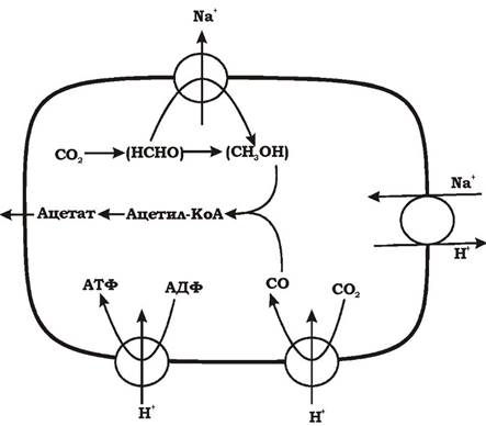
Fig. 6.32. Proton gradient formation in homoacetogens during carbon respiration
Homoacetogens are also capable of synthesizing ATP via the reductive acetyl-CoA pathway during The conversion of acetyl-CoA to acetate via acetyl phosphate. These reactions are catalyzed by the enzymes phosphotransacetylase and acetate kinase and constitute substrate-level phosphorylation.
Thus, in homoacetogens, the acetyl-CoA pathway is multifunctional, being used simultaneously in both energy metabolism and biosynthesis.
Homoacetogenic bacteria can also grow on a variety of organic substrates, thereby exhibiting chemoorganoheterotrophic metabolism. Under anaerobic conditions, most of these substrates serve as poor Energy Sources (Table 6.5).
Table 6.5. Substrates supporting the growth of homoacetogens
Substrate |
Microorganisms |
Н2+СО2 |
Most homoacetogens |
СО |
Peptostreptococcus productus, Butyribacterium me- thylotrophicum, Eubacterium limosum, Clostridium thermoaceticum, Clostridium thermoautotrophicum, Sporomusa termitida, Acetobacterium woodii, Aceti- tomaculum ruminis |
Methanol |
Eubacterium limosum, Butyribacterium methylotro- phicum, Clostridium formiaceticum, Acetobacterium carbinolicum, Sporomusa spp. |
Substrate |
Microorganisms |
Aliphatic alcohols |
Acetobacterium carbinolicum, Sporomusa spp. |
2,3-butanediol |
Acetobacterium carbinolicum, Sporomusa spp. |
Ethylene glycol |
Acetobacterium carbinolicum |
Betaine |
Eubacterium limosum, Sporomusa spp. |
Hexamethylenetetramine |
Acetobacterium spp. |
Malonate |
Sporomusa malonica |
2-methoxyethanol |
Acetobacterium malicum |
Phenyl methyl ethers |
Acetobacterium woodii, Clostridium thermoaceticum, Sporomusa termitida, Clostridium pfennigii, Syntro- phococcus sucromutans, Acetobacterium carbinolicum, Eubacterium limosum, Clostridium thermo- autotrophicum |
Mandelate |
Acetobacterium spp. |
First, glucose is converted into two pyruvate molecules via The Glycolytic Pathway, reducing 2 NAD+ (where 2 NADH2 is equivalent to 4H):
Glucose → 2 Pyruvate + 2 NADH2
Next, the two pyruvate molecules are oxidized to two molecules of acetate:
2 Pyruvate → 2 Acetate + 2СО2 + 4Н
The chemoorganoheterotrophic metabolism of homoacetogenic bacteria is examined using glucose utilization as an example (Fig. 6.33).
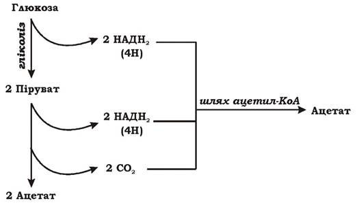
Fig. 6.33. Chemoorganoheterotrophic metabolism of homoacetogenic bacteria
Homoacetogens form the third acetate molecule by reducing 2СО2 (released in the previous reaction) via the acetyl-CoA pathway. Hydrogen, which also originates from glucose, is used to reduce СО2. The overall equation for glucose oxidation by homoacetogens is:
Glucose → 3 Acetate
Energetics of acetate formation in homoacetogens. Most homoacetogens can grow on a gas mixture of СО2 and Н2, using hydrogen as their sole electron source. This implies that the reduction of СО2 to acetate must be coupled with ATP synthesis. One mole of ATP is generated via the acetyl-CoA pathway during the conversion of acetyl phosphate to acetate. At the same time, formate activation coupled with formyltetrahydrofolate formation stoichiometrically consumes 1 ATP. Therefore, the branch of the acetyl-CoA pathway leading to cellular carbon synthesis does not serve as a source of substrate-level phosphorylation ATP. ATP accumulation during СО2 reduction to acetate is further hindered by the Energy Expenditure required for CO formation from СО2. It has been demonstrated for Acetobacterium woodii that the reduction of СО2 to CO is coupled to the utilization of a proton gradient. Under standard conditions, the Free energy change for the reduction of СО2 to CO is +20 kJ/mol. Calculations indicate that to energize this reaction, at least one proton must return into the cell across the membrane. In other words, reducing 1СО2 to 1СО "costs" the cell 1/3 of an ATP molecule. A similar phenomenon has been described in methanogens when they utilize the acetyl-CoA pathway for СО2 assimilation. This represents one of the few reactions in bacterial catabolism associated with Reverse Electron Transport.
Since CO formation and formate activation are endergonic processes, the cell must rely on one or more exergonic reactions. The only sufficiently exergonic reaction is the reduction of methylene-THF to methyl-THF (see above). It has been found that cell Suspensions of Acetobacterium woodii grown on СО2+Н2 generate a sodium ion concentration gradient. Research has shown that in methanogenic bacteria, the reduction of methylenetetrahydromethanopterin to methyl derivatives during methanogenesis from СО2 is accompanied by sodium translocation across the membrane. By analogy with methanogens, it was concluded that sodium ion translocation out of the cell must also occur during the reduction of methylene-THF to methyl-THF. This mechanism can generate an electrochemical sodium gradient. How this sodium gradient is converted into ATP remains unclear. Most likely, it is converted into a proton gradient via a sodium/proton (Na+/H+) antiporter, the presence of which has been demonstrated in Clostridium thermoaceticum. The proton gradient established by the Na+/H+ antiporter can then drive ATP synthesis via ATP synthase.
Owing to their broad substrate utilization spectrum, homoacetogens inhabit virtually all anoxic freshwater (and some marine) ecological niches, including freshwater sediments, sewage, the gut of ruminants, termites, and rodents, animal and human feces, and cyanobacterial mats. A crucial environmental requirement for homoacetogens is an adequate supply of molecular hydrogen, for which they compete successfully with methanogens, gaining a competitive edge under psychrophilic conditions below 20 0C. In ecological niches rich in organic matter (such as feces), homoacetogens preferentially utilize energetically more favorable substrates over molecular hydrogen.
Last update: 13/08/2026
Editorial and Educational Adaptation: This material has been compiled based on the primary/original source text. The project team performed an editorial review, corrected technical inaccuracies, structured sections, and adapted the content for an educational format.
What was processed:
- elimination of formatting defects (OCR errors, structural breaks, corrupted characters);
- editorial organization of content;
- standardization of terminology in accordance with academic sources;
- verification of factual statements against the original source text.
All mentions of the author, publication year, and origin of the primary text have been preserved in accordance with the source.