MICROBIOLOGY - M.H. Serhiichuk - 2008
Chapter 6. PHYSIOLOGICAL CHARACTERISTICS OF ANAEROBIC MICROORGANISMS
Methanogenesis
Organisms capable of producing methane belong to a distinct group of living entities known as archaea. Let us examine their specific features in greater detail.
In the late 1970s, phylogenetic studies based on rRNA demonstrated that archaea (Archaea), previously referred to as archaebacteria, constitute a unique group of living organisms separate from Bacteria (Eubacteria) and Eukarya.
Archaea are divided into two main phylogenetic groups: Crenarchaea and Euryarchaea (Fig. 6.34). All Crenarchaea are thermophiles that utilize sulfur compounds in their METABOLISM, whereas Euryarchaea comprise several thermophiles (sulfur-utilizing), methanogens, and extreme halophiles.
Class="center">
Fig. 6.34. Phylogeny of archaea (selected genera are shown; halophiles and methanogens are indicated by bold lines)
Archaea exhibit an extraordinary diversity of lifestyles and metabolic capabilities. Many species are adapted to extreme environmental conditions, such as high salinity, temperatures near the boiling point of Water, and extreme pH levels.
Phenotypically, archaea closely resemble bacteria. They are primarily small (0.5–5 µm) rods, cocci, spirilla, or filamentous forms, and they reproduce by fission. However, most archaea are thermophiles, and many are extreme thermophiles. They frequently possess an autotrophic metabolism or respire sulfur. Much like eukaryotes, archaea contain a high proportion of histone-like Proteins and possess DNA organized into nucleosome-like structures.
Methanogens (methanogenic archaea) are a group of archaea that produce methane (CH4) as a byproduct of their metabolism. Methanogenesis is the exclusive pathway by which methanogenic archaea obtain energy for growth, and they are the only known organisms capable of generating methane as a catabolic end product.
Taxonomically, all methanogens belong to the ancient kingdom Euryarchaeota and are classified into five orders: Methanobacteriales, Methanococcales, Methanomicrobiales, Methanosarcinales, and Methanopyrales. Among these, only Methanosarcinales can ferment acetate to CH4 and CO2, and grow on methanol, methylthiols, and methylamines as sole Energy Sources. Hyperthermophilic species are found exclusively within the orders Methanobacteriales, Methanococcales, and Methanopyrales.
In addition to the cytoplasmic membrane and Cell wall characteristics common to all archaea, methanogens contain unique Coenzymes involved in The transfer of hydrogen and C1 compounds during methanogenesis: coenzyme F420, methanofuran, tetrahydromethanopterin, coenzyme M, factor B, coenzyme F430, and methanophenazine.
Coenzyme F420 is a flavin derivative whose Structure resembles the common coenzyme FMN, but it differs in The structure of the R-group, lacks a nitrogen atom in the middle ring, and lacks a methyl group on the benzene ring (Fig. 6.35).
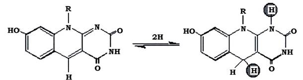
Fig. 6.35. Oxidized and reduced forms of factor F420
The Redox Potential of F420 is quite low (E0' = -380 mV). It interacts with several distinct methanogenic Enzymes, including Hydrogenase and NADP+ reductase. The oxidized form of F420 absorbs light at 420 nm and fluoresces blue-green, allowing methanogens to be readily identified using Fluorescence Microscopy. Upon reduction, F420 loses its color. Within The Cell, reduced F420 performs the following Functions:
1. Transfers 2[H] to NADP+ and ferredoxin in biosynthetic reactions unrelated to methane formation.
2. Transfers 2[H] to carbon compounds during methanogenesis.
Methanofuran (MRF, MF) is a low-molecular-weight coenzyme that serves as
the primary acceptor of CO2 in methanogenesis. Following the initial CO2 reduction step, formylmethanofuran is produced. Methanofuran is composed of a phenol moiety, two glutamic acid residues, a long fatty acid chain, and a furan ring (Fig. 6.36).
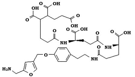
Fig. 6.36. Structure of methanofuran
Tetrahydromethanopterin (THMP, MP, H4MPT) is a coenzyme containing a substituted pterin ring, structurally similar to the vitamin Folic acid (Fig. 6.37). It acts as a carrier of C1 units throughout most stages of methanogenesis, ranging from the formyl to the methyl level.
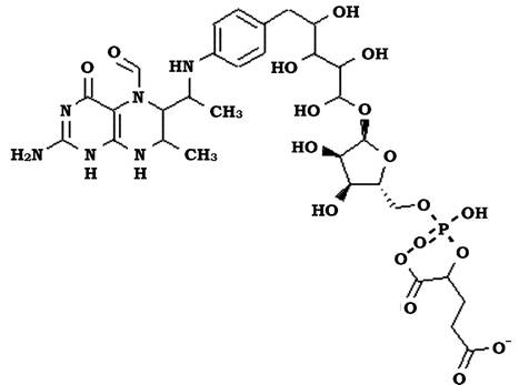
Fig. 6.37. Structure of tetrahydromethanopterin
Coenzyme M (2-mercaptoethanesulfonic acid, (H-S-CoM)) functions as a methyl group carrier. It features a very simple structure (Fig. 6.38).

Fig. 6.38. Structure of cofactor M
Coenzyme M is a highly specific carrier in the methyl reduction reaction, which proceeds via the methylreductase-F30 complex during The final stage of methanogenesis. Certain microorganisms, notably Methanobrevibacter ruminantium, cannot synthesize this compound independently and therefore require it as a growth factor. The coenzyme M analogue, bromoethanesulfonic acid, acts as a potent inhibitor of methanogenesis and can be used experimentally to selectively suppress methanogenic archaea when studying anaerobic processes. It is the smallest of all known coenzymes, found exclusively in methanogens.
Factor B (coenzyme B, H-S-CoB), or 7-mercaptoheptanoylthreonine phosphate (HTP-SH, HS-HTP), is a phosphorylated derivative of The amino acid Threonine, structurally similar to the vitamin pantothenic acid (Fig. 6.39).
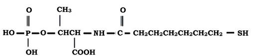
Fig. 6.39. Structure of factor B
Much like coenzyme M, it participates in the terminal reactions of methanogenesis and also catalyzes the methylreductase system by serving as an electron donor for it. Factor B reacts with methyl-CoM to form the CoM-S-S-HTP disulfide.
Coenzyme F430 is a yellow-colored, nickel-containing soluble tetrapyrrole (Fig. 6.40). It absorbs light at 430 nm, but unlike F420, it is non-fluorescent. The high nickel content in methanogen Cells is associated with the presence of F430. This coenzyme is of great importance in the final stage of methanogenesis, as it forms part of the methylreductase system.

Fig. 6.40. Structure of coenzyme F430
Methanophenazine (MP) is a low-molecular-weight, membrane-bound electron and hydrogen carrier (Fig. 6.41). It is reduced by F420 dehydrogenase or hydrogenase and, in turn, reduces the heterodisulfide during the final stages of methanogenesis. It has been detected only in methanogens growing on methylated compounds.
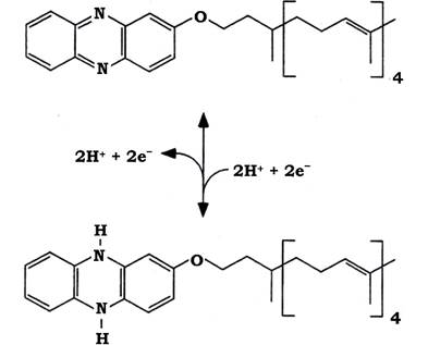
Fig. 6.41. Structure of methanophenazine in its oxidized and reduced states
Metabolism of methanogens. Methanogens convert the following substrates into methane:
1. Compounds containing oxidized carbon: carbon dioxide CO2, formate HCOOH, and carbon monoxide CO.
2. Compounds containing a methyl group: methanol CH3OH, methylamine CH3NH3+, dimethylamine (CH3)2NH2+, trimethylamine (CH3)3NH+, methyl mercaptan CH3SH, and dimethyl sulfide (CH3)2S.
3. Acetic acid CH3COOH.
These substrates are converted by methanogens with the release of energy According to the following equations:
СО2 + 4Н2 → СН4 + 2Н2О ΔG0'= -134 kJ/reaction
4НСООН → СН4 + 3СО2 + 2Н2О ΔG0'= -281 kJ/reaction
4СО + 2Н2О → СН4 + 3СО2 ΔG0'= -209 kJ/reaction
4СН3ОН → 3СН4 + СО2 + 2Н2О ΔG0'= -323 kJ/reaction
СН3ОН + Н2 → СН4 + Н2О ΔG0'= -133 kJ/reaction
4СНзКНзОІ + 2НзО → зСН4 + СО2 + 4NH4Cl ΔG0'= -230 kJ/reaction
СН3СООН → СН4 + СО2 ΔG0'= -130 kJ/reaction
Methanogenesis is sometimes viewed as a process of Anaerobic Respiration and referred to as carbonate respiration, because when methanogens grow on a mixture of СО2 and Н2, molecular hydrogen serves as the electron donor, while carbon dioxide acts as the acceptor. At the same time, archaea lack a traditional
Respiratory Chain with the classical set of electron carriers. The term "methane Fermentation" is frequently found in literature. When methanogens grow on methanol, one substrate molecule is oxidized while another is reduced, resembling the amino acid fermentation process. However, it is known that during methanogenesis, ATP is synthesized through the generation of a proton and sodium ion gradient, which is not typical for classical fermentation. Methanogens lack substrate-level phosphorylation. Thus, methanogenesis can be regarded neither as anaerobic respiration nor as fermentation. It is a unique, specific energy-yielding process characteristic only of ancient living forms—archaea.
METHANE FORMATION DURING the growth of methanogens on a mixture of СО2 and Н (Fig. 6.42). The formation of methane from a mixture of СО2 and Н2 involves eight reactions listed below:
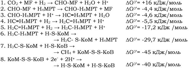
As can be seen from the equations, СО2 is reduced stepwise, and the resulting C1 compounds are continuously bound to one of three carriers: methanofuran (MF), tetrahydromethanopterin (Н4МРТ), and coenzyme M (H-S-KoM).
Reaction 1. The initial acceptor of СО2 is methanofuran (MF). Carbon dioxide activated by methanofuran is reduced to the formyl level with the formation of formylmethanofuran. The reduction proceeds with hydrogen, although the hydrogen carrier in this reaction has not yet been precisely identified. The formation reaction of formyl-MF is endergonic.
Reaction 2. The formyl group is transferred from methanofuran to tetrahydromethanopterin.
Reaction 3. Dehydration of formyltetrahydromethanopterin (CHO-H4МРТ) with the formation of methenyltetrahydromethanopterin (HC= Н4МРT).
Reaction 4. Reduction of methenyltetrahydromethanopterin (H2C=ЩМРT) to methylenetetrahydromethanopterin (H2C=Н4МРT). The hydrogen carrier in this reaction is coenzyme F420.
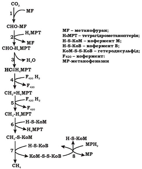
Fig. 6.42. Scheme of methane formation during the growth of methanogens on a mixture of Н2 and СО2
Reaction 5. Reduction of methylenetetrahydromethanopterin (Н2С=Н4МРТ) to methyltetrahydromethanopterin (Н3С-Н4МРТ). The hydrogen carrier in this reaction is coenzyme F420.
Reaction 6. The methyl group is transferred to coenzyme M. The process is accompanied by energy release (ΔG0' = -29.7 kJ/mol) and the export of Na+ ions into the periplasm. The pump for Na+ transport is the enzyme methyl-H4MPT:CoM methyltransferase, which is part of the central metabolic pathway of methanogens. Therefore, the export of Na+ into the periplasm is obligatory during methanogenesis and leads to the generation of a transmembrane Na+ gradient. The methyltransferase contains a cofactor (factor III or Co(III)) that participates in methyl conversion.
The Co(III) cofactor in its superreduced state is designated as Co(I). The Mechanism of sodium ion gradient generation (ΔμNa+) is shown in Fig. 6.43.
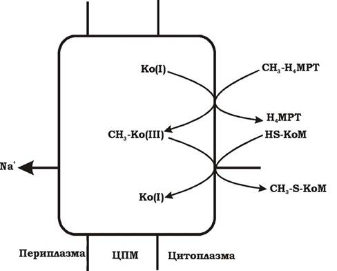
Fig. 6.43. Scheme of the sodium ion transport mechanism involving methyl-Н4МРТ: CoM methyltransferase
The transfer of the methyl group from Н4МРТ to CoM occurs in two steps. In the first step, Co(I) attaches the methyl group, being thereby oxidized to Co(III) and forming the intermediate product СН3-Co(III). In the second step, СН3-Co(III) transfers the СН3 group to coenzyme M, forming СН3-S-CoM. It is precisely in the second step of the process that the translocation of sodium ions occurs, coupled with the demethylation reaction of the methyltransferase cofactor.
Reaction 7. Methyl-CoM is reduced to methane. The reduction and demethylation process is catalyzed by methyl-CoM reductase and proceeds with the participation of electrons supplied from reduced coenzyme B; cofactor F430 is part of the methylreductase system. As a result, methane and the heterodisulfide of CoM and CoB (CoM-S-S-KoB) are formed.

Reaction 8. The CoM-S-S-KoB heterodisulfide is reduced by the heterodisulfide reductase enzyme complex. Electrons for its reduction are supplied from reduced methanophenazine (MP), a membrane-bound low-molecular-weight carrier.

This reaction is essential for completing the cycle and is critical for energy conservation. Heterodisulfide reductase is membrane-bound and functions as the terminal segment of several Electron Transport Chains.
Fig. 6.44 illustrates the relationship between reactions 7 and 8.

Fig. 6.44. Final reactions (7, 8) of methanogenesis:
7 — reaction of methane formation; 8 — reduction of the heterodisulfide CoM-S-S-CoB
The amount of energy released as a result of the reduction of methyl-CoM to CH4 is sufficient for the translocation of protons into the periplasm, i.e., for the generation of a proton gradient (ΔμΗ+).
The Biosynthesis of Organic compounds in methanogens growing on a mixture of CO2 and H2 proceeds via a modified acetyl-CoA pathway (Fig. 6.45).
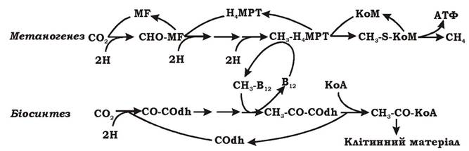
Fig. 6.45. Scheme of autotrophic metabolism in methanogens:
MF — methanofuran; H4MPT — tetrahydromethanopterin; CoM — coenzyme M; COdh — CO dehydrogenase
During autotrophic growth on a gas mixture of CO2 and H2, methanogens assimilate CO2 via the modified acetyl-CoA pathway, the end metabolite of which is acetyl-CoA, which is incorporated into the biosynthesis of all essential cellular components. Since the methyl group of acetyl-CoA is formed during methanogenesis, these microorganisms do not need to synthesize it de novo. Therefore, the modified acetyl-CoA pathway contains only a single branch in which CO2 is reduced to a carbonyl group. The methyl group, in turn, is formed as an intermediate during Catabolism and, following activation by a Vitamin B12-containing coenzyme, enters the acetyl-CoA pathway.
STRUCTURE OF THE Electron Transport Chain in methanogens. The electron transport chain of methanogens may include the following enzymes and carriers:
- Hydrogenase — the entry point for electrons derived from molecular hydrogen into the respiratory chain when methanogens utilize H2 as an electron donor.
- F420 dehydrogenase — the entry point for electrons released during The oxidation of F420H2, a universal hydrogen carrier in methanogens.
- Heterodisulfide reductase — an enzyme involved in the reduction of the heterodisulfide CoM-S-S-CoB.
- Methanophenazine (MP) — a low-molecular-weight, membrane-bound electron and hydrogen carrier. It is reduced by F42o dehydrogenase or hydrogenase and, in turn, reduces the heterodisulfide CoM-S-S-CoB.
- Cytochromes of the b and c types.
Based on the composition of their membrane-bound electron carriers and the mechanism of proton gradient generation, methanogens can be divided into two groups:
1. Methylotrophic organisms, such as Methanosarcina barkeri, M. mazei (possessing various b- and c-type cytochromes and the methanophenazine carrier).
2. Hydrogen-utilizing methanogens, such as Methanobacterium thermoautotrophicum (lacking cytochromes, but containing polyferredoxins and flavoprotein analogs).
Methanogens utilize various mechanisms for proton gradient generation depending on the substrate and the composition of electron carriers. Possible variants of their respiratory chains are shown in Fig. 6.46.
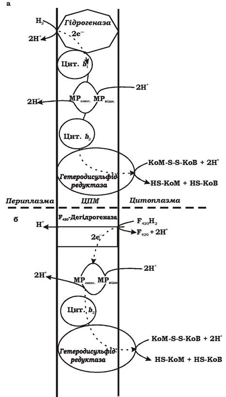
Fig. 6.46. Hypothetical scheme of proton gradient generation in methanogens at the stage of heterodisulfide CoM-S-S-CoB reduction during growth on various substrates: MP — Methanophenazine
ATP Synthesis in methanogens. Methanogens are organisms that generate two ion gradients: a proton gradient (ΔμΗ+) and a sodium gradient (ΔμNa+). Both of these gradients are involved in ATP synthesis. Protons flow back into the cell via ATP synthase (H+-ATPase), which is coupled to ATP synthesis. Regarding the Mechanism of ATP synthesis driven by the sodium ion gradient, two hypotheses exist. According to the first hypothesis, some methanogens, such as Methanobacterium thermoautotrophicum, possess a Na+-ATPase in addition to the H+-ATPase, which ensures ATP synthesis using the Na+ gradient. According to the second hypothesis, sodium ions re-enter the cell via a Na+/H+ antiporter, which drives the extrusion of protons. In this case, ATP synthesis is carried out solely by the H+-ATPase, while the Na+ gradient serves as an additional energy source.
Methane formation during growth of methanogens on methanol. Methanol (CH3OH) occupies an intermediate oxidation state of carbon between CO2 and CH4, and therefore it can be both oxidized and reduced. The oxidation of methanol to carbon dioxide releases three pairs of electrons, 3[2H]. The anaerobic oxidation of the methyl group is a thermodynamically unfavorable process because electrons from the methyl group ($E^{0'}_{\text{methanol/formaldehyde}} = -180\text{ mV}$) must be transferred to the F420 carrier ($E^{0'} = -340\text{ mV}$); thus, growth on methanol imposes an additional energy cost on methanogens. To balance the redox processes, the three pairs of electrons generated during the oxidation of the methyl group must reduce a suitable acceptor. Other methanol molecules serve as such acceptors. The reduction of one molecule of methanol to methane consumes one pair of electrons. Consequently, for the oxidation of one molecule of methanol, three molecules must be reduced.

The reaction of methane formation coupled with the reduction of the methyl group is linked to the generation of a proton gradient and ATP synthesis. The metabolic pathway of methanogens growing on methanol is illustrated in Fig. 6.47.
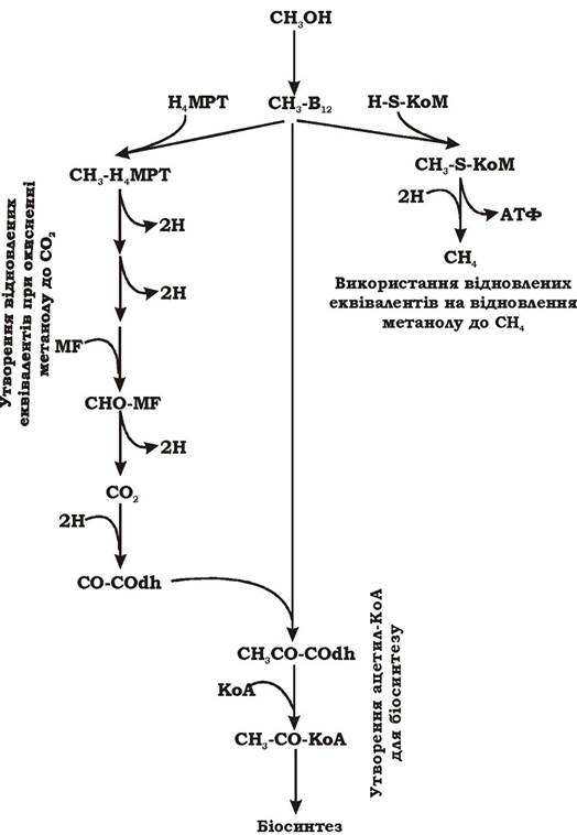
Fig. 6.47. Scheme of methanogenic metabolism during growth on methanol:
MF - methanofuran; H4MPT - tetrahydromethanopterin; COdh - CO dehydrogenase; H-S-CoM - coenzyme M; B12 - coenzyme with vitamin B12
As shown in the figure, The conversion of methanol begins with the activation of the methyl group by a vitamin B12-containing coenzyme. Then, one activated methyl group is oxidized to CO2, generating three pairs of reducing equivalents and switching the carbon carrier to H4MPT and MF (left branch). The subsequent activated methyl groups are reduced to methane utilizing the reducing equivalents produced during the oxidation of the methyl group. The release of methane is coupled with ATP synthesis (right branch). Part of the methyl group from methanol and the CO2 formed
during the oxidation reactions is channeled into biosynthesis via a modified acetyl-CoA pathway.
Methane production during growth of methanogens on acetate. Acetate is cleaved by methanogens using a large enzyme complex into CO and a methyl group, which at various Stages of the process is bound to different carriers. Subsequently, CO is oxidized to CO2 by CO dehydrogenase. The released electrons reduce methyl-CoM to methane, establishing a proton gradient and ultimately ATP (Fig. 6.48). The overall reaction equation is as follows:

Since the Cleavage products of acetate act as electron Donors (CO) and acceptors (methyl-CoM), acetate-dependent methanogenesis resembles fermentation. However, ATP in this case is synthesized not via substrate-level phosphorylation, but rather driven by ΔμH+.
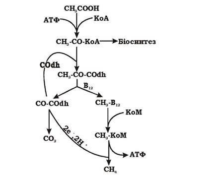
Fig. 6.48. Scheme of methanogenic metabolism during growth on acetate: CoM - coenzyme M; COdh - CO dehydrogenase; B12 - coenzyme with vitamin B12
Last update: 13/08/2026
Editorial and Educational Adaptation: This material has been compiled based on the primary/original source text. The project team performed an editorial review, corrected technical inaccuracies, structured sections, and adapted the content for an educational format.
What was processed:
- elimination of formatting defects (OCR errors, structural breaks, corrupted characters);
- editorial organization of content;
- standardization of terminology in accordance with academic sources;
- verification of factual statements against the original source text.
All mentions of the author, publication year, and origin of the primary text have been preserved in accordance with the source.