Molecular Biology of the Cell - Volume 3 - Alberts B., Bray D., Lewis J., Raff M., Roberts K., Watson J. 1994
From Cells to Multicellular Organisms
The Nervous System
Non-channel-linked Receptors and Synaptic Modulation
At synapses utilizing channel-linked receptors, Neurotransmitters mediate rapid, simple, and brief effects with a high degree of spatial precision. A neurotransmitter released by a single axon terminal affects only one postsynaptic Cell. In contrast, non-channel-linked receptors can trigger slow, complex, and prolonged effects that are frequently spatially diffuse. In such cases, a transmitter released from a single terminal can influence multiple neighboring Cells simultaneously. These slow effects are often cited as Examples of neuromodulation, as they modulate the rapid responses mediated by channel-linked receptors on the same cell. The mechanisms underlying non-channel-linked receptors are identical to those mediating the actions of Hormones and local chemical mediators outside The Nervous system; indeed, many receptors in both systems are likely identical.
As discussed in Chapter 12 (Section 12.3.1), cell-surface receptors for signaling molecules that are not coupled to Ion Channels fall into two major categories: (1) catalytic receptors, which are predominantly receptor Tyrosine Kinases that become activated upon Ligand binding and phosphorylate tyrosine residues on intracellular Proteins; and (2) G protein-coupled receptors, which transmit signals across the membrane by activating a regulatory GTP-binding protein (G protein), which in turn activates or inhibits a membrane-bound enzyme or ion channel. Evidently, the majority of known neurotransmitter receptors not linked to channels operate via G proteins, participating in signal Transduction through one of three pathways:
1. The G protein can stimulate or inhibit adenylyl cyclase, thereby regulating cyclic AMP levels within the postsynaptic cell. In turn, cyclic AMP modulates The activity of cAMP-dependent protein kinase (Protein Kinase A—see Section 12.4.1), which, alongside other target proteins, can phosphorylate Plasma Membrane ion channels and alter their properties. Cyclic AMP can also directly gate certain ion channels by binding to them.
2. The G protein can trigger the Inositol phospholipid pathway (Section 12.3.9), activating protein kinase C (Protein Kinase C) and inducing the release of Ca2+ from intracellular calcium stores into the postsynaptic Cytosol. Protein Kinase C regulates ion channel function through phosphorylation. Ca2+ ions can similarly affect ion channel behavior, either directly or indirectly via a Ca2+-dependent protein kinase that phosphorylates the channel (Section 12.4.3).
3. The G protein can interact directly with ion channels to promote their opening or closing. In each case, specific molecules act within the postsynaptic cell as links, or intracellular messengers, diffusing through the Cytoplasm to Relay the signal from the receptor to other cellular components. The more steps involved in this intracellular messenger cascade, the greater the potential for signal Amplification and regulation (Section 12.4.6).
Class="center">
Figure 19-36. A small bundle of autonomic motor axons innervating smooth Muscle Cells of the Ureter. Scanning electron micrograph. The varicosities contain synaptic vesicles packed with the neurotransmitter norepinephrine. Synapses are poorly defined here; the distance between the site of neurotransmitter release and the membrane of the nearest target muscle cell can reach 0.2 µm. (S. Tachibana et al., J. Urol., 134, 582-568, 1985. Copyright by Williams & Wilkins, 1985.)
Over 50 neurotransmitters have been identified that interact with non-channel-linked receptors to elicit diverse and complex effects. Some of these, such as acetylcholine, can also bind to channel-linked receptors, whereas others, like Neuropeptides (see below), apparently cannot.
19.5.1. Non-Channel-Linked Receptors Mediate Slow and Diffuse Effects [32]
While channel-linked receptors induce electrical Changes in the postsynaptic cell within milliseconds or less, non-channel-linked receptors require hundreds of milliseconds or longer to produce their effects. This temporal lag is expected, as a cascade of enzymatic reactions must occur between the initial signal and the ultimate cellular response. Furthermore, the signal itself is frequently dispersed both temporally and spatially.
A striking example is the innervation of smooth muscle by axons releasing norepinephrine, which activates adenylyl cyclase via a G protein-coupled receptor. In this system, the neurotransmitter is not released from specialized nerve terminals, but rather from varicosities or swellings distributed along the axon (Figure 19-36). Although these varicosities contain synaptic vesicles, they lack active zones that define precise sites of release. Moreover, the varicosities do not form tight junctions with specialized receptive areas on the postsynaptic cell; instead, the neurotransmitter diffuses over considerable distances, affecting multiple neighboring cells simultaneously (much like a local chemical mediator—see Section 12.7). It is likely that many signaling molecules interacting with catalytic or G-protein-coupled receptors in the Central Nervous System operate in this paracrine fashion. Indeed, many neurotransmitters also serve as hormones or local chemical mediators outside the nervous system: for example, norepinephrine, along with its close relative epinephrine, is secreted as a hormone from the Adrenal Glands.
Epinephrine and norepinephrine belong to the monoamine family of neurotransmitters, which fulfill diverse Functions in both vertebrates and invertebrates and are of major clinical importance (Figure 19-37A). Pharmacological agents can be engineered to interfere with the synthesis, reuptake, or degradation of specific monoamines, or to interact selectively with receptor subtypes. Some of these drugs have proven invaluable in treating neurological and psychiatric disorders. For instance, medications that block specific dopamine receptors are frequently used with great success in treating Schizophrenia, whereas drugs that elevate Brain dopamine levels dramatically improve motor function in Parkinson's disease (Figure 19-37B). Similarly, medications that increase the concentration of norepinephrine and/or serotonin in synapses are often effective in treating major depression.

Figure 19-37. (A) Monoamine neurotransmitters. (B) Distribution of dopamine-containing Neurons in the human brain. The motor coordination deficits observed in Parkinson's disease stem from the degeneration of a specific group of dopaminergic neurons (located in the substantia nigra). The symptoms can be alleviated with drugs that promote dopamine synthesis and inhibit its degradation. The distribution of monoaminergic neurons can be visualized by treating tissue sections with formaldehyde, which reacts with monoamines to yield fluorescent products.
19.5.2. Neuropeptides Constitute the Largest Group of Neurotransmitters [32, 33]
Most signaling molecules utilized throughout the body are also employed by neurons. This is particularly true for small protein molecules or Peptides that function as hormones and local mediators to regulate processes such as Blood pressure maintenance, digestive enzyme secretion, and cell proliferation.
Significant advances in The Study of neuropeptides have been achieved over the past decade, largely driven by immunocytochemistry. Antibodies can be raised against a peptide isolated from a particular tissue and then used to screen for the same or structurally related peptides elsewhere in the Organism. This approach has led to the discovery of peptides in neurons that were previously unknown in the nervous system, alongside many novel peptide families. In most cases, the evidence that these neuropeptides (Figure 19-38) function as neurotransmitters is compelling yet incomplete. For example, antibodies against a specific peptide can be shown to bind to particular neurons and their axon terminals, and local application of the peptide can mimic the Physiological effects of neuronal activity. More convincingly, it is sometimes possible to demonstrate that active neurons secrete a specific peptide and that the resulting physiological response is blocked by antibodies directed against that peptide. Neuropeptides appear to play a particularly prominent role in modulating sensations and drives such as pain, pleasure, hunger, thirst, and Sexual Behavior.
Non-peptide neurotransmitters are synthesized via enzymatic machinery typically located in both The Cell body and the axon terminal, allowing synaptic stores to be rapidly replenished even in lengthy axons. In contrast, neuropeptides are synthesized on Ribosomes of the rough Endoplasmic reticulum in the cell body and transported to axon terminals via fast axonal transport—a journey that can take a day or longer in long axons. Neuropeptides are generated from larger precursor proteins through proteolytic Cleavage. Often, the cleavage of a single precursor molecule yields multiple biologically active peptides; in such cases, the precursor is referred to as a polyprotein. Synaptic vesicles packed with neuropeptides can usually be identified by their Morphology: they are larger than vesicles containing acetylcholine, amino acid Transmitters, or monoamines.
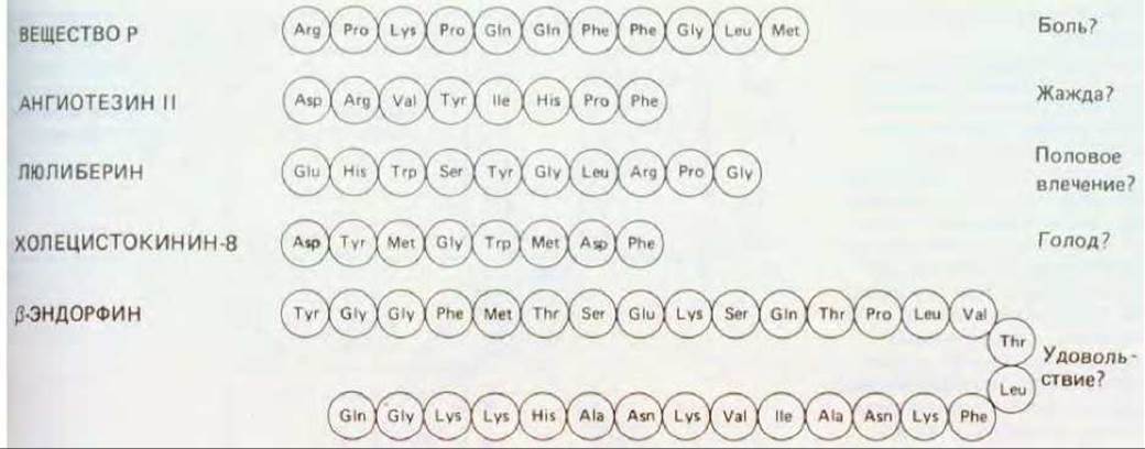
Figure 19-38. Selected neuropeptides, along with the sensations and motivational states they are thought to modulate.
At many synapses, neuropeptides are secreted alongside non-peptide transmitters, acting co-ordinately but through distinct mechanisms. For instance, presynaptic axon terminals in certain bullfrog autonomic ganglia contain, In addition to acetylcholine, a peptide structurally related to luteinizing hormone-releasing hormone (LHRH). The postsynaptic membrane contains at least three receptor types: (1) nicotinic (channel-linked) acetylcholine receptors, which mediate rapid signaling; (2) muscarinic (G protein-coupled) acetylcholine receptors, which mediate slower responses; and (3) receptors (likely also G protein-coupled) for the LHRH-like peptide, which mediate the slowest responses (Figure 19-39A). The action of the LHRH-like peptide is not only slower than that of acetylcholine but also more diffuse, causing peptide molecules released at a synapse on one postsynaptic cell to evoke postsynaptic potentials in adjacent neighboring cells as well (Figure 19-39B).
If (as seems likely) other neuropeptide transmitters share these properties, it becomes easy to understand why such a vast array of diverse neuropeptides is required. Because peptides diffuse readily, their sites of action are not restricted to their precise points of release. Therefore, to ensure that peptides released from distinct neighboring presynaptic terminals act selectively on appropriate postsynaptic targets, the peptides and their corresponding receptors must be chemically distinct.
19.5.3. Long-Lasting Behavioral Changes Involve Modifications at Specific Synapses [34]
Effects mediated by non-channel-linked receptors are distinguished by both their prolonged duration and delayed onset. This largely accounts for their specialized role in behavioral regulation: they induce enduring alterations in the nervous system's immediate responsiveness to incoming external signals and likely constitute the cellular basis for at least some forms of memory. This has been most clearly demonstrated in studies of the marine gastropod mollusk Aplysia (see Figure 19-49). In this organism, acquired behavioral modifications can be traced to specific neural circuits, allowing researchers to decipher the underlying molecular mechanisms.

Fig. 19-39. Responses to a peptide neurotransmitter.
A. Three Components of the postsynaptic potential generated in a frog ganglion cell following stimulation of the presynaptic nerve. Two neurotransmitters are released from the presynaptic axon terminal: acetylcholine and a peptide very similar to luliberin (luteinizing hormone-releasing hormone). Typically, a complex PSP is the sum of responses mediated by Three types of receptors—two variants of acetylcholine receptors and one receptor for the luliberin-like peptide. THE CONTRIBUTION OF each of the three components can be assessed by selectively blocking the receptors responsible for the other two components using specific toxins. Only the fast excitatory PSP, mediated by channel-coupled acetylcholine receptors, is large enough to trigger an Action Potential. The two slow components, likely mediated by non-channel-coupled receptors, alter cell excitability, making the cell more responsive to stimulation immediately following the initial stimulus. B. Experimental setup demonstrating the diffuse Nature of the neuro peptide's effect (similar to luliberin) on the same ganglion. This peptide is released simultaneously with acetylcholine at synapses formed on a single group of cells (C-cells), but diffuses over a distance of tens of micrometers to elicit a delayed slow PSP in other cells (B). (A - after Y.N. Jan et al., Cold Spring Harbor Symp. Quant. Biol., 48, 363-374, 1983.)
Aplysia withdraws its gill in response to siphon touching (Fig. 19-40). Following repeated touches, the animal exhibits habituation and the response disappears. In its biological function, habituation resembles adaptation, but it develops more slowly and, as we will see, involves a different part of the neural pathway. Any harsh stimulus, such as a strong jolt or an electric Shock, reverses the habituation effect and, conversely, heightens the animal's sensitivity, causing it to react with particular vigor to subsequent touches to the siphon. This sensitization effect persists for many minutes or even hours, depending on the strength of the eliciting stimulus, and represents a simple form of short-term memory. If the noxious stimulus is repeated over several days, sensitization (i.e., memory expression) becomes long-lasting and persists for several weeks. These behavioral modifications have been linked to changes occurring in a specific set of synapses within the neural circuit responsible for the gill-withdrawal reflex. Aplysia possesses very large (100 µm) neurons that are relatively few in number (~105) and can be individually identified by their appearance and Location. Touching the siphon leads to the excitation of a group of sensory neurons. These neurons form excitatory synapses on the motor neurons responsible for gill withdrawal. Changes at these synapses underlie these behavioral phenomena. During habituation, the amplitude of the postsynaptic potential in these motor neurons decreases upon repeated stimulation by their sensory cells. During sensitization, the opposite effect is observed—the postsynaptic potential increases. In both cases, The change in potential amplitude results from an alteration in The amount of neurotransmitter released from the presynaptic terminals of the activated sensory neurons. Thus, the problem reduces to the question of how neurotransmitter release is regulated at these synapses.

Рис. 19-40. Морская улитка Aplysia punctata (вид сверху). Лежащий сверху лоскут Ткани отогнут, что позволяет видеть жабру, защищенную мантией и раковиной. (По J. Giart, Mem. Soc. Zool. France, 14, 219, 1901.)
19.5.4. G protein-coupled receptors are responsible for sensitization in Aplysia [35]
As noted in Section 19.3.2, the amount of neurotransmitter released at synapses is regulated by the influx of Ca2+ ions into the nerve terminal during an action potential. In habituation, repetitive excitation of sensory cells modifies channel proteins at their axon terminals in such a way that Ca2+ influx into the cell is reduced, thereby decreasing the amount of neurotransmitter released. Conversely, in sensitization, these proteins are altered such that Ca2+ entry increases and neurotransmitter secretion is enhanced. The molecular mechanisms underlying these sensitization-related changes have been studied most thoroughly.

Рис. 19-41. Упрощенная схема нейронных путей, участвующих в привыкании и сенситизации по отношению к рефлексу втягивания жабры у аплизии. Показано только по одному нейрону из каждой группы.
During sensitization induced, for example, by blows to the HEAD, the amount of neurotransmitter released by sensory neurons changes As a result of the excitation of an additional group of neurons that respond to the noxious stimulus. These facilitating neurons form synapses on the presynaptic terminals of the sensory neurons (Fig. 19-41) that release serotonin (along with certain neuropeptides). The Effect of facilitating neurons can be mimicked by applying serotonin directly to the membrane of sensory neurons, whose presynaptic axon terminals bear serotonin receptors. The action of these receptors is G protein-mediated: binding of serotonin to the receptors activates adenylate cyclase, resulting in an elevation of intracellular cyclic AMP concentration, which in turn activates A-kinase (Section 12.4.1). It is this protein kinase that alters the electrical Properties of the sensory neuron membrane by phosphorylating a specific group of potassium channels (Fig. 19-42).

Рис. 19-42. Механизмы, лежащие в основе адаптации и сенситизации рефлекса втягивания жабры у аплизии. Электрически активные Нейроны выделены цветом. На верхней схеме представлен нормальный механизм передачи от сенсорного нейрона к мотонейрону, участвующему во втягивании жабры. На обеих нижних схемах справа объяснен механизм возникновения в окончании сенсорного нейрона стойких изменений, лежащих в основе памяти, а слева показано, каким образом эти изменения влияют на синаптическую передачу от сенсорного нейрона к мотонейрону, ответственному за втягивание жабры. Представленное объяснение более достоверно для сенситизации, чем для адаптации.

Fig. 19-43. A. Chain of events occurring during sensitization of the gill-withdrawal reflex, leading to the inactivation of a specific class of potassium channels (so-called S-channels) in the sensory nerve terminal (see Fig. 19-42). B. Patch-clamp recordings of currents passing through these channels during transitions between open and closed states. An excised patch of membrane contains four channels that remain open most of the time under control conditions. When the catalytic subunit of cAMP-dependent protein kinase (A-kinase) is added to the cytoplasmic face of the medium, two of the four channels are phosphorylated and consequently close, whereas the remaining two channels continue to spend most of their time open; as a result, the current flowing through the membrane patch is halved compared to the control. (Patch-clamp data reprinted with permission from M.J. Schuster, J.S. Camardo, S.A. Siegelbaum, E.R. Kandel, Nature, 313, 392-395, 1985. Copyright 1985 Macmillan Journals' Limited.)
The behavior of these potassium channels, termed S-channels, can be examined in detail using the patch-clamp technique (see Section 6.4.17). Upon binding of serotonin to Membrane Receptors, these channels close (Fig. 19-43). Potassium channels close in an identical manner if the membrane patch containing them is transferred to a bath with an artificial medium where the channels undergo direct phosphorylation by the catalytic subunit of A-kinase. This suggests that phosphorylation of S-channels (or closely associated proteins) can maintain the channels in a closed state for a prolonged period. Since normally the potassium ion current helps restore the Resting Potential, blockade of S-channels prolongs the action potentials arriving at the axon terminal. These "prolonged" action potentials keep voltage-gated calcium channels open for a longer duration, thereby increasing calcium ion influx, which in turn leads to the discharge of a larger number of synaptic vesicles; consequently, a significantly larger postsynaptic potential is generated in the motor neuron, resulting in a more vigorous gill withdrawal.
These experiments demonstrate how G protein-coupled receptors can transmit short-term signals leading to persistent changes in the electrical properties of a synapse and, consequently, in the animal's behavior. Phosphorylation of S-channels represents a form of memory, but it is merely a short-term memory that is easily erased by phosphoprotein Phosphatases (which dephosphorylate S-channels) and is limited by the lifespan of the proteins comprising the S-channels. The Mechanism of long-term memory arising from repeated exposure to a noxious stimulus is not fully understood, but it differs from short-term memory: it requires the synthesis of new RNA and new proteins, as well as likely changes in both the Structure and chemistry of presynaptic terminals (Section 19.8.12).
It is believed that cyclic AMP and A-kinase also mediate such changes, likely through the phosphorylation of other cellular proteins capable of altering Gene Expression. Although this mechanism has not yet been elucidated in detail, one of the intermediate steps in establishing long-term memory traces apparently involves the sustained Activation of a-kinase resulting from a decrease in the concentration of Regulatory Subunits that inhibit this enzyme (see Section 12.4.1). It is thought that
at high cyclic AMP levels, these regulatory subunits are degraded because upon binding cyclic AMP they dissociate from the catalytic subunits and undergo proteolysis.
19.5.5. Ca2+ and cyclic AMP are important intracellular messengers involved in associative learning in vertebrates [36]
The phenomena of habituation and sensitization discussed above represent only very simple forms of learning. The primary feature of more complex types of learning, most widely studied by psychologists, is their associative nature. For instance, in Pavlov's famous experiments, a dog learned to associate the sound of a bell with the delivery of food. Aplysia is also capable of associative learning. For example, if a sensitizing stimulus (a strong electric shock) is consistently applied simultaneously with a specific mild stimulus that normally elicits only a weak gill-withdrawal reflex, the animal begins to behave as if it has learned that this specific mild stimulation is associated with the electric shock, becoming strongly and specifically sensitized to the weak stimulus. It is believed that the same groups of neurons are involved here as in the simple sensitization described earlier. Simultaneous stimulation of different PARTS OF THE body leads to the simultaneous excitation of sensory and facilitating neurons. Thus, when an action potential arrives at the sensory axon terminals, opening voltage-gated calcium channels within them, the facilitating neurons release serotonin (or a neuropeptide), which causes an increase in the intracellular concentration of cyclic AMP within the axon. Cyclic AMP alone triggers simple sensitization; influx of Ca2+ is thought to enhance this effect, resulting in a much stronger sensitization than would occur from the excitation of facilitating neurons in the absence of sensory neuron activity.
It remains unclear how far data obtained from Aplysia can be extrapolated. The question of whether memory traces in other animals are encoded as pre- or postsynaptic changes in the chemical nature or structure of synapses—or indeed within synapses at all—remains open. However, experiments on mutants of the fruit fly Drosophila suggest that molecular mechanisms similar to those described in Aplysia underlie many Other forms of learning. For instance, wild-type Drosophila can learn to avoid a specific odor if that odor is repeatedly paired with an electric shock. Flies that rapidly forget the association or fail to acquire it altogether can be easily isolated by virtue of their failure to avoid areas with a pungent specific odor. This approach has allowed researchers to isolate "dunced" and "forgetful" mutants. Two variants of such mutants, dunce (dnc) and rutabaga (rut), are capable of learning but possess a strikingly short memory—on the order of a few tens of seconds in the case of dunce. It turns out that the mutation in dunce affects the phosphodiesterase that degrades cyclic AMP, whereas in rutabaga it affects the Ca2+-dependent adenylate cyclase that synthesizes this substance. Apparently, both excessively high and excessively low concentrations of cyclic AMP hinder the consolidation of memory traces. Other mutants, termed Ddc, appear incapable of learning at all: they lack the gene encoding DOPA decarboxylase, which catalyzes a crucial step in the synthesis of serotonin and dopamine. All Mutants with Impaired associative learning also exhibit defective sensitization. Evidently, these two processes share common mechanisms that operate—much like sensitization in Aplysia—via a monoaminergic neurotransmitter (initially) and protein phosphorylation (regulated by cyclic AMP and Ca2+ ions) to produce a lasting effect.
19.5.6. Learning in mammals is accompanied by changes in the hippocampus caused by Ca2+ influx through dually gated channels [37]
Virtually all animals are capable of learning, but mammals are presumably exceptionally adept at it (or we simply like to think so!). Unique molecular mechanisms may underlie these processes. In mammals, the hippocampus—a specialized region of the Cerebral Cortex—is thought to play a crucial role: if it is destroyed in both hemispheres, The ability to memorize new events drops sharply, although pre-existing long-term memory traces are preserved. In certain hippocampal synapses, repeated excitation produces pronounced functional changes. While random single action potentials leave no noticeable trace in the postsynaptic cell, a short burst of sequential discharges leads to long-term potentiation, such that subsequent single impulses arriving at the presynaptic terminal elicit a vastly stronger response in the postsynaptic cell. Depending on the number and intensity of the bursts, the effect persists for hours, days, or weeks. Potentiation occurs only in activated synapses: resting synapses on the same cell remain unaltered. However, if a single action potential is transmitted through another synapse on the same cell simultaneously with a burst of sequential impulses delivered to the first group of synapses, long-term potentiation also develops at this second synapse, even though an isolated impulse arriving there at a different time would leave no lasting trace. Undoubtedly, this mechanism underlies associative learning.
The rule governing processes occurring in the hippocampus is as follows: long-term potentiation takes place at synapses where the presynaptic cell is excited at a time when the postsynaptic membrane is strongly depolarized (as a result of repetitive excitation of the same presynaptic cell or due to other causes). There is compelling evidence that this rule reflects the behavior of specific ion channels in the postsynaptic membrane. The depolarizing current responsible for the excitatory PSP is generated primarily through standard ligand-Gated ion channels that bind glutamate. However, the depolarizing current also features a second, more enigmatic component mediated by a distinct subclass of glutamate receptor-linked channels: these are termed NMDA receptors because they are selectively activated by the synthetic glutamate analog N-methyl-D-aspartate. NMDA receptor-linked channels possess "double" gates that open only when two conditions are met simultaneously: the membrane must be strongly depolarized (these are specialized voltage-sensitive channels regulated by extracellular Mg2+), and the neurotransmitter glutamate must bind to the receptor. NMDA receptors play a pivotal role in long-term potentiation. If these channels are selectively blocked with a specific inhibitor, long-term potentiation fails to occur, although conventional synaptic transmission remains unimpaired. Under METABOLISM/18.html">The Influence of such an inhibitor, the animal loses the ability to perform the type of learning presumed to depend on the hippocampus, while otherwise behaving almost normally.
How do NMDA receptors produce such a striking effect? The answer apparently lies in the fact that these channels, when open, freely permeate Ca2+, which acts locally near its entry site in the postsynaptic cell as an intracellular messenger, triggering local modifications that result in long-term potentiation. Potentiation can be prevented by drastically reducing Ca2+ levels through the Introduction of the calcium-chelating agent EGTA into the postsynaptic cell, and conversely, it can be induced by artificially elevating calcium ion concentrations. The exact nature of the long-term changes induced by these ions is not fully known, but alterations in synaptic structure are widely suspected.
Despite the differences in memory mechanisms between invertebrates and mammals (see the examples discussed above), a common thread can be identified here. Neurotransmitters released at synapses can not only transmit short-term signals but also alter the concentration of intracellular second messengers that trigger cascades of enzymatic reactions, leading to long-term changes in the efficacy of synaptic transmission. However, A number of important unresolved questions remain. It is still unknown how such modifications persist for weeks, months, or even a lifetime amidst the normal turnover of cellular components. As we will see later, similar questions arise when studying The Development of the nervous system.
Unlike channel-linked receptors, non-channel-linked neurotransmitter receptors, upon ligand binding, initiate a cascade of enzymatic reactions within the postsynaptic cell. In most cases studied, the initial reaction of this cascade leads to the activation of a G protein, which either directly interacts with ion channels or regulates The production of intracellular messengers such as cyclic AMP and Ca2+. These messengers, in turn, either directly affect ion channels or activate kinases that phosphorylate various proteins, including ion channel proteins. Many synapses possess both channel-linked and non-channel-linked receptors that bind the same or different transmitters. When a receptor is not linked to a channel, its mediated effect is typically delayed and prolonged, and it can modulate the efficacy of subsequent synaptic transmission, which forms the basis for at least some forms of memory. Channel-linked receptors that allow Ca2+ entry into the cell (such as the NMDA receptor) may also be responsible for the expression of long-term memory.
Last update: 12/08/2026
Editorial and Educational Adaptation: This material has been compiled based on the primary/original source text. The project team performed an editorial review, corrected technical inaccuracies, structured sections, and adapted the content for an educational format.
What was processed:
- elimination of formatting defects (OCR errors, structural breaks, corrupted characters);
- editorial organization of content;
- standardization of terminology in accordance with academic sources;
- verification of factual statements against the original source text.
All mentions of the author, publication year, and origin of the primary text have been preserved in accordance with the source.