Molecular Biology of the Cell - Vol. 3 - Alberts B., Bray D., Lewis J., Raff M., Roberts K., Watson J. 1994
From Cells to Multicellular Organisms
The Immune System
Cellular Basis of Immunity
18.1.1. The human immune system consists of trillions of lymphocytes [1]
Lymphocytes, a specific type of WHITE Blood Cell, are directly responsible for the Specificity of the Immune Response. They are found in large numbers in the blood, in the Lymph (the clear fluid within Lymphatic vessels that interconnect Lymph Nodes), and in specialized Lymphoid Organs such as the Thymus, lymph nodes, Spleen, and Appendix (Fig. 18-1).
The total number of lymphocytes in The Human Body is approximately 2-1012; in terms of cell mass, The Immune System is comparable to the Liver or the Brain. Although lymphocytes were long recognized as a major cellular component of blood, their central role in Immunity was only demonstrated in the late 1950s. In decisive experiments, mice or rats were subjected to heavy irradiation, which killed most of their white Blood Cells, including lymphocytes. These irradiated animals, incapable of mounting an immune response, were then given various types of cells to determine which ones could restore immune reactivity. Only lymphocytes possessed this restorative property (Fig. 18-2). Because both cellular immune responses and antibody production were restored, these findings proved that lymphocytes are responsible for both classes of immune responses. When these experiments were conducted, lymphocytes were among the least understood vertebrate cell types; today, they are among the most thoroughly studied.
Class="center">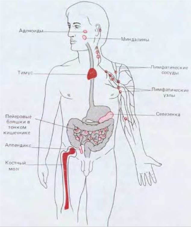
Fig. 18.1. Human lymphoid organs. Lymphocytes develop in the thymus and Bone Marrow (darkly shaded areas on the diagram), which are therefore designated as Primary lymphoid organs. Newly formed lymphocytes migrate from these primary organs into Secondary Lymphoid Organs (lightly shaded areas), where they can encounter and react with Antigens. Only selected secondary lymphoid organs are shown.
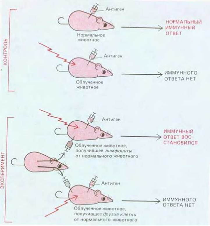
Fig. 18-2. A classic experiment demonstrating that lymphocytes are responsible for recognizing and reacting against foreign antigens. An essential prerequisite for all such cell-transfer experiments is that the donor and recipient belong to the same inbred strain and are therefore genetically identical. If donor lymphocytes are transferred to a genetically distinct, irradiated animal, they will react against the recipient's "foreign" antigens and may cause the recipient's death.
18.1.2. B lymphocytes mediate humoral immune responses, whereas T lymphocytes mediate cell-mediated immune responses [2]
During the 1960s, it was established that the two Major Classes of immune reactions are mediated by two distinct classes of lymphocytes: T cells, which develop in the thymus, are responsible for cell-mediated immunity, whereas B cells, which in mammals mature in the adult bone marrow or fetal liver, synthesize and secrete Antibodies. This dichotomy of the lymphoid system was initially uncovered in animals with experimentally induced immunodeficiencies. Neonatal thymectomy was shown to severely impair cell-mediated immune responses while having a much lesser effect on antibody production. The reverse effect could be demonstrated in birds, because in avian species B lymphocytes develop in the bursa of Fabricius (a gut-associated lymphoid organ unique to birds). Removal of the bursa in chicks abolishes antibody production with little impact on cell-mediated immunity. Studies of children born with immune deficiencies revealed that some are unable to produce antibodies yet have normal cell-mediated immunity, whereas others exhibit the exact opposite pattern. Children with selective defects in cell-mediated immunity almost invariably show developmental abnormalities of the thymus.
Investigations of animals with T-cell deficiencies (resulting from early thymectomy or thymic damage) uncovered a puzzling phenomenon: these animals not only lacked cell-mediated immune responses, but also exhibited a somewhat reduced capacity to produce antibodies. As we now understand, this occurs because certain T cells play a pivotal regulatory role in immunity, acting as helper cells for B cells during the humoral response.
Indeed, the majority of T LYMPHOCYTES PLAY regulatory roles in immunity, either enhancing or suppressing the responses of other white blood cells. These cells, designated as T helper cells and T suppressor cells, respectively, are collectively referred to as regulatory T cells. Other T lymphocytes, known as cytotoxic T cells, destroy cells infected by Viruses. Because both cytotoxic T cells and B cells participate directly in defending the Organism against infection, these two lymphocyte types are grouped together as effector cells.
18.1.3. Lymphocytes develop in primary lymphoid organs and react with foreign antigens in secondary lymphoid organs [3]
Lymphocytes originate from pluripotent stem cells that give rise to all blood cells, including erythrocytes, leukocytes, and platelets (Section 17.5.4). These stem cells reside primarily in hematopoietic Tissues—the fetal liver and adult bone marrow. In mammals, B cells differentiate from stem cells within the hematopoietic tissues themselves, whereas in birds they develop in the bursa of Fabricius from precursor cells that migrate there via the bloodstream from hematopoietic tissues. In all vertebrates, T cells develop in the thymus, which precursor cells enter via the bloodstream from hematopoietic tissues. Because hematopoietic tissues, the bursa of Fabricius, and the thymus serve as sites where lymphocytes are generated from precursors, they are termed primary lymphoid organs (see Fig. 18-1). Although many lymphocytes die shortly after differentiation in a primary lymphoid organ (Section 18.6.17), some migrate via the bloodstream to secondary lymphoid organs—principally the lymph nodes, spleen, and various Regions of the digestive tract (the appendix, Tonsils, adenoids, and Peyer's patches in the Small Intestine; see Fig. 18-1). It is primarily within these secondary lymphoid organs that T AND B cells encounter and react with foreign antigens (Fig. 18-3).

Fig. 18-3. Development of T and B lymphocytes. In both mammals and birds, a small number of precursor cells migrate via the blood into the thymus, where they differentiate into thymic lymphocytes. Most of these lymphocytes die within the thymus, but some migrate to secondary lymphoid organs and become thymus-derived lymphocytes (T cells). In birds, precursor cells home to the bursa of Fabricius, where they differentiate into bursal lymphocytes; many of these lymphocytes die, while others migrate to secondary lymphoid organs to become bursa-derived lymphocytes (B cells). In mammals, precursor cells destined to become B cells differentiate into lymphocytes within the hematopoietic tissue itself, after which they populate secondary lymphoid organs as mature B cells. The terms "T cells" and "T lymphocytes" are frequently used to denote thymus-derived lymphocytes, just as "B cells" and "B lymphocytes" denote bursa- or bone marrow-derived lymphocytes. At what stage of development precursor cells become committed to the T- or B-lymphocyte Lineage remains unclear. Later in this chapter, we will discuss why so many lymphocytes die within primary lymphoid organs (Sections 18.4.5 and 18.6.17).
Because the migration of lymphocytes from the thymus and bursa of Fabricius occurs predominantly during early development, surgical removal of these organs in an adult animal has relatively little impact on immune responses; this is precisely why their immunological role remained obscure for so long. By contrast, the mammalian bone marrow continues to generate vast numbers of new B cells throughout life (approximately 5-107 per day in the mouse).
18.1.4. Cell-surface markers allow T and B cells to be distinguished and separated [4]
T and B lymphocytes are morphologically indistinguishable until they are stimulated by an antigen. Unstimulated ("resting") T and B cells look strikingly similar even under an Electron microscope: they are small cells—only slightly larger than a red blood cell—whose volume is dominated by The Nucleus (Fig. 18-4, A). Both types are activated by antigens, which trigger their proliferation and subsequent maturation. Activated B lymphocytes ultimately differentiate into antibody-producing cells. The most mature of these are plasma cells, which feature an extensively developed granular Endoplasmic reticulum (Fig. 18-4, B). By contrast, activated T lymphocytes contain very few reticular elements and do not secrete antibodies (Fig. 18-4, C).
Because both T and B lymphocytes are present in all secondary lymphoid organs, Methods had to be developed to distinguish and separate these two cell types and their various subsets in order to study their individual properties. Fortunately, numerous Plasma Membrane Glycoproteins characteristic of distinct lymphocyte lineages serve as differential markers. For example, antibodies directed against the Thy-1 glycoprotein (Section 18.6.20), which is present on mouse T cells but absent from B cells, are widely used to deplete or purify T cells from mixed mouse lymphocyte populations. Similarly, antibodies against the CD4 and CD8 glycoproteins (Section 18.6.5) are extensively used to distinguish and isolate helper T cells and cytotoxic T cells from mice and humans.
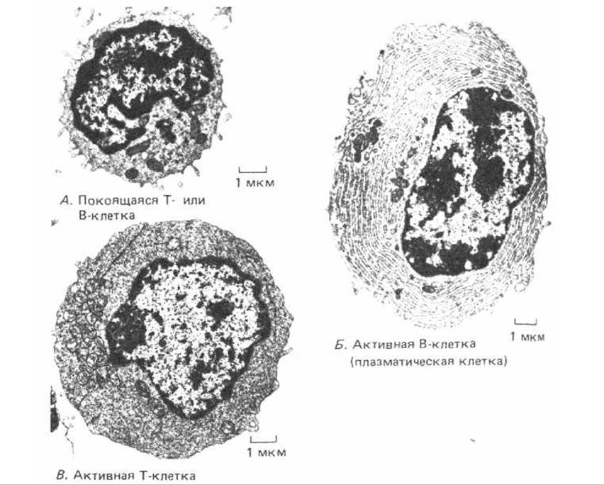
Fig. 18-4. Cytology/cytology/93.html">ELECTRON MICROGRAPHS OF a resting lymphocyte (A), an activated B cell (B), and an activated T cell (C). A resting lymphocyte can be either a T or a B cell, since these two lymphocyte populations are difficult to differentiate morphologically prior to activation. An activated B cell (plasma cell) is packed with granular endoplasmic reticulum whose cisternae are distended with antibody molecules, whereas an activated T cell contains relatively little granular reticulum but an Abundance of free Ribosomes. All three cells are shown at the same magnification. (Courtesy of: A - Dorothy Zucker-Franklin, B - Carlo Grossi, C - Stefanello de Petris. A and B are from D. Zucker-Franklin et al., Atlas of Blood Cells: Function and Pathology, 2nd ed. Milan, Italy: Edi. Ermies, 1988.)
18.1.5. The operation of the immune system is based on THE PRINCIPLE OF clonal Selection [5]
The most remarkable feature of the immune system is its ability to mount a highly specific response against millions of foreign antigens—for instance, by generating antibodies that specifically interact with the very antigen that triggered their formation. How can the immune system ensure such an extraordinary diversity of specific antibodies? One hypothesis, highly popular until the 1940s, proposed that antibodies are synthesized as unfolded polypeptide chains, and their final conformation is dictated by the antigen around which they fold. At the time, this seemed the simplest explanation for the fact that animals can produce specific antibodies against man-made molecules that do not exist in nature. However, this instructive hypothesis had to be abandoned once protein chemists established that the three-dimensional Structure of a folded protein molecule, such as an antibody, is determined solely by its Amino Acid Sequence. Indeed, a denatured (unfolded) antibody molecule can spontaneously refold to regenerate its original antigen-binding site even in the complete absence of antigen.

Fig. 18-5. The clonal selection theory. An antigen activates only those T- and B-cell clones that are already capable of responding to it. The immune system is postulated to consist of millions of distinct lymphocyte clones, hundreds of which may be activated by a given antigen. As we will see later, individual lymphocyte clones rarely respond to an antigen autonomously, as depicted in this simplified diagram and originally assumed by the clonal selection theory. Their responses are typically regulated through interactions with other lymphocyte clones (Section 18.6.12). Furthermore, T cells do not respond to free antigen as shown here; they respond exclusively to antigen presented On the surface of a host cell (Section 18.6).
In the 1950s, the instructive hypothesis gave way to The Theory of clonal selection. According to this theory, each lymphocyte during its development acquires The ability to react with a specific antigen even before ever encountering it. This occurs because receptor Proteins appear on The Cell surface that specifically match a certain antigen. If a cell encounters such an antigen, its binding to the receptors activates the cell, triggering its proliferation and the maturation of its progeny. Thus, a foreign antigen selectively stimulates those cells that happen to carry complementary specific receptors and therefore will inevitably react precisely to this antigen—which is why immune responses are antigen-specific (Fig. 18-5).
This selection is called "clonal" because, according to this theory, the immune system is formed by millions of different cell "families," or clones, each consisting of T or B lymphocytes derived from a common precursor. Since each precursor cell is already determined (or, as it is said, committed) to produce one specific antigen-specific receptor protein, all cells of a clone share the same antigenic specificity. Thus, According to the clonal selection theory, the working principle of the immune system can be compared to a ready-to-wear clothing factory rather than a custom tailor shop. Consequently, the question of how an animal's organism can produce such a vast array of different antibodies becomes a problem of genetics rather than Protein Chemistry.
The fundamental tenets of the clonal selection theory have received compelling confirmation. For example, if lymphocytes from an unimmunized animal are incubated in vitro with any of several labeled antigens, such as A, B, C, and D, only a very small fraction (<0.01%) of the lymphocytes will bind a given antigen. This means that only a few cells carry specific receptors for A, B, C, or D. This interpretation is supported by another experiment. Antigen A is made so highly radioactive that any cell binding it receives a lethal dose of radiation; the remaining lymphocyte population is no longer able to respond to antigen A, while continuing to respond normally to B, C, and D. The same effect can be achieved by packing an affinity Column (Section 4.4.3) with Glass beads coated with antigen A and then passing lymphocytes through this column. In such an experiment, cells with receptors for A bind to the beads, whereas the remaining cells pass through the column; the cells that have passed through no longer interact with A, but interact normally with other antigens (Fig. 18-6). These two experiments demonstrate that 1) lymphocytes are committed to react with a specific antigen even before they are exposed to it, and 2) committed lymphocytes possess surface receptors that specifically bind that antigen. Thus, two main predictions of the clonal selection theory are confirmed. Although most experiments of this kind studied B cells and antibody-mediated responses, other experiments have shown that cell-mediated immunity is also based on the principle of clonal selection.
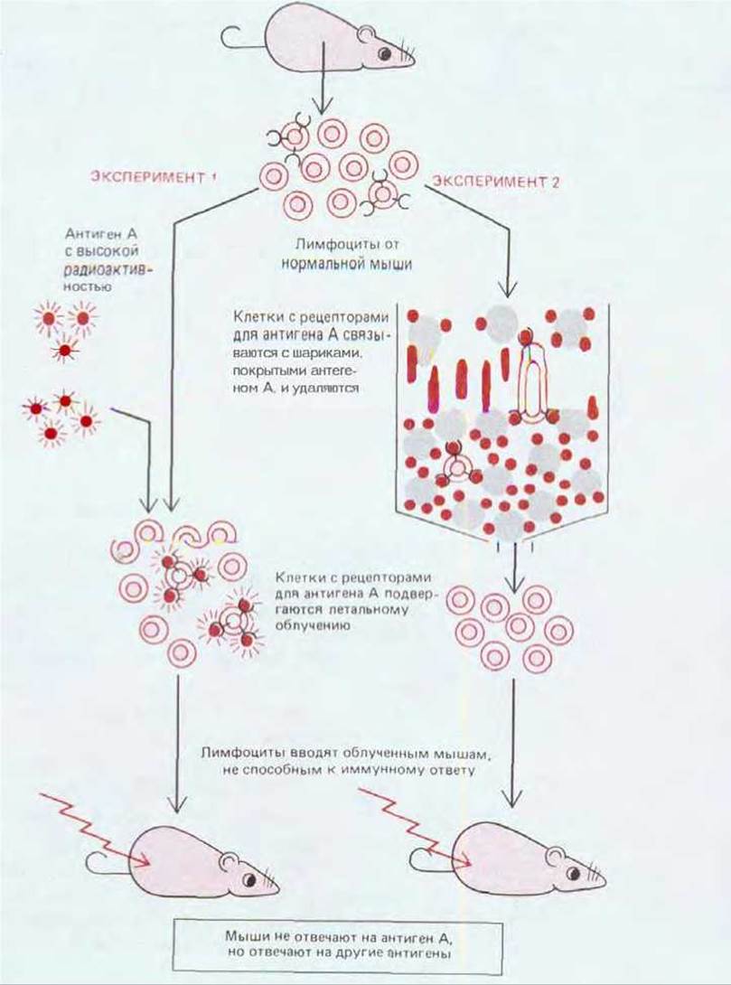
Fig. 18-6. Two Types of experiments supporting the theory of clonal selection. For simplicity, cell-surface receptors are shown only on lymphocytes capable of responding to antigen A; in reality, however, all T and B lymphocytes bear antigen-specific receptors on their surfaces. The experiments schematically presented here were performed mainly with B cells, since T cells recognize antigen only when it is bound to The surface of a host cell (Section 18.6).
18.1.6. In Most Cases, a Single Antigen Stimulates Many Different Lymphocyte Clones [6]
Most macromolecules, including practically all proteins and the majority of Polysaccharides, can serve as antigens. Those regions of an antigen that interact with the antigen-binding site of an antibody molecule or a lymphocyte receptor are called antigenic determinants (or epitopes). Molecules that bind specifically to such antigen-binding sites but fail to induce an immune response are called haptens. Haptens are typically small organic molecules; they are too small by themselves to elicit a response, but they become fully functional antigens when attached to a suitable carrier macromolecule. Haptens serve as important tools in experimental immunology. One of the most frequently used haptens is the dinitrophenyl (DNP) group, which is typically conjugated to a protein to render it antigenic (Fig. 18-7).
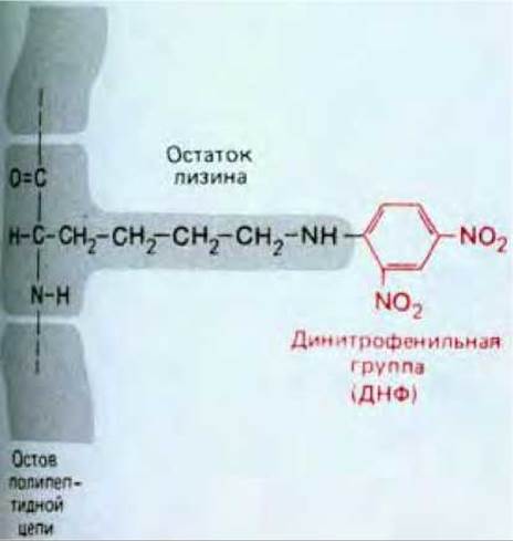
Fig. 18-7. A simple DNP hapten covalently linked to a Lysine side chain in a protein. Haptens can induce an immune response only when conjugated to such a macromolecular carrier.
Most antigens bear a whole set of different antigenic determinants that stimulate antibody production or T-cell responses. Some determinants are more immunogenic (i.e., induce immunity more effectively) than others, and the reaction to them may dominate the overall response; such determinants are called immunodominant.
As one would expect from a system operating on the principle of clonal selection, even a single antigenic determinant will generally activate multiple clones, each bearing surface receptors with its own distinct, individual affinity for that determinant. For example, even the relatively simple STRUCTURE OF THE DNP group permits many different interactions, and when this group is attached to a carrier protein, it typically stimulates The production of hundreds of types of anti-DNP antibodies, each produced by a separate clone of B cells. Such a response is termed polyclonal. When only a few clones react, the response is called oligoclonal, and if the entire response reduces to the reaction of just a single clone of B or T cells, it is termed monoclonal. Responses to most antigens are polyclonal.
Even an antigen that activates many clones affects only a tiny fraction of the total lymphocyte population. To ensure that an antigen encounters these scarce lymphocytes, antigens accumulate in secondary lymphoid organs, through which T and B lymphocytes continuously circulate. Antigens entering the body via the digestive tract are captured by associated lymphoid tissues; those penetrating through the Skin or respiratory tract are transported via lymph to local lymph nodes; and antigens entering the bloodstream are filtered out in the spleen.
18.1.7. Most Lymphocytes Are in Continuous Circulation [7]
The majority of T and B lymphocytes constantly traffic from the blood to secondary lymphoid organs and back. For example, in a lymph node, lymphocytes leave the bloodstream by squeezing between specialized endothelial cells. Having passed through the node, they collect in small lymphatic vessels that exit the node and join other lymphatic vessels, which then pass through downstream lymph nodes (Fig. 18-8). Passing into progressively larger vessels, the lymphocytes eventually reach the main lymphatic duct (the Thoracic duct), through which they return to the blood. This constant circulation not only ensures that the appropriate lymphocytes encounter their antigen, but also allows required lymphocytes to meet one another: as we shall see, interactions between specific lymphocytes play a crucial role in most immune responses. Lymphocyte circulation depends on specific interactions between the lymphocyte surface and the surface of specialized endothelial cells lining small Veins (postcapillary venules) in secondary lymphoid organs: of all blood cells coming into contact with these endothelial cells, only lymphocytes temporarily attach to them and then migrate across the postcapillary venules. Monoclonal Antibodies (Section 4.5.4), by binding to the lymphocyte surface and inhibiting their ability to adhere to specialized endothelial cells in tissue sections as well as to circulate in vivo, help identify various "homing receptors" that dictate lymphocyte migration pathways. The surface of most T and B cells bears two types of glycoproteins: one for circulation through LYMPH NODES AND another for circulation through Peyer's patches. Some lymphocytes possess only the second type of glycoprotein and selectively circulate through Peyer's patches; they essentially constitute an intestinal-tract-specific subsystem of lymphocytes specialized for responding to antigens entering the body through the gut. Other homing receptors apparently mediate the segregation of T and B cells into distinct regions within a lymphoid organ (see Fig. 18-8). When lymphocytes are activated by an antigen, they lose the homing receptors that mediate circulation through lymphoid organs and acquire new receptors that direct the activated cells to sites of inflammation.

Fig. 18-8. Highly simplified diagram of a human lymph node. B lymphocytes are located mainly in the cortex, where they are gathered into structures called lymphoid follicles. T lymphocytes reside primarily in the paracortical area. Lymphocytes of both types enter the lymph node from the blood via small specialized venules in the paracortical region (not shown); T cells remain in this zone of the node, whereas B cells migrate into the lymphoid follicles. Over time, both T and B cells migrate into the medullary sinuses and leave the node via the efferent lymphatic vessel. This vessel eventually empties into the bloodstream, allowing lymphocytes to begin the next cycle of circulation through a secondary lymphoid organ. Foreign antigens entering the lymph node end up on the surface of specialized antigen-presenting cells: cells of one type present the antigen (in the form of an antigen-antibody complex) to B cells in the lymphoid follicles, while cells of another type present it to T cells in the paracortical area (Section 18.6.10).
18.1.8. Immunological Memory Results from Clonal Expansion and Lymphocyte Maturation [8]
The immune system, much like The Nervous system, possesses memory. This is why we can acquire lifelong immunity to many viral diseases after being exposed to a virus just once. A similar phenomenon can be demonstrated in experimental animals. If an animal is injected once with antigen A, after a lag period of several days it mounts an immune response (either antibodies or a cell-mediated response) that rises rapidly (exponentially) and then declines more gradually. This is the characteristic time course of a primary immune response, observed following an animal's first encounter with an antigen. If weeks, months, or even years later the animal is injected with antigen A again, it triggers a secondary immune response that differs substantially from the primary one: the lag period is shorter, and the reaction is stronger and more sustained (Fig. 18-9). These differences indicate that the animal has "remembered" its initial encounter with antigen A. If, instead of a booster injection of antigen A, the animal is administered a different antigen (e.g., B), the reaction takes the form of a primary rather than a secondary immune response; consequently, the secondary response reflects specific immunological memory for antigen A.
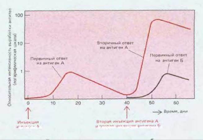
Fig. 18-9. Primary and secondary humoral responses (antibody production) elicited by the First and Second administrations of antigen A, respectively. Note that the secondary response is faster and stronger than the primary one and is specific for A. This demonstrates that the immune system specifically "remembered" the previously administered antigen A. Such immunological memory is revealed in The Study of both B-cell and T-cell immune responses.
The theory of clonal selection provides the conceptual framework for understanding the cellular mechanism of immunological memory. In the secondary lymphoid organs of an adult animal, populations of T and B lymphocytes simultaneously contain cells at at least three stages of maturation: naive cells, memory cells, and active cells. When naive cells encounter an antigen for the first time, some of them are stimulated to proliferate and become active cells, which we define as cells actively participating in mounting an immune response (active T cells mediate cellular responses, whereas B cells secrete antibodies). Other naive cells are instead stimulated to proliferate and mature into memory cells, which do not respond themselves but readily differentiate into active cells upon subsequent encounter with the same antigen (Fig. 18-10). Naive lymphocytes are thought to be relatively short-lived in secondary lymphoid organs and probably die within a few days unless they encounter their specific antigen. Memory cells, by contrast, can live for many months or even years without dividing, continuously recirculating between the blood and secondary lymphoid organs. Furthermore, memory cells exhibit a greater readiness to respond to antigen than do naive cells. Later we will see (Section 18.4.4) that the enhanced responsiveness of memory B cells is explained, in part, by the higher affinity of these cells' receptors for the antigen.
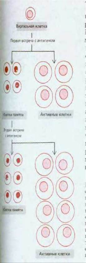
Fig. 18-10. Naive T or B cells, when stimulated by a specific antigen, divide and mature. Some of them then begin to mount an immune response, whereas others differentiate into memory cells. Upon subsequent encounter with the antigen, memory cells respond to it more "readily" than naive cells do: they proliferate and give rise to active cells and new memory cells. According to this model, an individual naive cell can, depending on conditions, give rise to either a memory cell or an activated cell. According to an alternative model not shown here, memory cells and activated cells originate from distinct naive cells. Which of these models is correct remains unknown.
In accordance with this scheme, immunological memory is established during the primary response As a result of the following: 1) proliferation of antigen-activated naive cells multiplies the number of memory cells (clonal expansion); 2) memory cells have a much longer lifespan than naive cells and continuously recirculate between the blood and secondary lymphoid organs; 3) each memory cell is capable of responding to antigen more "readily" than a naive cell. The changes occurring during the primary response result in the majority of long-lived cells within the recirculating lymphocyte pool now being "tailored" to the animal's antigenic environment and poised for immediate action.
18.1.9. Unresponsiveness to the Body's Own Antigens Results from Acquired Immunological Tolerance [9]
How does the immune system distinguish between "self" and "non-self"? One possibility is that an animal inherits genes encoding receptors for foreign antigens rather than its own, and therefore its immune system is genetically programmed to respond exclusively to foreign antigens. Another possibility is that the immune system might initially be capable of responding to both self and foreign antigens, but during early development "learns" not to respond to self. It has been shown that the second hypothesis is correct. The first evidence for this came from an observation made in 1945. As a rule, when tissue is transplanted from one individual to another, the graft is recognized as foreign by the immune system and rejected. However, it turned out that this does not occur during skin grafts between dizygotic (developed from two fertilized eggs, i.e., non-identical) twin cows, which could exchange blood cells during intrauterine development due to spontaneous fusion of their placentas. These results were later reproduced in chickens (by connecting the Blood Vessels of two different embryos) and in mice (by injecting newborn mice with spleen cells from mice of another strain—these cells survived for the major part of the recipient mouse's life). In both cases, once the animals reached adulthood, it was possible to transplant tissue into them either from the temporarily attached individual or from the donor individual, and the graft survived (Fig. 18-11), whereas tissues transplanted from other, control animals were rejected. Thus, the continuous presence of foreign antigens from a time when the immune system has not yet matured leads to long-term unresponsiveness to these antigens. This state of induced antigen-specific failure to mount an immune response was named acquired immunological tolerance.
There is compelling evidence that the inability of an animal's immune system to react to its own macromolecules (natural immunological tolerance) is acquired in the very same way—it is not innate. For example, normal mice do not mount an Immune Response to their own blood protein, Complement component C5 (Section 18.5.1). However, mutant mice lacking the C5-encoding Gene (otherwise genetically identical to normal mice) can mount an immune response against this protein. Thus, it is clear that the immune system is potentially capable of reacting to the body's own antigens, but "learns" not to do so. At least in some cases, the "learning" process involves the elimination of lymphocytes that react to "self" (Section 18.6.17), though we do not know how this occurs. It is believed that many such lymphocytes are eliminated in primary lymphoid organs upon encountering an antigen. Such a negative response to an antigen could be due to the specific microenvironment within these organs or the exceptional activity of newly formed lymphocytes. Perhaps because new self-reactive lymphocytes continue to be generated from stem cells throughout life, the maintenance of natural tolerance requires the continuous presence of self-antigens. If such an antigen as C5 is removed, the animal's organism regains the ability to mount an immune response to it weeks or months later.
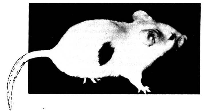
Fig. 18-11. The skin graft shown here, transplanted from an adult brown mouse to an adult white mouse, survived for many weeks only because the white mouse was made immunologically tolerant by injecting it immediately after birth with blood cells from the brown mouse. (Courtesy of Leslie Brent, from I. Roitt, Essential Immunology, 6th ed. Oxford, U.K.: Blackwell Scientific, 1988.)
Tolerance to self-antigens is sometimes broken, leading to T- or B-cell (or both) reactions against the body's own tissue antigens. An example of such autoimmune diseases is myasthenia gravis. In this disease, antibodies are produced against acetylcholine receptors on Skeletal Muscle fibers (Section 6.4.18); these antibodies impair the normal functioning of the receptors. Such patients suffer from muscle weakness and may die due to respiratory muscle failure.
18.1.10. Immunological tolerance to foreign antigens can also be induced in adult animals [10]
In an adult animal, establishing immunological tolerance to foreign antigens is generally much more difficult than at an early stage of development. However, for certain antigens this can be achieved experimentally by administering the antigen 1) in very high doses, 2) repeatedly in very small doses, 3) together with an immunosuppressant, or 4) intravenously after chemical cross-linking of the antigen to the surface of B lymphocytes or ultracentrifugation of the antigen to remove all aggregates (this renders normal antigen-presentation mechanisms ineffective, see Section 18.6.10). Thus, the binding of an antigen to its complementary receptors on T or B lymphocytes can either stimulate the lymphocyte to divide and mature, turning it into an active or memory cell, or eliminate or inactivate the lymphocyte, leading to tolerance. The molecular mechanisms determining the outcome are insufficiently understood. Whether an antigen causes activation or induces tolerance depends primarily on 1) the degree of lymphocyte maturity, 2) The Nature and concentration of the antigen, and 3) complex interactions between different classes of lymphocytes and between lymphocytes and specialized antigen-presenting cells, which will be described later.
The immune system evolved in vertebrates to protect against infections. It consists of millions of lymphocyte clones. Lymphocytes of each clone carry a receptor on their surface that enables them to bind a particular "antigenic determinant"—a specific grouping of atoms within the antigen molecule. There are two classes of lymphocytes: B cells, which produce antibodies, and T cells, which mediate cell-mediated immune responses.
Already at early Stages of development, many lymphocytes reacting with antigenic determinants of the body's own macromolecules are eliminated or inactivated; as a result, the immune system normally responds only to foreign antigens. The binding of a foreign antigen to a lymphocyte triggers an immune response directed against this antigen. In the process, some of the lymphocytes proliferate and mature into long-lived memory cells, so that upon a secondary encounter of the organism with the same antigen, the immune response develops faster and is stronger.
Last update: 12/08/2026
Editorial and Educational Adaptation: This material has been compiled based on the primary/original source text. The project team performed an editorial review, corrected technical inaccuracies, structured sections, and adapted the content for an educational format.
What was processed:
- elimination of formatting defects (OCR errors, structural breaks, corrupted characters);
- editorial organization of content;
- standardization of terminology in accordance with academic sources;
- verification of factual statements against the original source text.
All mentions of the author, publication year, and origin of the primary text have been preserved in accordance with the source.