Human Anatomy - Kotsan I. Ya. 2009
The Lymphatic System
Along with the Circulatory system, humans and vertebrates possess another Vascular System known as The Lymphatic system (systema lymphaticum). It acts essentially as an auxiliary bed of the Venous system, developing in close connection with it and sharing similar structural features (such as Valves and the flow of Lymph from Tissues toward The Heart). The lymphatic system performs the following Functions:
✵ trophic (delivering nutrients and Hormones to tissues and Cells along with lymph);
✵ participation in Water-salt, protein, and METABOLISM/26.html">Energy Metabolism;
✵ hematopoiesis (producing lymphocytes and monocytes);
✵ barrier (trapping foreign particles, microbial bodies, and tumor cells and neutralizing them);
✵ immune (producing immune bodies responsible for Immunity);
✵ drainage (performing organ drainage alongside the venous system via lymph outflow);
✵ Homeostasis and detoxification.
The lymphatic system comprises lymphatic capillaries, Lymphatic vessels, lymphatic trunks, and ducts branched throughout Organs and tissues, through which lymph flows from its site of formation to the point of entry into the venous system, passing through a series of Lymph Nodes (Fig. 219).
Lymph (lympha) is a transparent, greenish-yellow or gray fluid. In its composition, lymph resembles Blood Plasma, but unlike plasma, it contains a lower concentration of Proteins. Lymph contains white Blood Cells—lymphocytes and a small number of monocytes and eosinophils. There are 2,000 to 20,000 lymphocytes per 1 mm3 of lymph. Red blood cells are absent in lymph. Although the exact volume of lymph in the body is difficult to determine, it is estimated to be 1–2 liters. Lymph is formed from the liquid portion of blood plasma that filters from blood capillaries into tissues, and then from tissues into lymphatic capillaries.
Lymphatic capillaries (lymphocapillary vessels) (vasa lymphocapillaria) are the initial link and roots of the lymphatic system. They are present in all organs and Tissues of the human body, except for the Brain AND SPINAL cord and their membranes, the Eyeball, the Inner ear, the epithelial lining of the Skin and mucous membranes, Cartilage, the parenchyma of the Spleen, Bone Marrow, and the Placenta.
Unlike blood capillaries, lymphatic capillaries have a large diameter (up to 0.2 mm), irregular contours, lateral protrusions, and dilations. The walls of lymphatic capillaries are formed by a single layer of endothelium. The lumen of lymphatic capillaries is wider than that of blood capillaries, and the endothelial cells lining them are 3–4 times larger than those in blood capillaries. A basement membrane is absent in lymphatic capillaries, allowing the endothelium to come into direct contact with the intercellular substance of Connective Tissue, which accounts for the high permeability of lymphatic capillaries. They take up not only dissolved substances but also larger particles, such as dust, smoke, dead cells, and others.
Class="center">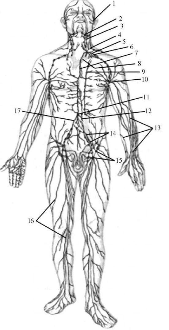
Fig. 219. Diagram of The Structure of the human lymphatic system
1 — lymphatic Vessels of the face; 2 — submandibular lymph nodes; 3 — lateral cervical lymph nodes; 4 — left jugular trunk; 5 — left subclavian trunk; 6 — subclavian vein; 7 — Thoracic duct; 8 — left brachiocephalic vein; 9 — parasternal nodes; 10 — axillary lymph nodes; 11 — cisterna chyli; 12 — intestinal trunk; 13 — superficial lymphatic vessels of the upper extremity; 14 — common and external iliac lymph nodes; 15 — superficial inguinal lymph nodes; 16 — superficial lymphatic vessels of the lower extremity; 17 — right lumbar trunk
By interconnecting, lymphatic capillaries form closed lymphocapillary networks (rete lymphocapillaria), the loops of which lie in one or multiple planes depending on the structure (architecture) of the organ in which they run. The orientation of the capillaries is determined by the direction of the connective tissue bundles in which the lymphatic capillaries lie, as well as the position (shape) of the Structural elements of the organ. Thus, in volumetric organs (Muscles, Lungs, Liver, Kidneys, large glands, etc.), networks of lymphatic capillaries have a three-dimensional structure. Their lymphatic capillaries run in various directions, lying between the Structural and functional elements of the organ: bundles of Muscle fibers, groups of glandular cells, renal corpuscles, and hepatic lobules. In flat organs (fasciae, serous membranes, skin, layers of hollow organ walls), the lymphocapillary network is located in a single plane parallel to The surface of the organ. In some organs, the networks of lymphatic capillaries form long, finger-shaped blind projections (for example, lymphatic sinuses in the villi of the Small Intestine).
Lymphatic vessels (vasa lymphatica) are formed by the fusion of lymphatic capillaries. The walls of lymphatic vessels are somewhat thicker than those of lymphatic capillaries. In intra-organ, and often extra-organ, lymphatic vessels, external to the endothelium lies only a thin connective tissue membrane (non-muscular vessels). The walls of larger lymphatic vessels consist of three layers: the inner layer, formed by the endothelium and a subendothelial layer of Collagen and elastic fibers; the middle layer, composed of collagen and elastic fibers and smooth muscle cells, the contraction of which facilitates the movement of lymph; and the outer connective tissue layer. Lymphatic vessels possess numerous valves formed by folds of the inner layer. Each valve consists of two cusps (folds) located opposite each other. The distance between adjacent valves ranges from 2–3 mm (in intra-organ lymphatic vessels) to 12–15 mm (in larger extra-organ vessels). The valves in lymphatic vessels are adapted to allow lymph to flow in one direction—from the periphery toward the lymph nodes, trunks, and ducts, thereby ensuring the flow of lymph toward the heart. Adjacent intra-organ lymphatic vessels anastomose with one another, forming networks—lymphatic plexuses (plexus lymphatici) with loops of various shapes and lengths.
Classification of lymphatic vessels. Lymphatic vessels are divided into deep and superficial. Deep lymphatic vessels (vasa lymphatica profunda) are vessels that emerge from Internal Organs and muscles. As a rule, they run alongside major Arteries and Veins. Superficial lymphatic vessels (vasa lymphatica superficialia) are located above the superficial fascia of certain body regions, run alongside or close to subcutaneous veins, and collect lymph from the skin and subcutaneous tissue. In mobile areas of the body and at bending points (near joints), lymphatic vessels fork, branch, and reconnect, forming collateral pathways that ensure the uninterrupted flow of lymph during changes in THE POSITION OF the body or its parts, as well as when the patency of certain lymphatic vessels is impaired during flexion and extension movements in joints.
Depending on their relationship to lymph nodes, afferent and efferent lymphatic vessels are distinguished.
Depending on their relationship to organs, intra-organ and extra-organ lymphatic vessels are distinguished.
Lymphatic trunks (trunci lymphatici). Efferent lymphatic vessels form the next stage of the lymphatic bed—the lymphatic trunks, through which lymph from specific body regions enters the lymphatic ducts or directly into the venous system. Each part of the body has its own lymphatic trunk. There are 9 such trunks in The Human Body: four paired (jugular, subclavian, bronchomediastinal, and lumbar) and one unpaired (intestinal). However, in 50% of cases, there may be 8 trunks.
The jugular trunk (right and left) (truncus jugularis [dexter et sinister]) is formed by the efferent lymphatic vessels of the lateral deep cervical (internal jugular) lymph nodes of the corresponding side. The right jugular trunk empties into the right venous angle, into the right Internal jugular vein, or participates in The formation of the Right lymphatic duct. The left jugular trunk empties directly into the left venous angle, into the left internal jugular vein, or, in most cases, into the cervical part of the thoracic duct. The jugular trunks collect lymph from the lymph nodes of the HEAD and Neck.
The subclavian trunk (right and left) (truncus subclavius [dexter et sinister]) is formed by the efferent lymphatic vessels of the axillary lymph nodes. The right subclavian trunk opens into the right venous angle, the right subclavian vein, or the right lymphatic duct; the left subclavian trunk opens into the left venous angle, the left subclavian vein, or, in half of the cases, into the terminal part of the thoracic duct. The subclavian trunks collect lymph from the lymph nodes of the upper extremity.
The bronchomediastinal trunks (right and left) (trunci bronchomediastinales [dexter et sinister]) are formed by efferent lymphatic vessels originating from the lymph nodes of the walls and Organs of the corresponding half of the thoracic cavity. The right bronchomediastinal trunk empties into the right lymphatic duct, or, if it is absent, directly into the right subclavian vein. The left bronchomediastinal trunk empties into the upper part of the thoracic duct.
The lumbar trunk (right and left) (truncus lumbalis [dexter et sinister]) is formed by the fusion of efferent lymphatic vessels from the corresponding right and left lumbar lymph nodes. The lumbar trunks collect lymph from the lymph nodes of the lower extremities, pelvis, and Abdominal cavity. The fusion of the lumbar trunks at the level of the 2nd lumbar vertebra gives rise to the thoracic duct.
The intestinal trunk (truncus intestinalis), described in textbooks as the third ROOT of the thoracic duct, is relatively uncommon. It empties into the beginning of the thoracic duct or into the left (more frequently) or right lumbar trunk. The intestinal trunk is formed by efferent lymphatic vessels carrying lymph from the mesenteric lymph nodes. It collects lymph from the abdominal organs, predominantly from the small intestine.
Lymphatic ducts (ductus lymphatici). Lymphatic trunks empty into the largest lymphatic vessels, known as lymphatic ducts. There are two lymphatic ducts: the thoracic and the right.
The thoracic duct (ductus thoracicus) originates in the abdominal cavity within the retroperitoneal tissue at the level of the 12th thoracic to 2nd lumbar vertebrae by the union of the right and left lumbar lymphatic trunks (Fig. 220). In approximately 25% of cases, 1 to 3 efferent lymphatic vessels from the mesenteric lymph nodes, which form the intestinal trunk, empty into the initial part of the thoracic duct.
The thoracic duct is the largest vessel in the lymphatic system, reaching a length of 30—41 cm and a diameter of 2—3 mm. It comprises an abdominal part (pars abdominalis), a thoracic part (pars thoracica), and a cervical part (pars cervicalis), as well as the arch of the thoracic duct (arcus ductus thoracici). In 75% of cases, the initial abdominal part features a dilation called the cisterna chyli (also known as the receptacle of chyle). In 25% of cases, the Origin of the thoracic duct appears as a reticular plexus formed by the efferent lymphatic vessels of the lumbar, abdominal, and mesenteric lymph nodes. The wall of the initial part of the thoracic duct is fused with the right crus of the Diaphragm, which compresses the thoracic duct during respiratory movements, facilitating the propulsion of lymph. Originating in the abdominal cavity, the thoracic duct passes through the aortic hiatus into the thoracic cavity, ascending anterior to THE Vertebral Column, positioned to the right of the Thoracic Aorta, posterior to the Esophagus, and subsequently posterior to the aortic arch. Upon reaching the aortic arch, at the level of the V—VII thoracic vertebrae, it begins to deviate to the left. At the level of the VII cervical vertebra, the thoracic duct enters the neck and, forming an arch, empties into the left internal jugular vein or the left venous angle—the junction of the left subclavian and internal jugular veins. In about 50% of cases, the thoracic duct features a dilation just before entering the vein. Sometimes, the terminal section of the thoracic duct divides and opens into the venous bed through multiple small trunks rather than a single one.
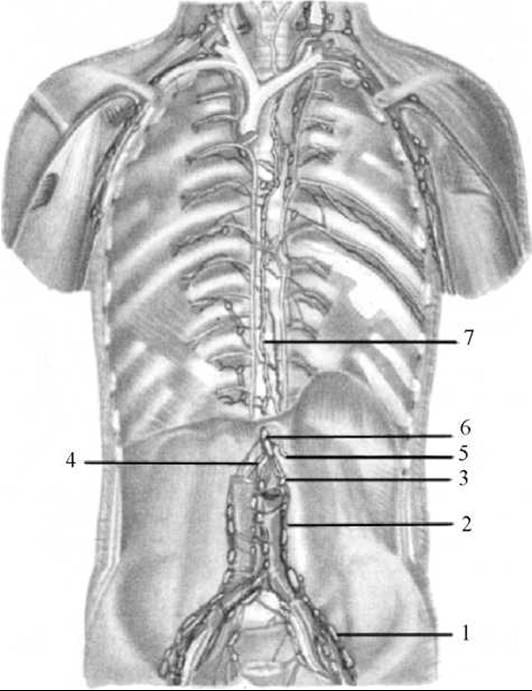
Fig. 220. Thoracic duct
1 — common iliac lymph nodes; 2 — lumbar lymph nodes; 3 — left lumbar trunk; 4 — right lumbar trunk; 5 — intestinal trunk; 6 — cisterna chyli; 7 — thoracic duct.
At the orifice of the thoracic duct, There is a paired valve formed by its inner lining, which prevents the backflow of blood from the vein. Along the entire length of the thoracic duct, there are 7—9 valves that prevent the backward flow of lymph. The wall of the thoracic duct features a well-developed middle (muscular) layer composed of smooth muscle cells, the contraction of which AIDS in the movement of lymph.
The upper part of the thoracic duct receives the left bronchomediastinal trunk, which collects lymph from the walls and organs of the left half of the Thorax; the left subclavian trunk, carrying lymph from the left upper limb; and the left jugular trunk, which collects lymph from the left half of the head and neck. Thus, the thoracic duct collects about % of all lymph, meaning from almost the entire body, with the exception of the right half of the head and neck, the right upper limb, the organs and walls of the right half of the thorax, and the lower lobe of the left lung. Lymph from these specified areas flows into the right lymphatic duct.
The right lymphatic duct (ductus lymphaticus dexter) is no more than 10—12 mm in length. It is formed by the union of three trunks: the right jugular trunk, which collects lymph from the right side of the head and neck; the right subclavian trunk, carrying lymph from the right upper limb; and the right bronchomediastinal trunk, which collects lymph from the walls and organs of the right half of the thorax and the lower lobe of the left lung. The right lymphatic duct empties into the right subclavian vein, the right venous angle, or the right jugular vein. Quite frequently, this duct is absent; in such cases, the aforementioned three trunks empty independently into the subclavian vein.
Lymph nodes (nodi lymphatici) are located along the course of lymphatic vessels. Within the lymph nodes, lymph is enriched with lymphocytes that multiply there. Here, toxic substances are neutralized, and microbes and various foreign particles brought from tissues via the lymphatic vessels are trapped and phagocytosed. Each lymph node, or group of nodes, collects and monitors lymph from a specific area, serving as its biological filter.
The human body contains approximately 800—900 lymph nodes. They are mainly arranged in groups ranging from 2—3 to 20 or more, or, less frequently, singly.
Taking into account their topographical distribution (the anatomical-topographical principle) as well as the direction of lymph flow from organs (THE PRINCIPLE OF regionality), about 150 regional groups of lymph nodes are distinguished (from Latin *regio* — region). Accordingly, lymph nodes are named after the region where they are located (e.g., axillary lymph nodes, lumbar lymph nodes). In other cases, a group of lymph nodes takes the name of the blood vessel alongside which they are situated (e.g., abdominal lymph nodes, iliac lymph nodes).
In certain areas of the human body, groups of lymph nodes are arranged in two layers (one group above the other). As a rule, a fascia lies between such groups. In these instances, the nodes lying superficial to the fascia are called superficial nodes, while those lying beneath the fascia are called deep nodes (for example, superficial inguinal lymph nodes, located on the fascia lata of the thigh, and deep inguinal lymph nodes, lying beneath the fascia lata of the thigh).
Lymph nodes that receive lymph from the organs of The Musculoskeletal System (popliteal, inguinal, cubital, axillary) or from the walls of Body Cavities (intercostal, upper diaphragmatic, lumbar, lower diaphragmatic) are called somatic (parietal) nodes. Those nodes that are regional exclusively to internal organs are termed visceral (visceral) lymph nodes. Nodes that receive lymph from both internal organs and muscles, fasciae, and skin are called mixed nodes (e.g., deep lateral cervical nodes).
Lymph nodes have a pinkish-gray color, a round, oval, or bean-like shape, and variable dimensions (ranging from 0.5 mm to 50 mm or more) (Fig. 221). Externally, each lymph node is covered by a connective tissue capsule (capsula nodi lymphatici), from which delicate partitions—capsular trabeculae—extend into the interior of the node. In addition to Cytology/practical/45.html">Dense connective tissue, the framework of the capsule and trabeculae contains scattered smooth muscle fibers, the contractions of which assist in lymph propulsion.
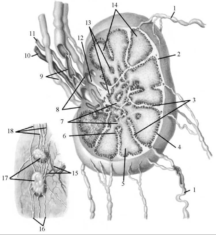
Fig. 221. SCHEMATIC STRUCTURE OF a lymph node (after R. D. Sinelnikov)
1 — afferent lymphatic vessels; 2 — capsule of the lymph node; 3 — trabeculae; 4 — subcapsular (marginal) sinus; 5 — lymphoid nodules; 6 — intermediate sinus; 7 — medullary cords; 8 — hilum of the lymph node; 9 — efferent lymphatic vessels (one cut open, valves visible); 10 — vein; 11 — artery; 12 — hilar (terminal) sinus; 13 — medulla; 14 — cortex; 15 — artery and vein of the lymph nodes; 16 — afferent vessels; 17 — lymph nodes; 18 — efferent vessels
On the surface of the node, there is an indentation called the hilum of the node (hilum nodi lymphatici). Somatic nodes feature a single hilum, whereas visceral nodes often have 3 to 4. Arteries and nerves enter the node through the hilum, while veins and efferent lymphatic vessels exit. Hilar trabeculae extend from the capsule into the parenchyma of the node in the region of the hilum. The hilar and capsular trabeculae interconnect, imparting a lobular architecture to the lymph node.
Inside the lymph node, between the trabeculae, lies a fine-meshed stroma consisting of reticular fibers and reticular cells, which form a three-dimensional network with meshes of various Sizes and Shapes. Cellular elements of the lymphoid series are located within the meshes of the reticular tissue. The parenchyma of the lymph nodes is divided into the cortex and medulla.
The cortex (cortex) is darker in color, lies closer to the capsule, and occupies the peripheral Regions of the node. The cortex contains round lymphoid nodules 0.5—1 mm in diameter, which represent accumulations of lymphoid cells, predominantly B lymphocytes. Diffuse lymphoid tissue surrounds the lymphoid nodules. Within it, the cortical plateau is distinguished, which includes areas of lymphoid tissue between the nodules—the internodular zone. The cortical plateau also incorporates tissue located externally to the lymphoid nodules, between them and the capsule. Internal to the nodules, directly bordering the medulla, lies a band of lymphoid tissue known as the paracortex (Thymus-dependent zone), which predominantly contains T lymphocytes. A characteristic feature of this part of the cortex is the presence of postcapillary venules, whose walls are lined with cuboidal endothelial cells through which lymphocytes migrate.
The medulla (medulla) is lighter in color, situated closer to the hilum and in the central part of the node. The medullary parenchyma is represented by elongated tracts of lymphatic tissue—the medullary cords—which extend from the inner sections of the cortex toward the hilum of the lymph node. The medullary cords are built of reticulocytes and reticular fibers. Interlacing with one another, the cords form a network that houses B lymphocytes, plasma cells, and macrophages.
Between the capsule, trabeculae, and the parenchyma of the node are clefts—lymphatic sinuses—through which lymph flows through the lymph node. Directly beneath the capsule lies the subcapsular (marginal) sinus. From it, intermediate sinuses (initially cortical, then medullary) extend into the parenchyma of the lymph node, transitioning into the hilar sinus in the region of the organ's hilum. The subcapsular sinus also opens into this sinus, enveloping the cortex peripherally and terminating in the area of the node's hilum.
Lymph reaches the lymph node via its afferent lymphatic vessels, numbering 2—4, which approach the convex side of the node, pierce the capsule, and empty into the subcapsular sinus. Then, through this sinus and the interconnected intermediate sinuses located within the node's parenchyma, the lymph reaches the hilar sinus. Emerging from the hilar sinus are 1—2 efferent lymphatic vessels that leave the lymph node. The lumen of the sinuses contains a fine-meshed network formed by reticular fibers and cells. As lymph passes through the sinusoidal System of the lymph node, foreign particles that have entered the lymphatic vessels from the tissues (microbial bodies, tumor cells, dust particles) may be trapped within the meshes of this network. Lymphocytes enter the lymph from the parenchyma of the lymph node.
Each lymph node has a rich blood supply, with arteries penetrating the node not only through the hilum but also through the capsule.
Three conventional types of lymph nodes are distinguished.
The first type is characterized by a cortical area smaller than the medullary area. Lymph nodes of this type fill rapidly and intensively with radiopaque medium.
Lymph nodes of the second type are compact. In these, conversely, the mass of the cortex exceeds that of the medulla. They are characterized by radiologically slow and weak opacification. The transport function of such nodes is minimal.
The most common are lymph nodes of the third type, which are intermediate. The mass of the cortical and medullary substances in them is approximately equal. They fill well with radiopaque medium. Their structure effectively ensures lymph Processing and transport function.
The observed variations in lymph nodes, individual structural features, and, consequently, functional capacities determine the varying survival rates of Cancer patients.
Last update: 08/08/2026
Editorial and Educational Adaptation: This material has been compiled based on the primary/original source text. The project team performed an editorial review, corrected technical inaccuracies, structured sections, and adapted the content for an educational format.
What was processed:
- elimination of formatting defects (OCR errors, structural breaks, corrupted characters);
- editorial organization of content;
- standardization of terminology in accordance with academic sources;
- verification of factual statements against the original source text.
All mentions of the author, publication year, and origin of the primary text have been preserved in accordance with the source.